
Shopping Cart
Remove All Your shopping cart is currently empty

Synonyms: Short-chain 3-hydroxyacyl-CoA dehydrogenase, short chain 3-hydroxyacyl-coa dehydrogenase, Short chain 3 hydroxyacyl CoA dehydrogenase mitochondrial, SCHAD, formerly, SCHAD, OTTHUMP00000219688, OTTHUMP00000162626, MSCHAD, mitochondrial, MGC8392, Medium and short-chain L-3-hydroxyacyl-coenzyme A dehydrogenase, Medium and short chain L 3 hydroxyacyl coenzyme A dehydrogenase, M SCHAD, L 3 hydroxyacyl Coenzyme A dehydrogenase short chain, hydroxyacyl-coenzyme A dehydrogenase, mitochondrial, Hydroxyacyl-coenzyme A dehydrogenase, Hydroxyacyl CoA dehydrogenase, HHF4, HCDH_HUMAN, HCDH, HADSC, formerly, HADHSC, formerly, HADHSC, HADH1, HADH, HAD, 3 hydroxyacyl Coenzyme A dehydrogenase
Anti-HADHSC Antibody
(1K241)
| Pack Size | Price | USA Stock | Global Stock | Quantity |
|---|---|---|---|---|
| 50 µL | $296 | 7-10 days | 7-10 days | |
| 100 µL | $427 | 7-10 days | 7-10 days |
| Description | Anti-HADHSC Antibody (1K241) is a Mouse antibody targeting HADHSC. Anti-HADHSC Antibody (1K241) can be used in ICC,IHC,WB. |
| Synonyms | Short-chain 3-hydroxyacyl-CoA dehydrogenase, short chain 3-hydroxyacyl-coa dehydrogenase, Short chain 3 hydroxyacyl CoA dehydrogenase mitochondrial, SCHAD, formerly, SCHAD, OTTHUMP00000219688, OTTHUMP00000162626, MSCHAD, mitochondrial, MGC8392, Medium and short-chain L-3-hydroxyacyl-coenzyme A dehydrogenase, Medium and short chain L 3 hydroxyacyl coenzyme A dehydrogenase, M SCHAD, L 3 hydroxyacyl Coenzyme A dehydrogenase short chain, hydroxyacyl-coenzyme A dehydrogenase, mitochondrial, Hydroxyacyl-coenzyme A dehydrogenase, Hydroxyacyl CoA dehydrogenase, HHF4, HCDH_HUMAN, HCDH, HADSC, formerly, HADHSC, formerly, HADHSC, HADH1, HADH, HAD, 3 hydroxyacyl Coenzyme A dehydrogenase |
| Clone | 1K241 |
| Reactivity | Human,Mouse,zebrafish |
| Verified Activity | 1. Western blot analysis of HADH on different lysates with HADH at 1/1,000 dilution. Lane 1: HepG2 cell lysate (20 ug/Lane), Lane 2: HeLa cell lysate (20 ug/Lane), Lane 3: HT-29 cell lysate (20 ug/Lane), Lane 4: HL-60 cell lysate (20 ug/Lane), Lane 5: A431 cell lysate (20 ug/Lane), Lane 6: K-562 cell lysate (20 ug/Lane), Lane 7: Human liver tissue lysate (40 ug/Lane), Lane 8: Mouse liver tissue lysate (40 ug/Lane), Lane 9: Mouse heart tissue lysate (40 ug/Lane), Lane 10: Rat liver tissue lysate (40 ug/Lane), Lane 11: Rat heart tissue lysate (40 ug/Lane), Lane 12: Zebrafish tissue lysate (40 ug/Lane), Predicted band size: 34 kDa, Observed band size: 34 kDa. 2. Immunocytochemistry analysis of HeLa cells labeling HADH at 1/100 dilution. Cells were fixed in 4% paraformaldehyde for 20 minutes at room temperature, permeabilized with 0.1% Triton X-100 in PBS for 5 minutes at room temperature, then blocked with 1% BSA in 10% negative goat serum for 1 hour at room temperature. Cells were then incubated with HADH at 1/100 dilution in 1% BSA in PBST overnight at 4 ℃. Goat Anti-Mouse IgG H&L (iFluor 488) was used as the secondary antibody at 1/1,000 dilution. PBS instead of the primary antibody was used as the secondary antibody only control. Nuclear DNA was labelled in blue with DAPI. 3. Immunohistochemical analysis of paraffin-embedded human kidney tissue with HADH at 1/1,000 dilution.The section was pre-treated using heat mediated antigen retrieval with Tris-EDTA buffer (pH 9.0) for 20 minutes. The tissues were blocked in 1% BSA for 20 minutes at room temperature, washed with ddH2O and PBS, and then probed with the primary antibody at 1/1,000 dilution for 1 hour at room temperature. The detection was performed using an HRP conjugated compact polymer system. DAB was used as the chromogen. Tissues were counterstained with hematoxylin and mounted with DPX. 4. Immunohistochemical analysis of paraffin-embedded mouse liver tissue with HADH at 1/1,000 dilution. The section was pre-treated using heat mediated antigen retrieval with Tris-EDTA buffer (pH 9.0) for 20 minutes. The tissues were blocked in 1% BSA for 20 minutes at room temperature, washed with ddH2O and PBS, and then probed with the primary antibody at 1/1,000 dilution for 1 hour at room temperature. The detection was performed using an HRP conjugated compact polymer system. DAB was used as the chromogen. Tissues were counterstained with hematoxylin and mounted with DPX. 5. Immunohistochemical analysis of paraffin-embedded rat liver tissue with HADH at 1/1,000 dilution. The section was pre-treated using heat mediated antigen retrieval with Tris-EDTA buffer (pH 9.0) for 20 minutes. The tissues were blocked in 1% BSA for 20 minutes at room temperature, washed with ddH2O and PBS, and then probed with the primary antibody at 1/1,000 dilution for 1 hour at room temperature. The detection was performed using an HRP conjugated compact polymer system. DAB was used as the chromogen. Tissues were counterstained with hematoxylin and mounted with DPX. 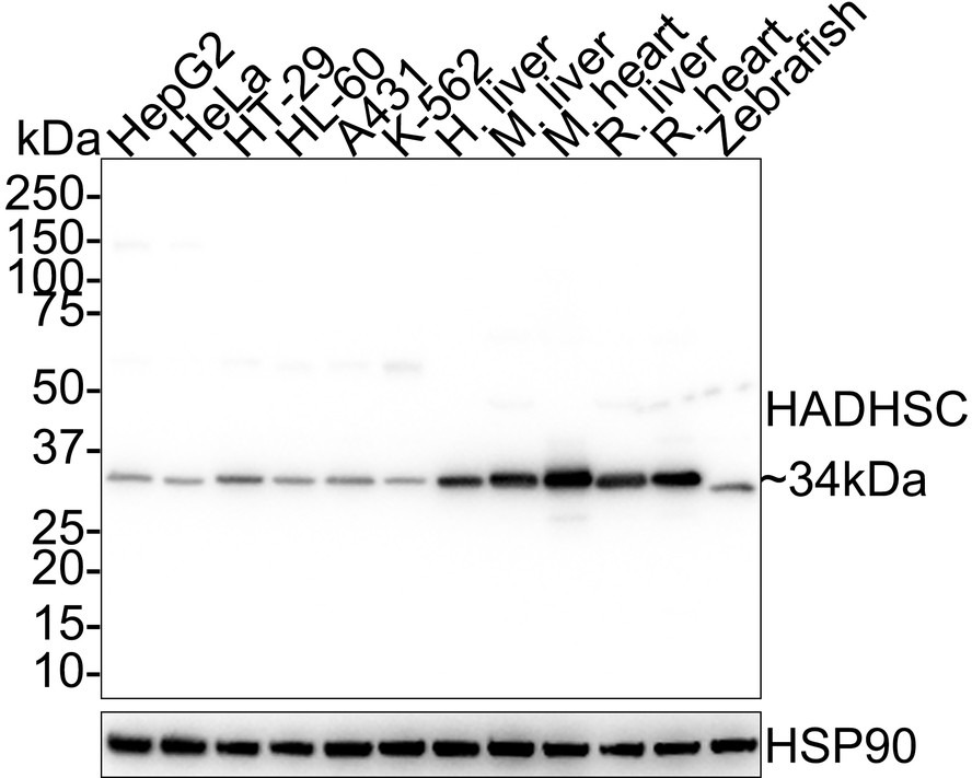 , ,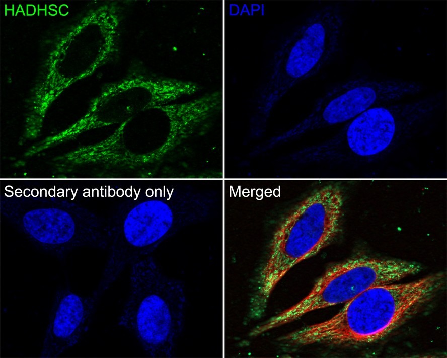 , ,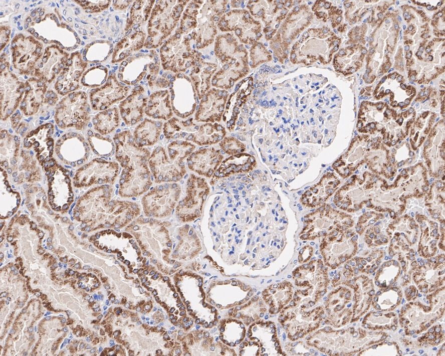 , ,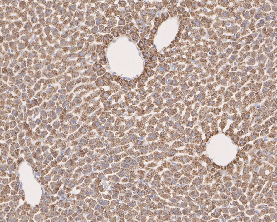 , ,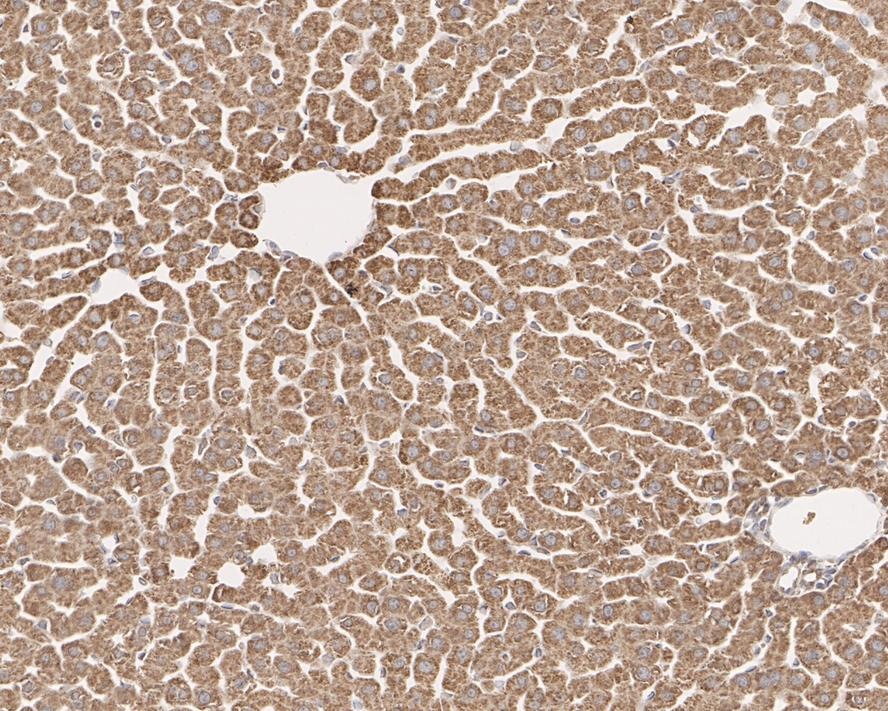 |
| Application | |
| Recommended Dose | WB: 1:1000-2000; ICC: 1:500 |
| Antibody Type | Monoclonal |
| Host Species | Mouse |
| Construction | Hybridoma Monoclonal Antibody |
| Purification | ProA affinity purified |
| Appearance | Liquid |
| Formulation | 1*TBS (pH7.4), 0.5%BSA, 40%Glycerol. Preservative: 0.05% Sodium Azide. |
| Research Background | Hydroxyacyl-Coenzyme A dehydrogenase also known as HADH is an enzyme which in humans is encoded by the HADH gene. This gene is a member of the 3-hydroxyacyl-CoA dehydrogenase gene family. The encoded protein functions in the mitochondrial matrix to catalyze the oxidation of straight-chain 3-hydroxyacyl-CoAs as part of the beta-oxidation pathway. Its enzymatic activity is highest with medium-chain-length fatty acids. Mutations in this gene cause one form of familial hyperinsulinemic hypoglycemia. A deficiency is associated with 3-hydroxyacyl-coenzyme A dehydrogenase deficiency. |
| Conjucates | Unconjugated |
| Immunogen | Peptide |
| Uniprot ID |
| Molecular Weight | Theoretical: 34 kDa. |
| Stability & Storage | Store at -20°C or -80°C for 12 months. Avoid repeated freeze-thaw cycles. |
| Transport | Shipping with blue ice. |
| Size | Quantity | Unit Price | Amount | Operation |
|---|
 Hello! How can I help you today?
 Copyright © 2015-2026 TargetMol Chemicals Inc. All Rights Reserved.
