
Shopping Cart
Remove All Your shopping cart is currently empty

Synonyms: PIG61, NISBD2, mENA, LEGFR, HER1, ERBB1, ERBB, EGFR, EC 2.7.10.1, EC 2.7.10
Anti-EGFR Antibody
(2W575)
| Pack Size | Price | USA Stock | Global Stock | Quantity |
|---|---|---|---|---|
| 50 µL | $298 | 7-10 days | 7-10 days | |
| 100 µL | $498 | 7-10 days | 7-10 days |
| Description | Anti-EGFR Antibody (2W575) is a Rabbit antibody targeting EGFR. Anti-EGFR Antibody (2W575) can be used in FCM,ICC/IF,IHC,IP,WB. |
| Synonyms | PIG61, NISBD2, mENA, LEGFR, HER1, ERBB1, ERBB, EGFR, EC 2.7.10.1, EC 2.7.10 |
| Ig Type | IgG |
| Clone | 2W575 |
| Reactivity | Human,Mouse,Rat |
| Verified Activity | 1. Western blot analysis of EGFR on different lysates using anti-EGFR antibody at 1/1,000 dilution. Positive control: Lane 1: Hela, Lane 2: 293T, Lane 3: Human skin. 2. Immunohistochemical analysis of paraffin-embedded human breast carcinoma tissue using anti-EGFR antibody. Counter stained with hematoxylin. 3. Immunohistochemical analysis of paraffin-embedded mouse brain tissue using anti-EGFR antibody. Counter stained with hematoxylin. 4. Immunohistochemical analysis of paraffin-embedded mouse skin tissue using anti-EGFR antibody. Counter stained with hematoxylin. 5. Immunohistochemical analysis of paraffin-embedded human tonsil tissue using anti-EGFR antibody. Counter stained with hematoxylin. 6. ICC staining EGFR in Hela cells (green). The nuclear counter stain is DAPI (blue). Cells were fixed in paraformaldehyde, permeabilised with 0.25% Triton X100/PBS. 7. ICC staining EGFR in A431 cells (green). The nuclear counter stain is DAPI (blue). Cells were fixed in paraformaldehyde, permeabilised with 0.25% Triton X100/PBS. 8. ICC staining EGFR in SHG-44 cells (green). The nuclear counter stain is DAPI (blue). Cells were fixed in paraformaldehyde, permeabilised with 0.25% Triton X100/PBS. 9. Flow cytometric analysis of A431 cells with EGFR antibody at 1/50 dilution (red) compared with an unlabelled control (cells without incubation with primary antibody; black). Alexa Fluor 488-conjugated goat anti rabbit IgG was used as the secondary antibody. 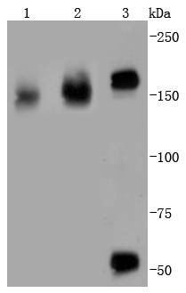 , ,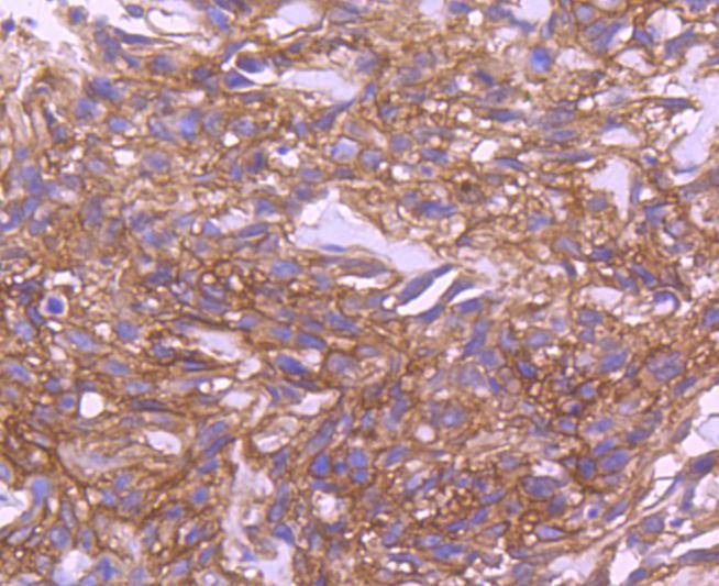 , ,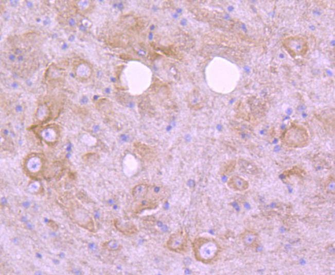 , ,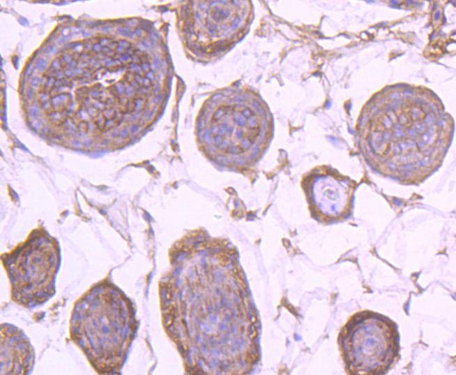 , ,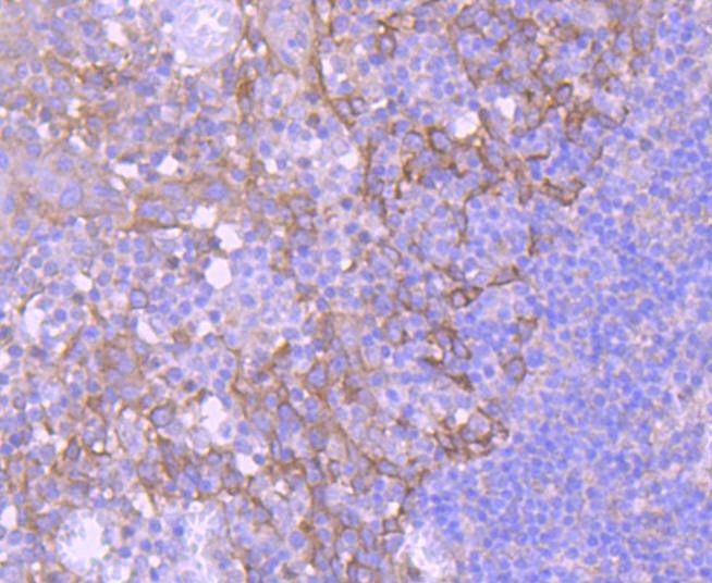 , ,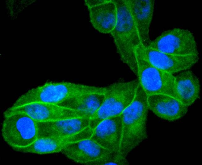 , ,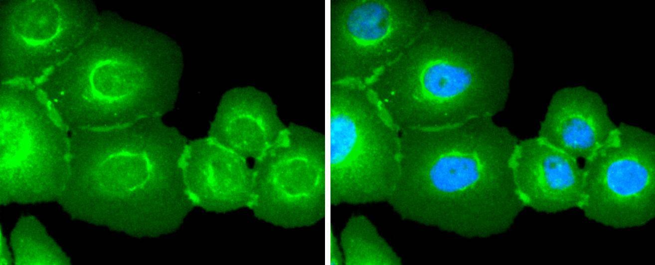 , ,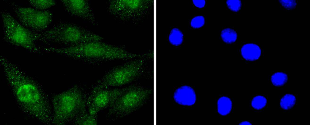 , ,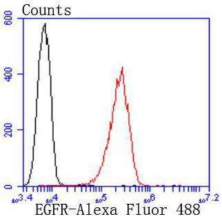 |
| Application | |
| Recommended Dose | WB: 1:1000-2000; IHC: 1:50-200; ICC/IF: 1:100-500; FCM: 1:50-100 |
| Antibody Type | Monoclonal |
| Host Species | Rabbit |
| Construction | Recombinant Antibody |
| Purification | ProA affinity purified |
| Appearance | Liquid |
| Formulation | 1*TBS (pH7.4), 1%BSA, 40%Glycerol. Preservative: 0.05% Sodium Azide. |
| Research Background | The EGF receptor family comprises several related receptor tyrosine kinases that are frequently overexpressed in a variety of carcinomas. Members of this receptor family include EGFR (HER1), Neu (ErbB-2, HER2), ErbB-3 (HER3) and ErbB-4 (HER4), which form either homodimers or heterodimers upon ligand binding. Exons in the EGFR gene product are frequently either deleted or duplicated to produce deletion mutants (DM) or tandem duplication mutants (TDM), respectively, which are detected at various molecular weights. EGFR binds several ligands, including epidermal growth factor (EGF), transforming growth factor α (TGFα), Amphiregulin and heparin binding-EGF (HB-EGF). Ligand binding promotes the internalization of EGFR via Clathrin-coated pits and its subsequent degradation in response to its intrinsic tyrosine kinase. EGFR is involved in organ morphogenesis and maintenance and repair of tissues, but upregulation of EGFR is associated with tumor progression. The oncogenic effects of EGFR include initiation of DNA synthesis, enhanced cell growth, invasion and metastasis. Abrogation of EGFR results in cell cycle arrest, apoptosis or dedifferentiation of cancer cells, suggesting that EGFR may be an effective therapeutic target. |
| Conjucates | Unconjugated |
| Immunogen | Recombinant Protein |
| Uniprot ID |
| Molecular Weight | Theoretical: 150/50 kDa. |
| Stability & Storage | Store at -20°C or -80°C for 12 months. Avoid repeated freeze-thaw cycles. |
| Transport | Shipping with blue ice. |
| Size | Quantity | Unit Price | Amount | Operation |
|---|
 Hello! How can I help you today?
 Copyright © 2015-2026 TargetMol Chemicals Inc. All Rights Reserved.
