
Shopping Cart
Remove All Your shopping cart is currently empty

Synonyms: Melanoma-associated chondroitin sulfate proteoglycan, Melanoma chondroitin sulfate proteoglycan, MCSP, CSPG4, Chondroitin sulfate proteoglycan NG2, Chondroitin sulfate proteoglycan 4
Anti-CSPG4 Polyclonal Antibody
| Pack Size | Price | USA Stock | Global Stock | Quantity |
|---|---|---|---|---|
| 50 µL | $221 | 7-10 days | 7-10 days | |
| 100 µL | $373 | 7-10 days | 7-10 days | |
| 200 µL | $527 | 7-10 days | 7-10 days |
| Description | Anti-CSPG4 Polyclonal Antibody is a Rabbit antibody targeting CSPG4. Anti-CSPG4 Polyclonal Antibody can be used in IF,IHC-Fr,IHC-P,WB. |
| Synonyms | Melanoma-associated chondroitin sulfate proteoglycan, Melanoma chondroitin sulfate proteoglycan, MCSP, CSPG4, Chondroitin sulfate proteoglycan NG2, Chondroitin sulfate proteoglycan 4 |
| Ig Type | IgG |
| Reactivity | Human,Mouse (predicted:Rat,Pig,Horse) |
| Verified Activity | 1. Paraformaldehyde-fixed, paraffin embedded (Human melanoma); Antigen retrieval by boiling in sodium citrate buffer (pH6.0) for 15 min; Block endogenous peroxidase by 3% hydrogen peroxide for 20 min; Blocking buffer (normal goat serum) at 37°C for 30 min; Antibody incubation with (NG2) Polyclonal Antibody, Unconjugated (TMAB-00488) at 1:400 overnight at 4°C, followed by operating according to SP Kit (Rabbit) instructionsand DAB staining. 2. Sample: Hippocampus (Mouse) Lysate at 40 μg Primary: Anti-NG2 (TMAB-00488) at 1/1000 dilution Secondary: IRDye800CW Goat Anti-Rabbit IgG at 1/20000 dilution Predicted band size: 247 kDa Observed band size: 350 kDa 3. Paraformaldehyde-fixed, paraffin embedded (Mouse brain); Antigen retrieval by boiling in sodium citrate buffer (pH6.0) for 15 min; Block endogenous peroxidase by 3% hydrogen peroxide for 20 min; Blocking buffer (normal goat serum) at 37°C for 30 min; Antibody incubation with (NG2) Polyclonal Antibody, Unconjugated (TMAB-00488) at 1:400 overnight at 4°C, followed by operating according to SP Kit (Rabbit) instructionsand DAB staining. 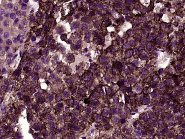 , ,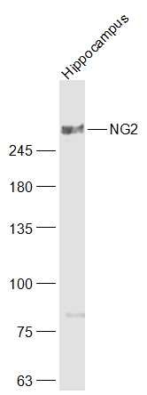 , ,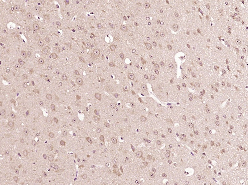 |
| Application | |
| Recommended Dose | WB: 1:500-2000; IHC-P: 1:100-500; IHC-Fr: 1:100-500; IF: 1:100-500 |
| Antibody Type | Polyclonal |
| Host Species | Rabbit |
| Subcellular Localization | Apical cell membrane. Cell projection > lamellipodium membrane. Localized at the apical plasma membrane it relocalizes to the lamellipodia of astrocytoma upon phosphorylation by PRKCA. Localizes to the retraction fibers. Localizes to the plasma membrane of oligodendrocytes. |
| Tissue Specificity | Detected only in malignant melanoma cells. |
| Construction | Polyclonal Antibody |
| Purification | Protein A purified |
| Appearance | Liquid |
| Formulation | 0.01M TBS (pH7.4) with 1% BSA, 0.02% Proclin300 and 50% Glycerol. |
| Concentration | 1 mg/mL |
| Research Background | Proteoglycans (PGs) are a family of proteins composed of different polypeptide chains containing glycosaminoglycan (GAG) modifications. They vary in their cellular locations and have diverse structure and functions. NG2 is a proteoglycan found in the nervous system, comprising a large integral membrane PG with a core protein of 300 kDa and at least one covalently attached chondroitin sulfate (CS) GAG chain. In the CNS this protein is expressed mainly on the surfaces of developing and adult oligodendrocyte precursor cells, but is also associated with developing chondrocytes, cardiomyocytes, pericytes and several human tumours. NG2 also stimulates alpha-4, beta-1 integrin-mediated adhesion and spreading by recruiting and activating a signaling cascade through CDC42, ACK1 and BCAR1. May activate FAK and ERK1/ERK2 signaling cascades. |
| Immunogen | KLH conjugated synthetic peptide: human NG2 |
| Antigen Species | Human |
| Gene Name | CSPG4 |
| Gene ID | |
| Protein Name | Chondroitin sulfate proteoglycan 4 |
| Uniprot ID | |
| Biology Area | Tumor Associated,Neurogenesis,Oligodendrocyte marker,Integrins |
| Function | Proteoglycan playing a role in cell proliferation and migration which stimulates endothelial cells motility during microvascular morphogenesis. May also inhibit neurite outgrowth and growth cone collapse during axon regeneration. Cell surface receptor for collagen alpha 2(VI) which may confer cells ability to migrate on that substrate. Binds through its extracellular N-terminus growth factors, extracellular matrix proteases modulating their activity. May regulate MPP16-dependent degradation and invasion of type I collagen participating in melanoma cells invasion properties. May modulate the plasminogen system by enhancing plasminogen activation and inhibiting angiostatin. Functions also as a signal transducing protein by binding through its cytoplasmic C-terminus scaffolding and signaling proteins. May promote retraction fiber formation and cell polarization through Rho GTPase activation. May stimulate alpha-4, beta-1 integrin-mediated adhesion and spreading by recruiting and activating a signaling cascade through CDC42, ACK1 and BCAR1. May activate FAK and ERK1/ERK2 signaling cascades. |
| Molecular Weight | Theoretical: 247 kDa. Actual: 350 kDa. |
| Stability & Storage | Store at -20°C or -80°C for 12 months. Avoid repeated freeze-thaw cycles. |
| Transport | Shipping with blue ice. |
| Size | Quantity | Unit Price | Amount | Operation |
|---|
 Hello! How can I help you today?
 Copyright © 2015-2026 TargetMol Chemicals Inc. All Rights Reserved.
