
Shopping Cart
Remove All Your shopping cart is currently empty

Synonyms: Bcl-2-like protein 4, Bcl2-L-4, BCL2L4, Apoptosis regulator BAX
Anti-BAX Polyclonal Antibody 2
| Pack Size | Price | USA Stock | Global Stock | Quantity |
|---|---|---|---|---|
| 50 µL | $222 | 7-10 days | 7-10 days | |
| 100 µL | $374 | 7-10 days | 7-10 days | |
| 200 µL | $528 | 7-10 days | 7-10 days |
| Description | Anti-BAX Polyclonal Antibody 2 is a Rabbit antibody targeting BAX. Anti-BAX Polyclonal Antibody 2 can be used in FCM, ICC/IF, IF, IHC-Fr, IHC-P, WB. |
| Synonyms | Bcl-2-like protein 4, Bcl2-L-4, BCL2L4, Apoptosis regulator BAX |
| Ig Type | IgG |
| Reactivity | Human,Mouse,Rat (predicted:Dog,Pig,Cow,Rabbit,Sheep) |
| Verified Activity | 1. Sample: Hela (Human) Cell Lysate at 30 μg Primary: Anti-Bax (TMAB-00188) at 1/1000 dilution Secondary: IRDye800CW Goat Anti-Rabbit IgG at 1/20000 dilution Predicted band size: 21 kDa Observed band size: 23 kDa 2. Paraformaldehyde-fixed, paraffin embedded (Human liver carcinoma); Antigen retrieval by boiling in sodium citrate buffer (pH6.0) for 15 min; Block endogenous peroxidase by 3% hydrogen peroxide for 20 min; Blocking buffer (normal goat serum) at 37°C for 30 min; Antibody incubation with (Bax) Polyclonal Antibody, Unconjugated (TMAB-00188) at 1:400 overnight at 4°C, followed by operating according to SP Kit (Rabbit) instructionsand DAB staining. 3. Paraformaldehyde-fixed, paraffin embedded (Rat brain); Antigen retrieval by boiling in sodium citrate buffer (pH6.0) for 15 min; Block endogenous peroxidase by 3% hydrogen peroxide for 20 min; Blocking buffer (normal goat serum) at 37°C for 30 min; Antibody incubation with (Bax) Polyclonal Antibody, Unconjugated (TMAB-00188) at 1:400 overnight at 4°C, followed by operating according to SP Kit (Rabbit) instructionsand DAB staining. 4. Overlay histogram showing HL 60 cells stained with TMAB-00188 (Green line). The cells were fixed with 90% methanol (5 min) and then permeabilized with 0.01M PBS-Tween for 20 min. The cells were then incubated in 1x PBS / 10% normal goat serum to block non-specific protein-protein interactions followed by the antibody (TMAB-00188,1 μg/1x10^6 cells) for 30 min at 22°C. The secondary antibody used was fluorescein isothiocyanate goat anti-rabbit IgG (H+L) (Brillant blue line) at 1/200 dilution for 30 min at 22°C. Isotype control antibody was rabbit IgG (polyclonal, Orange line) (1 μg/1x10^6 cells) used under the same conditions. Unlabelled sample (blue line) was also used as a control. Acquisition of 20,000 events were collected using a 20mW Argon ion laser (488nm) and 525/30 bandpass filter. 5. Sample: Lane 1: Testis (Mouse) Lysate at 40 μg Lane 2: Kidney (Rat) Lysate at 40 μg Lane 3: Testis (Rat) Lysate at 40 μg Primary: Anti-Bax (TMAB-00188) at 1/1000 dilution Secondary: IRDye800CW Goat Anti-Rabbit IgG at 1/20000 dilution Predicted band size: 21 kDa Observed band size: 21 kDa 6. Tissue/cell: SH-SY5Y cell; 4% Paraformaldehyde-fixed; Triton X-100 at room temperature for 20 min; Blocking buffer (normal goat serum) at 37°C for 20 min; Antibody incubation with (Bax) polyclonal Antibody, Unconjugated (TMAB-00188) 1:100, 90 minutes at 37°C; followed by a FITC conjugated Goat Anti-Rabbit IgG antibody at 37°C for 90 minutes, DAPI (blue) was used to stain the cell nucleus. 7. Tissue/cell: SH-SY5Y cell; 4% Paraformaldehyde-fixed; Triton X-100 at room temperature for 20 min; Blocking buffer (normal goat serum) at 37°C for 20 min; Antibody incubation with (Bax) polyclonal Antibody, Unconjugated (TMAB-00188) 1:100, 90 minutes at 37°C; followed by a FITC conjugated Goat Anti-Rabbit IgG antibody at 37°C for 90 minutes, DAPI (blue) was used to stain the cell nucleus. 8. Paraformaldehyde-fixed, paraffin embedded (Rat spinal cord); Antigen retrieval by boiling in sodium citrate buffer (pH6.0) for 15 min; Block endogenous peroxidase by 3% hydrogen peroxide for 20 min; Blocking buffer (normal goat serum) at 37°C for 30 min; Antibody incubation with (Bax) Polyclonal Antibody, Unconjugated (TMAB-00188) at 1:400 overnight at 4°C, followed by operating according to SP Kit (Rabbit) instructionsand DAB staining. 9. Sample: Cerebral cortex (Rat) Lysate at 40 μg Primary: Anti-Bax (TMAB-00188) at 1/1000 dilution Secondary: IRDye800CW Goat Anti-Rabbit IgG at 1/20000 dilution Predicted band size: 21 kDa Observed band size: 21 kDa 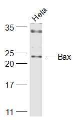 , ,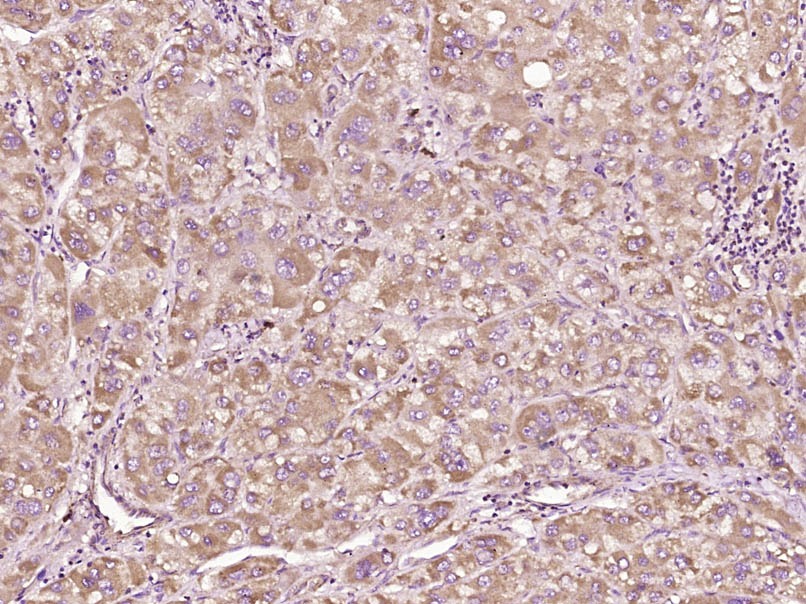 , ,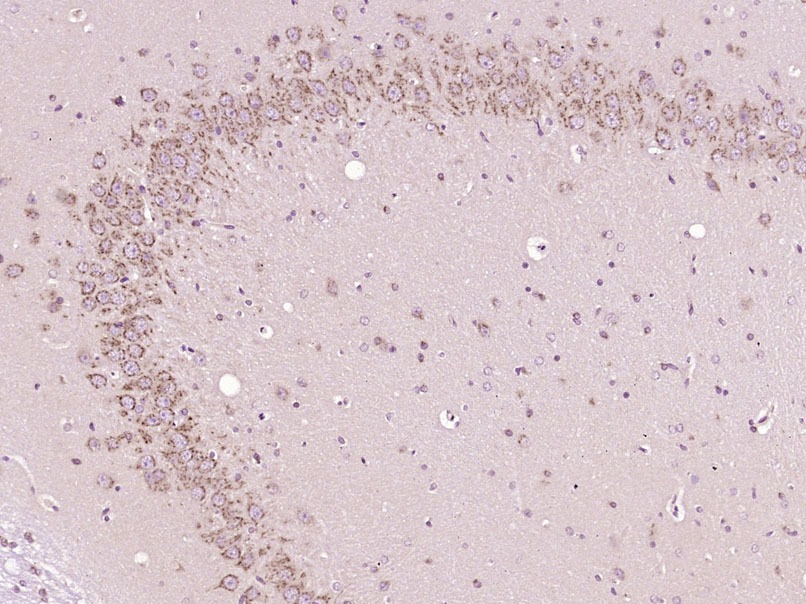 , ,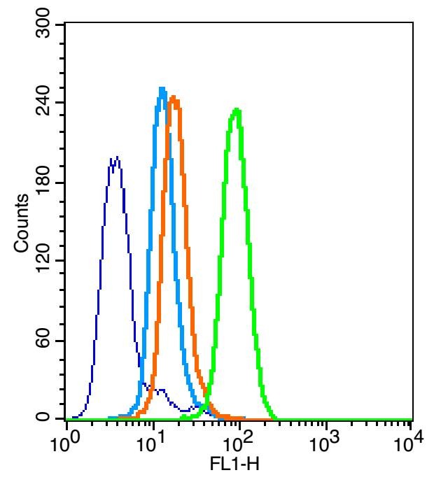 , ,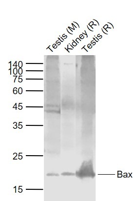 , ,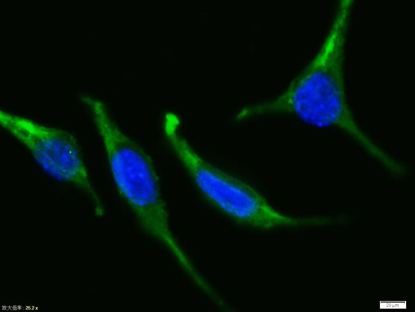 , ,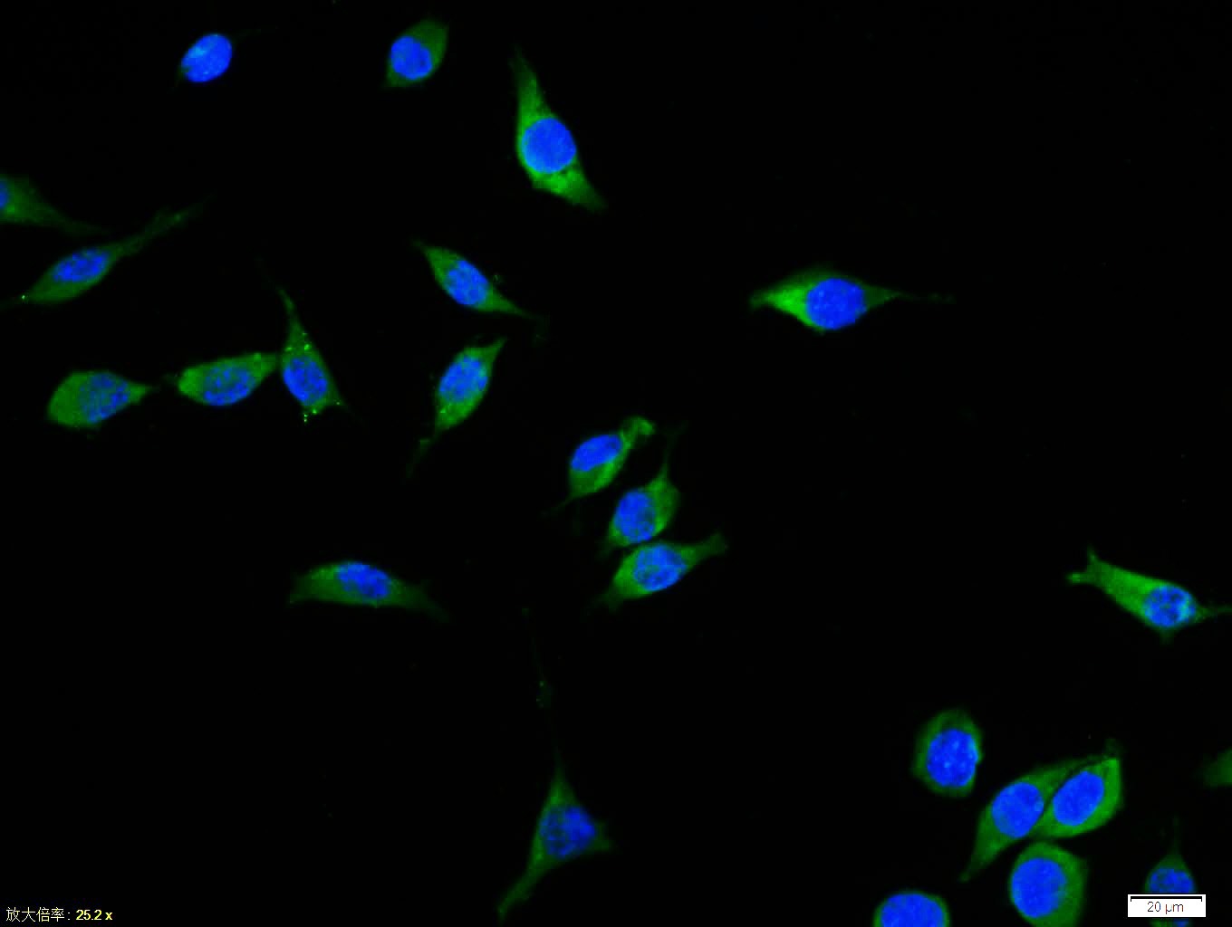 , ,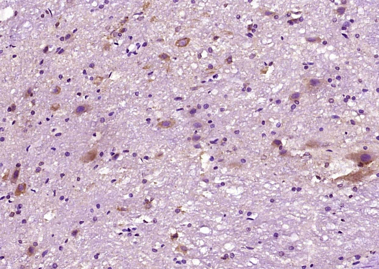 , ,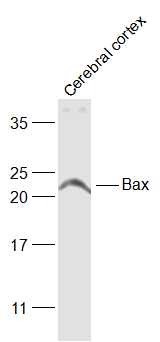 |
| Application | |
| Recommended Dose | FCM=1 μg/Test; ICC/IF=1:50-200; IF=1:100-500; IHC-Fr=1:100-500; IHC-P=1:100-500; WB=1:500-2000 |
| Antibody Type | Polyclonal |
| Host Species | Rabbit |
| Subcellular Localization | Isoform Alpha: Mitochondrion membrane; Single-pass membrane protein. Cytoplasm. Note=Colocalizes with 14-3-3 proteins in the cytoplasm. Under stress conditions, undergoes a conformation change that causes release from JNK-phosphorylated 14-3-3 proteins and translocation to the mitochondrion membrane. Isoform Beta: Cytoplasm. Isoform Gamma: Cytoplasm. Isoform Delta: Cytoplasm (Potential). |
| Tissue Specificity | Expressed in a wide variety of tissues. Isoform Psi is found in glial tumors. Isoform Alpha is expressed in spleen, breast, ovary, testis, colon and brain, and at low levels in skin and lung. Isoform Sigma is expressed in spleen, breast, ovary, testis, lu |
| Construction | Polyclonal Antibody |
| Purification | Protein A purified |
| Appearance | Liquid |
| Formulation | 0.01M TBS (pH7.4) with 1% BSA, 0.02% Proclin300 and 50% Glycerol. |
| Concentration | 1 mg/mL |
| Research Background | The protein encoded by this gene belongs to the BCL2 protein family. BCL2 family members form hetero- or homodimers and act as anti- or pro-apoptotic regulators that are involved in a wide variety of cellular activities. This protein forms a heterodimer with BCL2, and functions as an apoptotic activator. This protein is reported to interact with, and increase the opening of, the mitochondrial voltage-dependent anion channel (VDAC), which leads to the loss in membrane potential and the release of cytochrome c. The expression of this gene is regulated by the tumor suppressor P53 and has been shown to be involved in P53-mediated apoptosis. Multiple alternatively spliced transcript variants, which encode different isoforms, have been reported for this gene. [provided by RefSeq, Jul 2008]. |
| Immunogen | KLH conjugated synthetic peptide: human Bax |
| Antigen Species | Human |
| Gene Name | BAX |
| Gene ID | |
| Protein Name | Apoptosis regulator BAX |
| Uniprot ID | |
| Biology Area | Bcl 2 family,Metabolism,Bcl 2 family,Associated Proteins,Bcl2 Family,Apoptosis marker and proteins ELISA kits,Apoptosis |
| Function | Accelerates programmed cell death by binding to, and antagonizing the apoptosis repressor BCL2 or its adenovirus homolog E1B 19k protein. Under stress conditions, undergoes a conformation change that causes translocation to the mitochondrion membrane, leading to the release of cytochrome c that then triggers apoptosis. Promotes activation of CASP3, and thereby apoptosis. |
| Molecular Weight | Theoretical: 21 kDa. Actual: 20-23 kDa. |
| Stability & Storage | Store at -20°C or -80°C for 12 months. Avoid repeated freeze-thaw cycles. |
| Transport | Shipping with blue ice. |
| Size | Quantity | Unit Price | Amount | Operation |
|---|
 Hello! How can I help you today?
 Copyright © 2015-2026 TargetMol Chemicals Inc. All Rights Reserved.
