
Shopping Cart
Remove All Your shopping cart is currently empty

Synonyms: STK6, STK15, Serine/threonine-protein kinase Ayk1, Serine/threonine-protein kinase aurora-A, Serine/threonine-protein kinase 6, Serine/threonine-protein kinase 15, Ipl1- and aurora-related kinase 1, IAK1, BTAK, Breast tumor-amplified kinase, AYK1, Aurora/IPL1-related kinase 1 (ARK-1;Aurora-related kinase 1), Aurora kinase A, Aurora 2, AURKA, AURA, ARK1, AIRK1, AIK
Anti-AURKA Polyclonal Antibody
| Pack Size | Price | USA Stock | Global Stock | Quantity |
|---|---|---|---|---|
| 50 µL | $220 | 7-10 days | 7-10 days | |
| 100 µL | $374 | 7-10 days | 7-10 days | |
| 200 µL | $529 | 7-10 days | 7-10 days |
| Description | Anti-AURKA Polyclonal Antibody is a Rabbit antibody targeting AURKA. Anti-AURKA Polyclonal Antibody can be used in WB. |
| Synonyms | STK6, STK15, Serine/threonine-protein kinase Ayk1, Serine/threonine-protein kinase aurora-A, Serine/threonine-protein kinase 6, Serine/threonine-protein kinase 15, Ipl1- and aurora-related kinase 1, IAK1, BTAK, Breast tumor-amplified kinase, AYK1, Aurora/IPL1-related kinase 1 (ARK-1;Aurora-related kinase 1), Aurora kinase A, Aurora 2, AURKA, AURA, ARK1, AIRK1, AIK |
| Ig Type | IgG |
| Reactivity | Mouse |
| Verified Activity | Sample: Lane 1: Large intestine (Mouse) Lysate at 40 μg Lane 2: Lung (Mouse) Lysate at 40 μg Lane 3: Kidney (Mouse) Lysate at 40 μg Lane 4: NIH/3T3 (Mouse) Cell Lysate at 30 μg Primary: Anti-Aurora A (TMAB-00173) at 1/1000 dilution Secondary: IRDye800CW Goat Anti-Rabbit IgG at 1/20000 dilution Predicted band size: 48 kDa Observed band size: 48 kDa 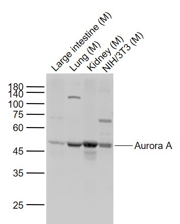 |
| Application | |
| Recommended Dose | WB: 1:500-2000 |
| Antibody Type | Polyclonal |
| Host Species | Rabbit |
| Subcellular Localization | Cytoplasm, cytoskeleton, centrosome. Cytoplasm, cytoskeleton, spindle pole. Note=Detected at the neurite hillock in developing neurons (By similarity). Localizes at the centrosome in mitotic cells from early prophase until telophase, but also localizes to the spindle pole MTs from prophase to anaphase. Moves to the midbody during both telophase and cytokinesis. Associates with both the pericentriolar material (PCM) and centrioles. |
| Tissue Specificity | Highly expressed in testis and weakly in skeletal muscle, thymus and spleen. Also highly expressed in colon, ovarian, prostate, neuroblastoma, breast and cervical cancer cell lines. |
| Construction | Polyclonal Antibody |
| Purification | Protein A purified |
| Appearance | Liquid |
| Formulation | 0.01M TBS (pH7.4) with 1% BSA, 0.02% Proclin300 and 50% Glycerol. |
| Concentration | 1 mg/mL |
| Research Background | Aurora A plays a role in cell cycle regulation during anaphase and/or telophase, in relation to the function of the centrosome/spindle pole region during chromosome segregation. Aurora A plays a key role during tumor development and progression and is overexpressed in many human cancers including breast, ovarian and colorectal. Aurora A is viewed as a potential target for anticancer drug treatment. Aurora B is a mitotic protein kinase that phosphorylates histone H3 (probably on Serine 10), behaves as a chromosomal passenger protein, and may regulate several stages of mitosis such as centrosome separation, chromosome segregation and cytokinesis. It localizes to the inner centromere region from prophase to anaphase. The Aurora kinases, members of the Ser/Thr protein kinase family, associate with microtubules during chromosome movement and segregation. Aurora kinase C may play a part in organizing microtubules in relation to the function of the centrosome/spindle pole during mitosis. This protein is localized to centrosome from anaphase to cytokinesis. Expression is limited to testis in normal cells. Elevated expression levels are seen only in a subset of cancer cells such as HepG2, HuH7 and HeLa cells. Aurora-C expression is maximum at M phase. |
| Immunogen | KLH conjugated synthetic peptide: mouse Auroa A |
| Antigen Species | Mouse |
| Gene Name | AURKA |
| Gene ID | |
| Protein Name | Aurora kinase A |
| Uniprot ID | |
| Biology Area | Cell division,Tumor biomarkers,Spindle,Aurora |
| Function | Contributes to the regulation of cell cycle progression. Required for normal mitosis. Associates with the centrosome and the spindle microtubules during mitosis and functions in centrosome maturation, spindle assembly, maintenance of spindle bipolarity, centrosome separation and mitotic checkpoint control. Phosphorylates numerous target proteins, including ARHGEF2, BRCA1, KIF2A, NDEL1, PARD3, PLK1 and BORA. Regulates KIF2A tubulin depolymerase activity (By similarity). Required for normal axon formation. Plays a role in microtubule remodeling during neurite extension. Important for microtubule formation and/or stabilization. |
| Molecular Weight | Theoretical: 48 kDa. Actual: 48 kDa. |
| Stability & Storage | Store at -20°C or -80°C for 12 months. Avoid repeated freeze-thaw cycles. |
| Transport | Shipping with blue ice. |
| Size | Quantity | Unit Price | Amount | Operation |
|---|
 Hello! How can I help you today?
 Copyright © 2015-2026 TargetMol Chemicals Inc. All Rights Reserved.
