
Shopping Cart
Remove All Your shopping cart is currently empty

Synonyms: Unc-51-like kinase 1, Unc-51 like kinase 1(C. elegans), UNC51, Unc 51(C. elegans) like kinase 1, Unc 51 like kinase 1, UNC 51, ULK1, ULK 1, Serine/threonine protein kinase Unc51.1, Serine/threonine protein kinase ULK1, hATG1, Autophagy-related protein 1 homolog, ATG1A, ATG1, ATG 1
Anti-ATG1 Polyclonal Antibody
| Pack Size | Price | USA Stock | Global Stock | Quantity |
|---|---|---|---|---|
| 50 µL | $221 | 7-10 days | 7-10 days | |
| 100 µL | $373 | 7-10 days | 7-10 days | |
| 200 µL | $529 | 7-10 days | 7-10 days |
| Description | Anti-ATG1 Polyclonal Antibody is a Rabbit antibody targeting ATG1. Anti-ATG1 Polyclonal Antibody can be used in FCM, ICC/IF, IF, IHC-Fr, IHC-P, WB. |
| Synonyms | Unc-51-like kinase 1, Unc-51 like kinase 1(C. elegans), UNC51, Unc 51(C. elegans) like kinase 1, Unc 51 like kinase 1, UNC 51, ULK1, ULK 1, Serine/threonine protein kinase Unc51.1, Serine/threonine protein kinase ULK1, hATG1, Autophagy-related protein 1 homolog, ATG1A, ATG1, ATG 1 |
| Ig Type | IgG |
| Reactivity | Human,Mouse,Rat (predicted:Chicken,Pig,Cow,Horse,Rabbit) |
| Verified Activity | 1. Positive control: RSC96 Isotype Control Antibody: Rabbit IgG; Secondary Antibody: Goat anti-rabbit IgG-FITC; Dilution: 1:200 in 1 X PBS containing 0.5% BSA Primary Antibody catalog number: TMAB-00162; Dilution: 0.5 μg in 100 μL 1X PBS containing 0.5% BSA 2. Tissue/cell: mouse pancreas tissue; 4% Paraformaldehyde-fixed and paraffin-embedded; Antigen retrieval: citrate buffer (0.01M, pH6.0), Boiling bathing for 15 min; Block endogenous peroxidase by 3% Hydrogen peroxide for 30 min; Blocking buffer (normal goat serum) at 37°C for 20 min; Incubation: Anti-ATG1/ULK1 Polyclonal Antibody, Unconjugated (TMAB-00162) 1:200, overnight at 4°C, followed by conjugation to the secondary antibody and DAb staining. 3. Sample: 293t (Human) Cell Lysate at 30 μg Primary: Anti-ATG1/ULK1 (TMAB-00162) at 1/500 dilution Secondary: IRDye800CW Goat Anti-Rabbit IgG at 1/20000 dilution Predicted band size: 116 kDa Observed band size: 116 kDa 4. SH-SY5Y cell; 4% Paraformaldehyde-fixed; Triton X-100 at room temperature for 20 min; Blocking buffer (normal goat serum) at 37°C for 20 min; Antibody incubation with (ATG1/ULK1) polyclonal Antibody, Unconjugated (TMAB-00162) 1:100, 90 minutes at 37°C; followed by a conjugated Goat Anti-Rabbit IgG antibody at 37°C for 90 minutes, DAPI (blue) was used to stain the cell nucleus. 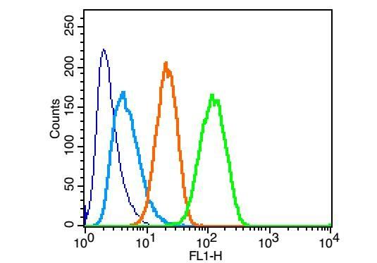 , ,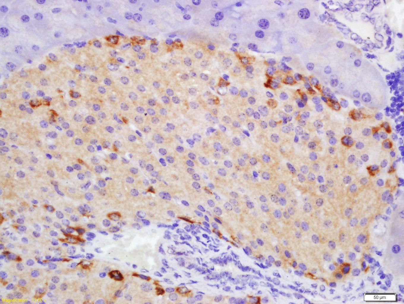 , ,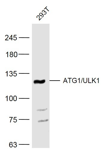 , ,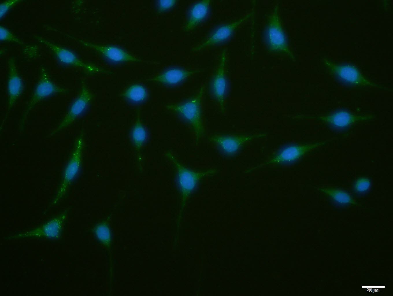 |
| Application | |
| Recommended Dose | FCM=0.5 μg/Test; ICC/IF=1:100-500; IF=1:100-500; IHC-Fr=1:100-500; IHC-P=1:100-500; WB=1:500-2000 |
| Antibody Type | Polyclonal |
| Host Species | Rabbit |
| Subcellular Localization | Cytoplasm, cytosol. Preautophagosomal structure. Note=Under starvation conditions, is localized to puncate structures primarily representing the isolation membrane that sequesters a portion of the cytoplasm resulting in the formation of an autophagosome. |
| Tissue Specificity | Ubiquitously expressed. Detected in the following adult tissues: skeletal muscle, heart, pancreas, brain, placenta, liver, kidney, and lung. |
| Construction | Polyclonal Antibody |
| Purification | Protein A purified |
| Appearance | Liquid |
| Formulation | 0.01M TBS (pH7.4) with 1% BSA, 0.02% Proclin300 and 50% Glycerol. |
| Concentration | 1 mg/mL |
| Research Background | ULK1 belongs to the serine/threonine protein kinase family. It is involved in axon growth and plays an essential role in neurite branching during sensory axon outgrowth. Knockdown of ULK1 results in impaired endocytosis of nerve growth factor (NGF), excessive axon arborization, and severely stunted axon elongation indicating that ULK1 mediates a non clathrin coated endocytosis in sensory growth cones. Knockdown of ULK1 also inhibits the autophagic response. It appears to act as a convergence point for multiple signals that regulate autophagy, and in turn interacts with a large number of autophagy related (Atg) proteins. |
| Immunogen | KLH conjugated synthetic peptide: human ATG1/ULK1 |
| Antigen Species | Human |
| Gene Name | ULK1 |
| Gene ID | |
| Protein Name | Serine/threonine-protein kinase ULK1 |
| Uniprot ID | |
| Biology Area | Other Kinases,Neurogenesis,Mitophagy fission and fusion,Autophagy |
| Function | Serine/threonine-protein kinase involved in autophagy in response to starvation. Acts upstream of phosphatidylinositol 3-kinase PIK3C3 to regulate the formation of autophagophores, the precursors of autophagosomes. Part of regulatory feedback loops in autophagy: acts both as a downstream effector and negative regulator of mammalian target of rapamycin complex 1 (mTORC1) via interaction with RPTOR. Activated via phosphorylation by AMPK and also acts as a regulator of AMPK by mediating phosphorylation of AMPK subunits PRKAA1, PRKAB2 and PRKAG1, leading to negatively regulate AMPK activity. May phosphorylate ATG13/KIAA0652 and RPTOR; however such data need additional evidences. Plays a role early in neuronal differentiation and is required for granule cell axon formation. |
| Molecular Weight | Theoretical: 116 kDa. Actual: 116 kDa. |
| Stability & Storage | Store at -20°C or -80°C for 12 months. Avoid repeated freeze-thaw cycles. |
| Transport | Shipping with blue ice. |
| Size | Quantity | Unit Price | Amount | Operation |
|---|
 Hello! How can I help you today?
 Copyright © 2015-2026 TargetMol Chemicals Inc. All Rights Reserved.
