
Shopping Cart
Remove All Your shopping cart is currently empty

Synonyms: TXREB, TAXREB67, Tax Responsive Enhancer Element B67, DNA binding protein TAXREB67, Cyclic AMP response element binding protein 2, Cyclic AMP dependent transcription factor ATF 4, CREB2, CREB 2, ATF4, ATF 4, Activating Transcription Factor 4
Anti-ATF-4 Polyclonal Antibody
| Pack Size | Price | USA Stock | Global Stock | Quantity |
|---|---|---|---|---|
| 50 µL | $222 | 7-10 days | 7-10 days | |
| 100 µL | $374 | 7-10 days | 7-10 days | |
| 200 µL | $529 | 7-10 days | 7-10 days |
| Description | Anti-ATF-4 Polyclonal Antibody is a Rabbit antibody targeting ATF-4. Anti-ATF-4 Polyclonal Antibody can be used in FCM, ICC/IF, IF, IHC-Fr, IHC-P, WB. |
| Synonyms | TXREB, TAXREB67, Tax Responsive Enhancer Element B67, DNA binding protein TAXREB67, Cyclic AMP response element binding protein 2, Cyclic AMP dependent transcription factor ATF 4, CREB2, CREB 2, ATF4, ATF 4, Activating Transcription Factor 4 |
| Ig Type | IgG |
| Reactivity | Human,Mouse,Rat (predicted:Dog,Pig,Cow,Horse,Rabbit,Sheep) |
| Verified Activity | 1. U2OS cell; 4% Paraformaldehyde-fixed; Triton X-100 at room temperature for 20 min; Blocking buffer (normal goat serum) at 37°C for 20 min; Antibody incubation with (ATF4) polyclonal Antibody, Unconjugated (TMAB-00159) 1:100, 90 minutes at 37°C; followed by a conjugated Goat Anti-Rabbit IgG antibody at 37°C for 90 minutes, DAPI (blue) was used to stain the cell nucleus. 2. Sample: Cerebrum (Rat) Lysate at 40 μg Cerebrum (Mouse) Lysate at 40 μg Primary: Anti-ATF4 (TMAB-00159) at 1/1000 dilution Secondary: IRDye800CW Goat Anti-Rabbit IgG at 1/20000 dilution Predicted band size: 37/50 kDa Observed band size: 50 kDa 3. Blank control (blue line): Hela Primary Antibody (green line): Rabbit Anti-Bid antibody (TMAB-00159) Dilution: 0.2 μg/10^6 cells; Isotype Control Antibody (orange line): Rabbit IgG. Secondary Antibody (white blue line): Goat anti-rabbit IgG-FITC Dilution: 1 μg/test. Protocol The cells were fixed with 70% ethanol (Overnight at 4°C) and then permeabilized with 90% ice-cold methanol for 30 min on ice. Cells stained with Primary Antibody for 30 min at room temperature. The cells were then incubated in 1 X PBS/2% BSA/10% goat serum to block non-specific protein-protein interactions followed by the antibody for 15 min at room temperature. The secondary antibody used for 40 min at room temperature. 4. Blank control: Mouse spleen. Primary Antibody (green line): Rabbit Anti-ATF4 Alpha antibody (TMAB-00159) Dilution: 2 μg/10^6 cells; Isotype Control Antibody (orange line): Rabbit IgG. Secondary Antibody: Goat anti-rabbit IgG-FITC Dilution: 1 μg/test. Protocol The cells were fixed with 4% PFA (10 min at room temperature) and then permeabilized with 90% ice-cold methanol for 20 min at-20°C. The cells were then incubated in 5% BSA to block non-specific protein-protein interactions for 30 min at room temperature. Cells stained with Primary Antibody for 30 min at room temperature. The secondary antibody used for 40 min at room temperature. 5. Hela cell; 4% Paraformaldehyde-fixed; Triton X-100 at room temperature for 20 min; Blocking buffer (normal goat serum) at 37°C for 20 min; Antibody incubation with (ATF4) polyclonal Antibody, Unconjugated (TMAB-00159) 1:100, 90 minutes at 37°C; followed by a conjugated Goat Anti-Rabbit IgG antibody at 37°C for 90 minutes, DAPI (blue) was used to stain the cell nucleus. 6. Paraformaldehyde-fixed, paraffin embedded (Human brain); Antigen retrieval by boiling in sodium citrate buffer (pH6.0) for 15 min; Block endogenous peroxidase by 3% hydrogen peroxide for 20 min; Blocking buffer (normal goat serum) at 37°C for 30 min; Antibody incubation with (ATF4) Polyclonal Antibody, Unconjugated (TMAB-00159) at 1:200 overnight at 4°C, followed by operating according to SP Kit (Rabbit) instructionsand DAB staining. 7. Paraformaldehyde-fixed, paraffin embedded (human breast carcinoma); Antigen retrieval by boiling in sodium citrate buffer (pH6.0) for 15 min; Block endogenous peroxidase by 3% hydrogen peroxide for 20 min; Blocking buffer (normal goat serum) at 37°C for 30 min; Antibody incubation with (ATF4) Polyclonal Antibody, Unconjugated (TMAB-00159) at 1:200 overnight at 4°C, followed by operating according to SP Kit (Rabbit) instructionsand DAB staining. 8. Paraformaldehyde-fixed, paraffin embedded (mouse skin); Antigen retrieval by boiling in sodium citrate buffer (pH6.0) for 15 min; Block endogenous peroxidase by 3% hydrogen peroxide for 20 min; Blocking buffer (normal goat serum) at 37°C for 30 min; Antibody incubation with (ATF4) Polyclonal Antibody, Unconjugated (TMAB-00159) at 1:100 overnight at 4°C, followed by operating according to SP Kit (Rabbit) instructionsand DAB staining. 9. Paraformaldehyde-fixed, paraffin embedded (mouse intestine); Antigen retrieval by boiling in sodium citrate buffer (pH6.0) for 15 min; Block endogenous peroxidase by 3% hydrogen peroxide for 20 min; Blocking buffer (normal goat serum) at 37°C for 30 min; Antibody incubation with (ATF4) Polyclonal Antibody, Unconjugated (TMAB-00159) at 1:100 overnight at 4°C, followed by operating according to SP Kit (Rabbit) instructionsand DAB staining. 10. Paraformaldehyde-fixed, paraffin embedded (rat skin); Antigen retrieval by boiling in sodium citrate buffer (pH6.0) for 15 min; Block endogenous peroxidase by 3% hydrogen peroxide for 20 min; Blocking buffer (normal goat serum) at 37°C for 30 min; Antibody incubation with (ATF4) Polyclonal Antibody, Unconjugated (TMAB-00159) at 1:100 overnight at 4°C, followed by operating according to SP Kit (Rabbit) instructionsand DAB staining. 11. Paraformaldehyde-fixed, paraffin embedded (rat intestine); Antigen retrieval by boiling in sodium citrate buffer (pH6.0) for 15 min; Block endogenous peroxidase by 3% hydrogen peroxide for 20 min; Blocking buffer (normal goat serum) at 37°C for 30 min; Antibody incubation with (ATF4) Polyclonal Antibody, Unconjugated (TMAB-00159) at 1:100 overnight at 4°C, followed by operating according to SP Kit (Rabbit) instructionsand DAB staining. 12. Paraformaldehyde-fixed, paraffin embedded (human lung carcinoma); Antigen retrieval by boiling in sodium citrate buffer (pH6.0) for 15 min; Block endogenous peroxidase by 3% hydrogen peroxide for 20 min; Blocking buffer (normal goat serum) at 37°C for 30 min; Antibody incubation with (ATF4) Polyclonal Antibody, Unconjugated (TMAB-00159) at 1:100 overnight at 4°C, followed by operating according to SP Kit (Rabbit) instructionsand DAB staining. 13. Paraformaldehyde-fixed, paraffin embedded (human cervical carcinoma); Antigen retrieval by boiling in sodium citrate buffer (pH6.0) for 15 min; Block endogenous peroxidase by 3% hydrogen peroxide for 20 min; Blocking buffer (normal goat serum) at 37°C for 30 min; Antibody incubation with (ATF4) Polyclonal Antibody, Unconjugated (TMAB-00159) at 1:100 overnight at 4°C, followed by operating according to SP Kit (Rabbit) instructionsand DAB staining. 14. Paraformaldehyde-fixed, paraffin embedded (human Abdominal skin); Antigen retrieval by boiling in sodium citrate buffer (pH6.0) for 15 min; Block endogenous peroxidase by 3% hydrogen peroxide for 20 min; Blocking buffer (normal goat serum) at 37°C for 30 min; Antibody incubation with (ATF4) Polyclonal Antibody, Unconjugated (TMAB-00159) at 1:100 overnight at 4°C, followed by operating according to SP Kit (Rabbit) instructionsand DAB staining. 15. Sample: Lane 1: Mouse Cerebrum tissue lysates Lane 2: Rat Cerebrum tissue lysates Lane 3: Human HeLa cell lysates Lane 4: Human Jurkat cell lysates Lane 5: Human HL-60 cell lysates Primary: Anti-ATF4 (TMAB-00159) at 1/1000 dilution Secondary: IRDye800CW Goat Anti-Rabbit IgG at 1/20000 dilution Predicted band size: 38 kDa Observed band size: 47 kDa 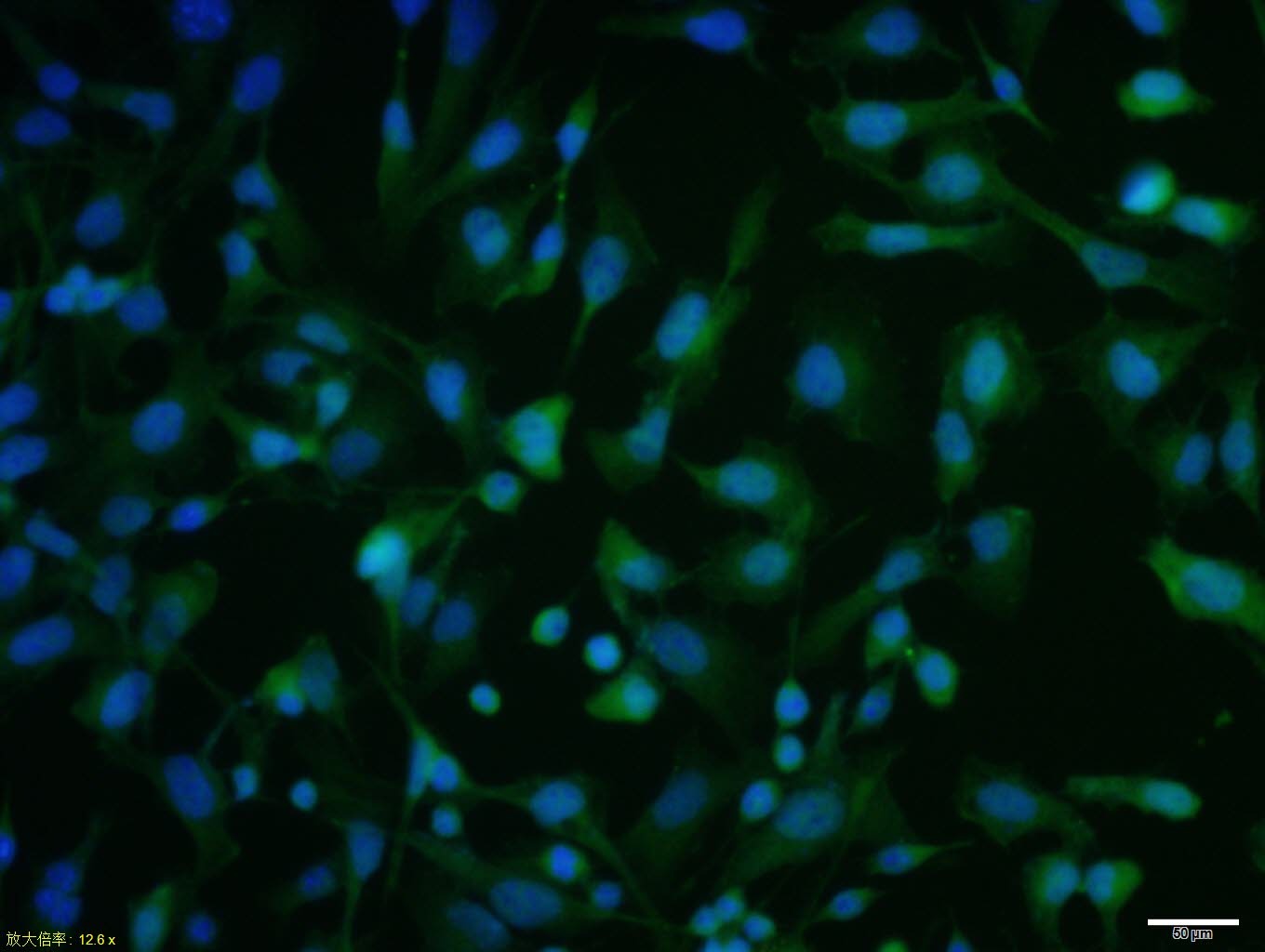 , ,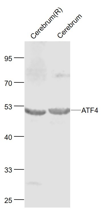 , ,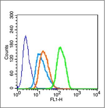 , ,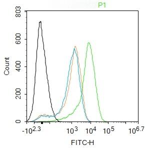 , ,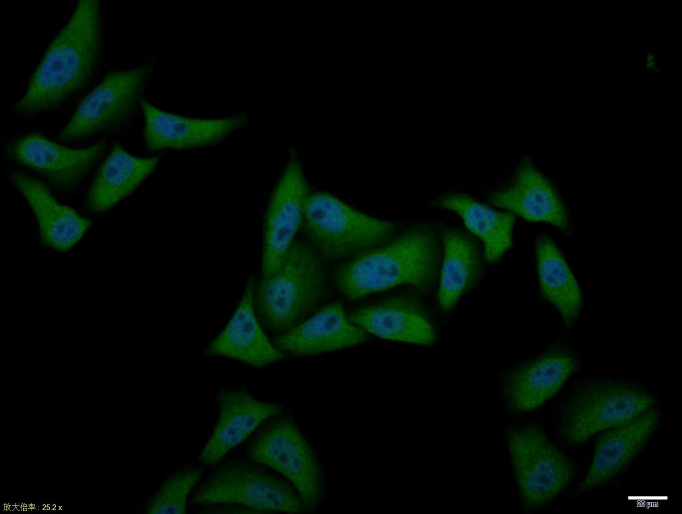 , ,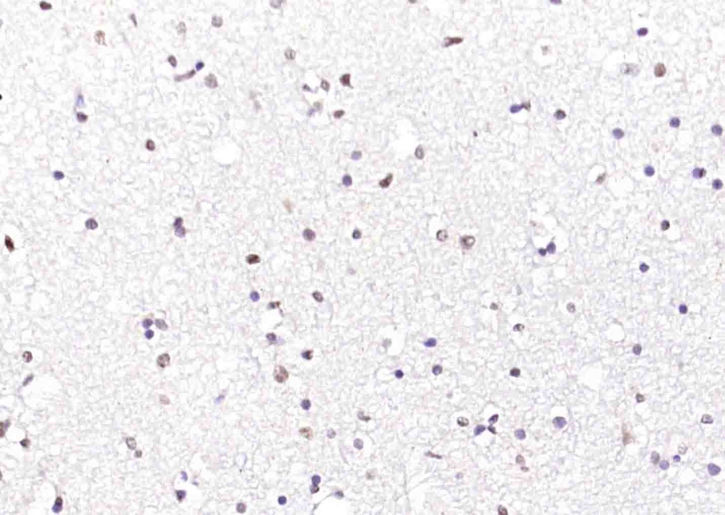 , ,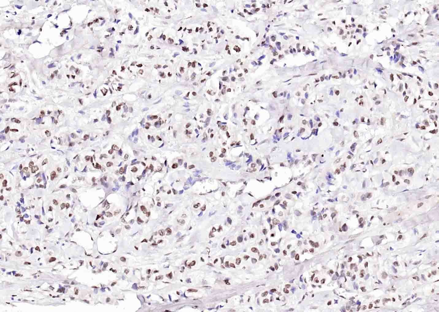 , ,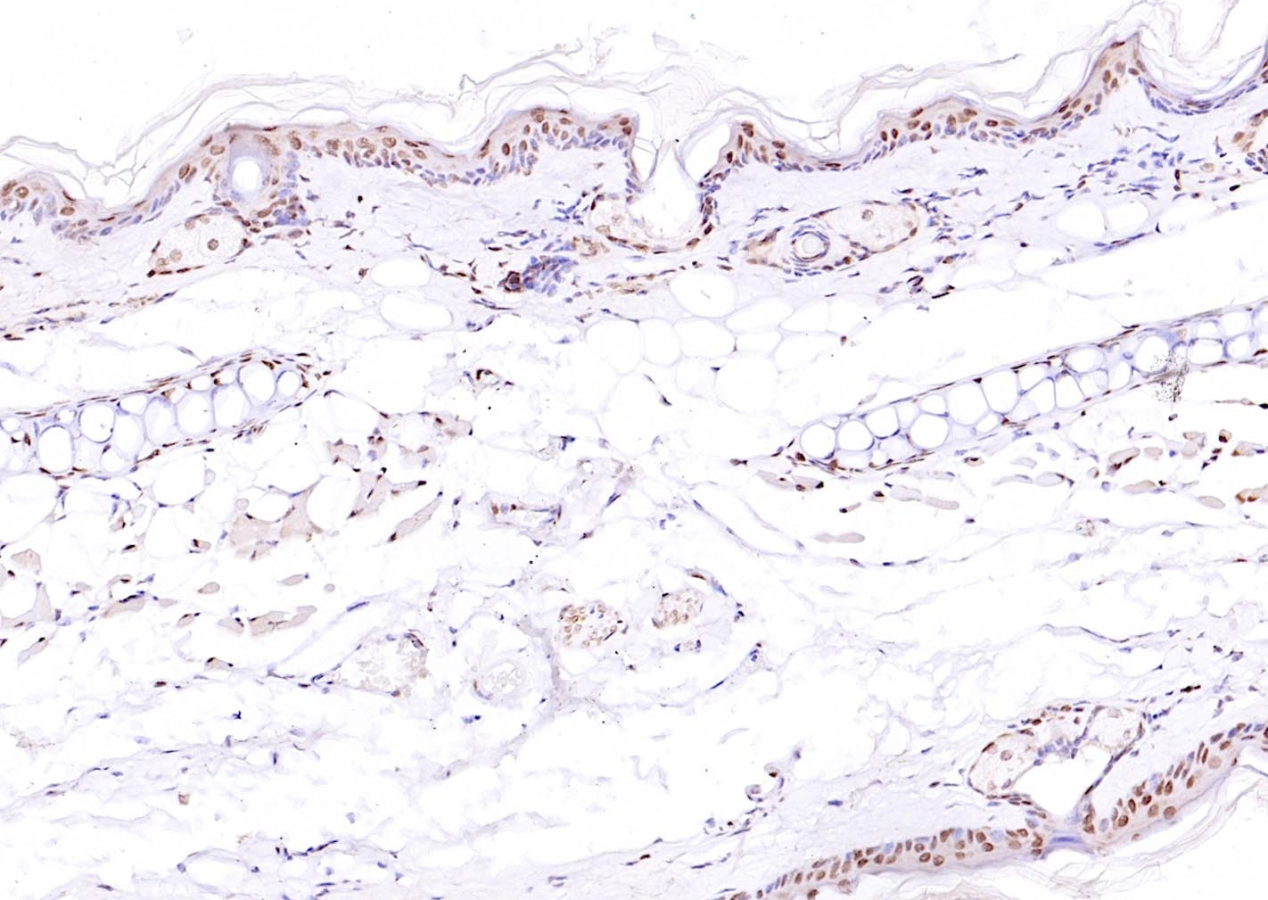 , ,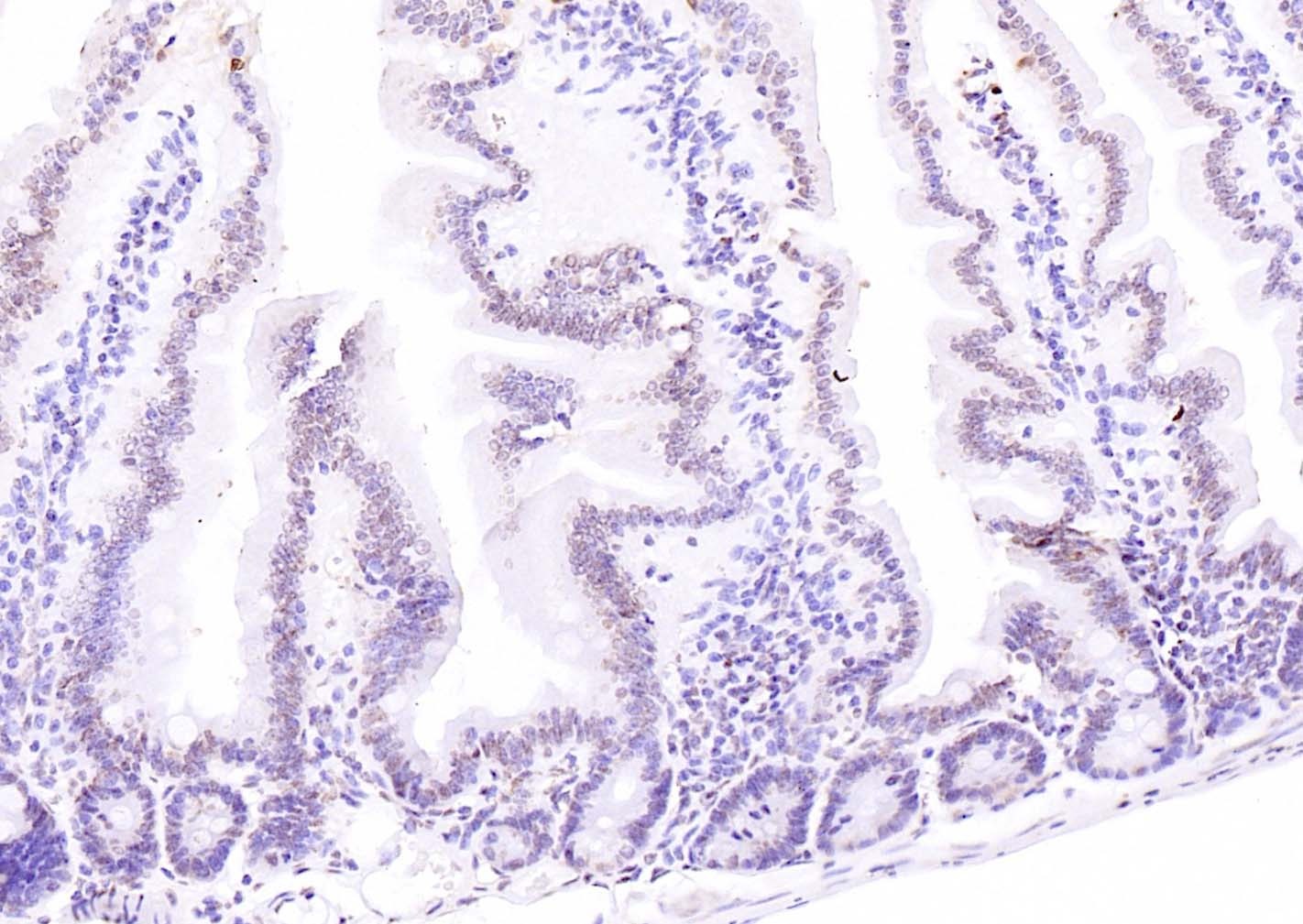 , ,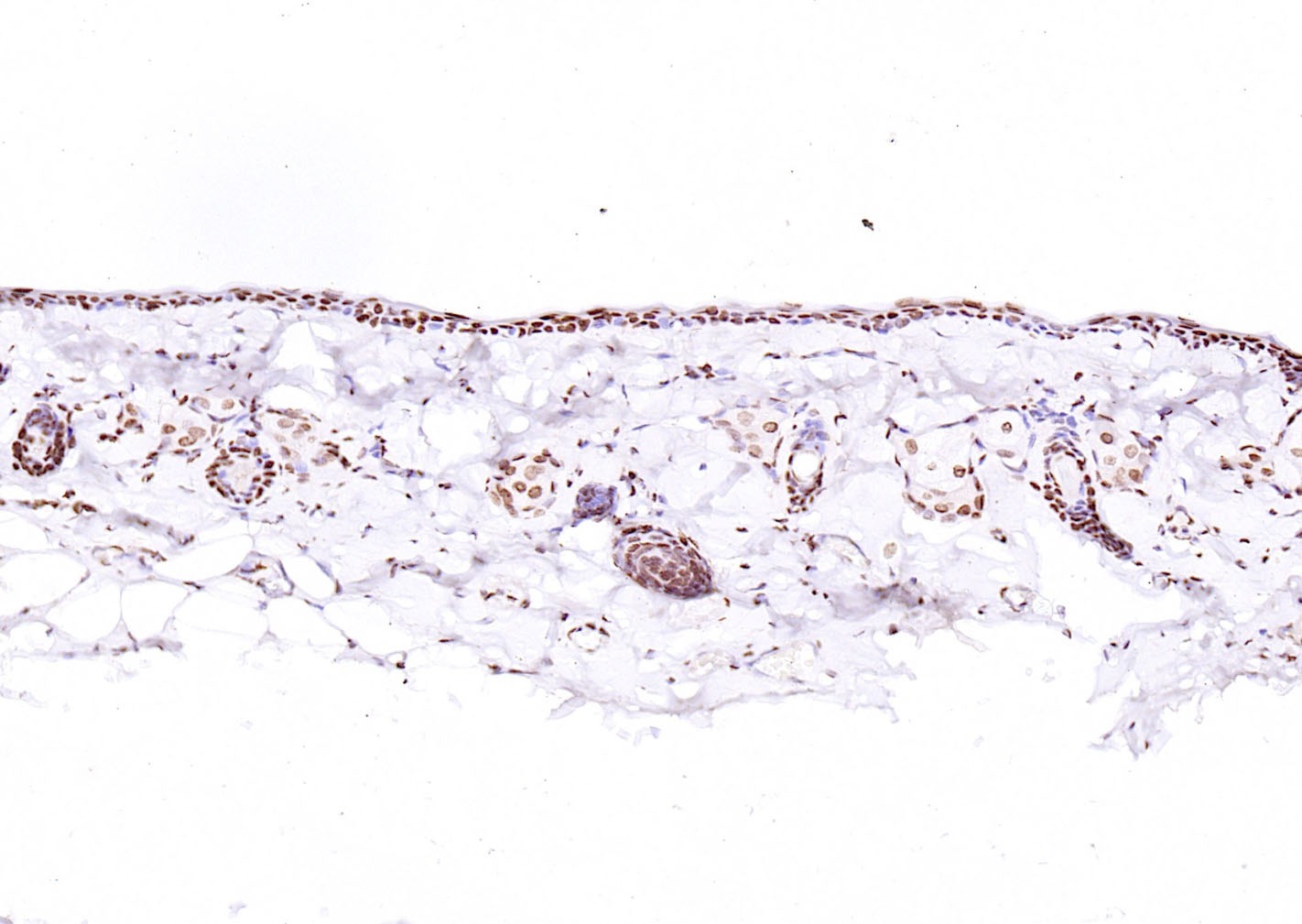 , ,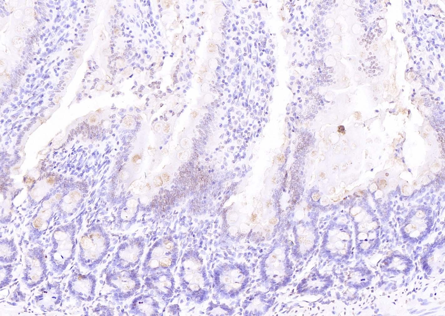 , ,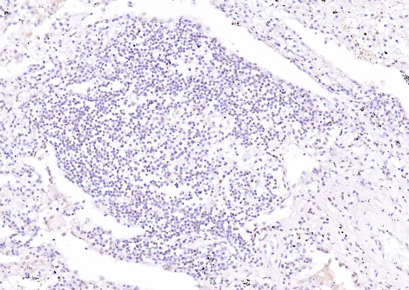 , ,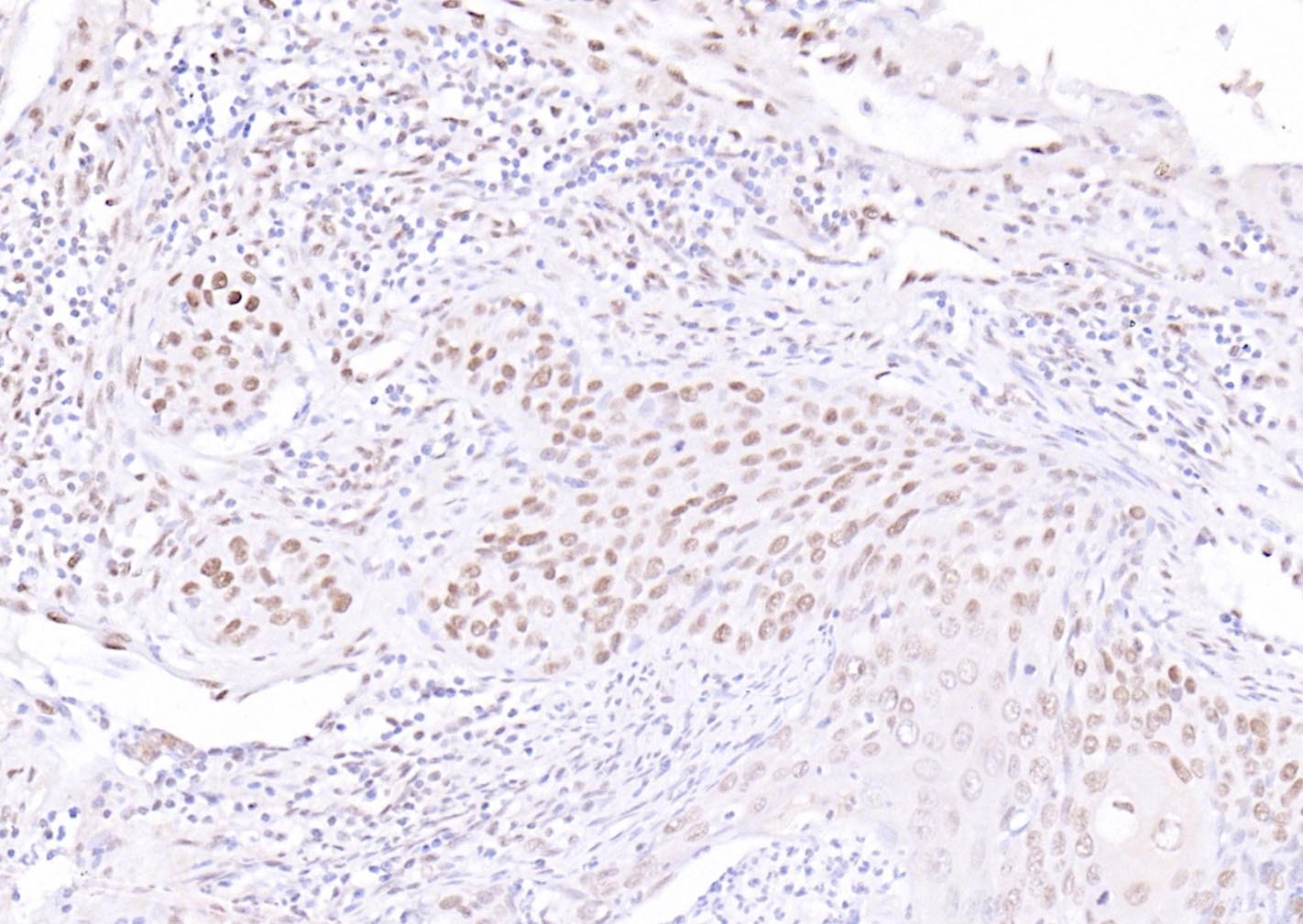 , ,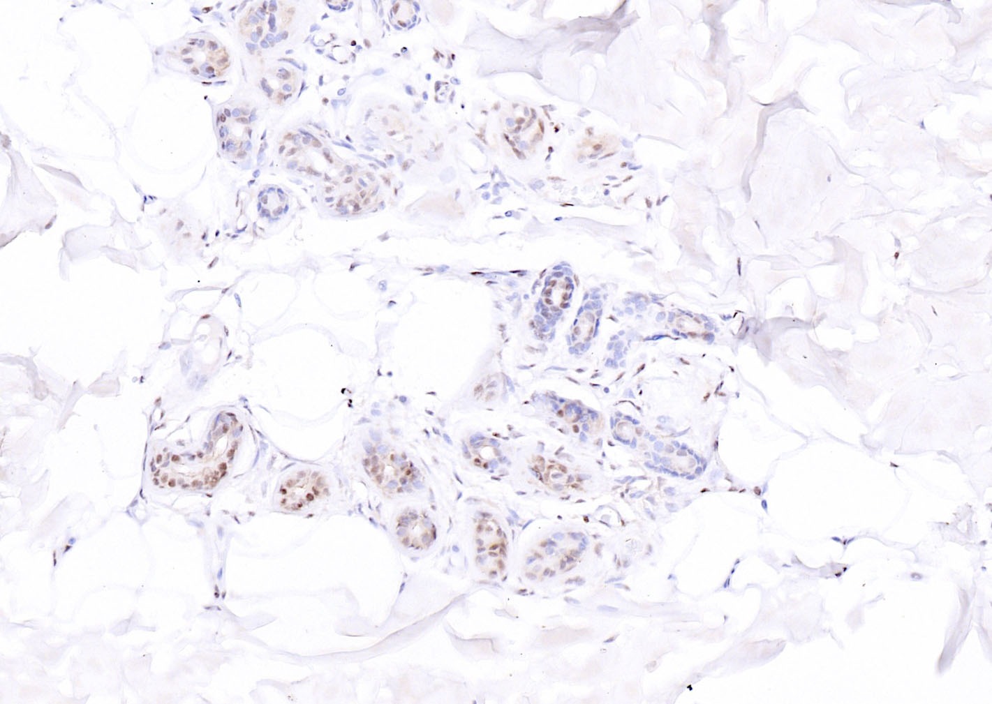 , ,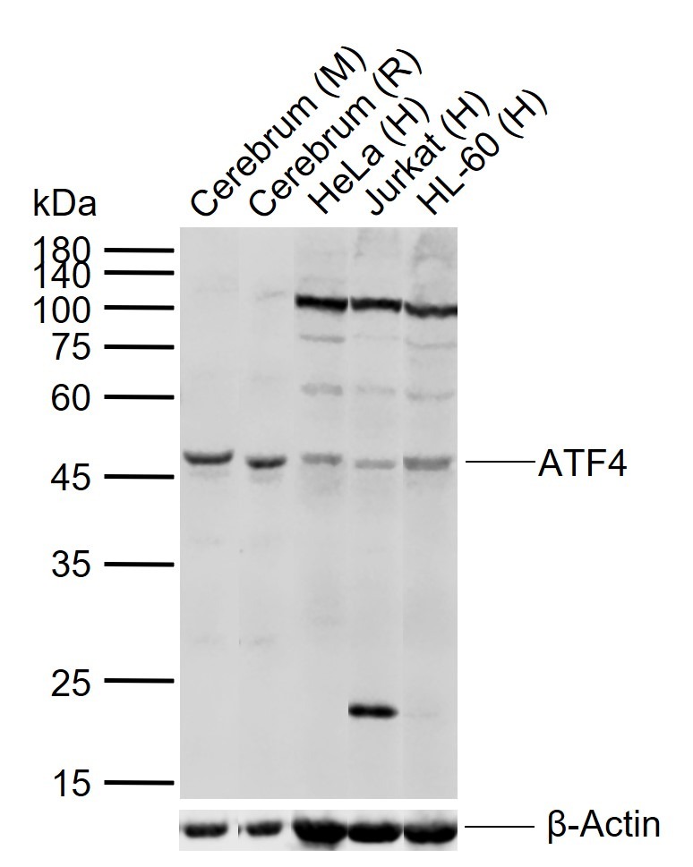 |
| Application | |
| Recommended Dose | FCM=0.2 μg/Test; ICC/IF=1:100-500; IF=1:100-500; IHC-Fr=1:100-500; IHC-P=1:100-500; WB=1:500-2000 |
| Antibody Type | Polyclonal |
| Host Species | Rabbit |
| Subcellular Localization | Cytoplasm. Cell membrane. Nucleus. Colocalizes with GABBR1 in hippocampal neuron dendritic membranes. |
| Construction | Polyclonal Antibody |
| Purification | Protein A purified |
| Appearance | Liquid |
| Formulation | 0.01M TBS (pH7.4) with 1% BSA, 0.02% Proclin300 and 50% Glycerol. |
| Concentration | 1 mg/mL |
| Research Background | ATF4 is a transcription factor that was originally identified as a widely expressed mammalian DNA binding protein that could bind a tax-responsive enhancer element in the LTR of HTLV1. The encoded protein was also isolated and characterized as the cAMP-response element binding protein 2 (CREB2). The protein encoded by this gene belongs to a family of DNA-binding proteins that includes the AP1 family of transcription factors, cAMP-response element binding proteins (CREBs) and CREB-like proteins. These transcription factors share a leucine zipper region that is involved in protein-protein interactions, located C-terminal to a stretch of basic amino acids that functions as a DNA binding domain (referenced from Entrez gene). |
| Immunogen | KLH conjugated synthetic peptide: human ATF4 |
| Antigen Species | Human |
| Gene Name | ATF4 |
| Gene ID | |
| Protein Name | Cyclic AMP-dependent transcription factor ATF-4 |
| Uniprot ID | |
| Function | Transcriptional activator. Binds the cAMP response element (CRE) (consensus: 5'-GTGACGT[AC][AG]-3'), a sequence present in many viral and cellular promoters. It binds to a Tax-responsive enhancer element in the long terminal repeat of HTLV-I. |
| Molecular Weight | Theoretical: 38 kDa. Actual: 47 kDa. |
| Stability & Storage | Store at -20°C or -80°C for 12 months. Avoid repeated freeze-thaw cycles. |
| Transport | Shipping with blue ice. |
| Size | Quantity | Unit Price | Amount | Operation |
|---|
 Hello! How can I help you today?
 Copyright © 2015-2026 TargetMol Chemicals Inc. All Rights Reserved.
