
Shopping Cart
Remove All Your shopping cart is currently empty

Synonyms: HASH1, Class A basic helix-loop-helix protein 46 (bHLHa46), bHLHa46, ASH-1, ASH1, ASCL1, Achaete-scute homolog 1
Anti-ASCL1 Polyclonal Antibody
| Pack Size | Price | USA Stock | Global Stock | Quantity |
|---|---|---|---|---|
| 50 µL | $220 | 7-10 days | 7-10 days | |
| 100 µL | $372 | 7-10 days | 7-10 days | |
| 200 µL | $529 | 7-10 days | 7-10 days |
| Description | Anti-ASCL1 Polyclonal Antibody is a Rabbit antibody targeting ASCL1. Anti-ASCL1 Polyclonal Antibody can be used in FCM, ICC/IF, IF, IHC-Fr, IHC-P, WB. |
| Synonyms | HASH1, Class A basic helix-loop-helix protein 46 (bHLHa46), bHLHa46, ASH-1, ASH1, ASCL1, Achaete-scute homolog 1 |
| Ig Type | IgG |
| Reactivity | Human,Mouse,Rat (predicted:Cow,Sheep) |
| Verified Activity | 1. Tissue/cell: human glioma tissue; 4% Paraformaldehyde-fixed and paraffin-embedded; Antigen retrieval: citrate buffer (0.01M, pH6.0), Boiling bathing for 15 min; Block endogenous peroxidase by 3% Hydrogen peroxide for 30 min; Blocking buffer (normal goat serum) at 37°C for 20 min; Incubation: Anti-MASH1 Polyclonal Antibody, Unconjugated (TMAB-00150) 1:200, overnight at 4°C, followed by conjugation to the secondary antibody and DAb staining. 2. Tissue/cell: rat brain tissue; 4% Paraformaldehyde-fixed and paraffin-embedded; Antigen retrieval: citrate buffer (0.01M, pH6.0), Boiling bathing for 15 min; Block endogenous peroxidase by 3% Hydrogen peroxide for 30 min; Blocking buffer (normal goat serum) at 37°C for 20 min; Incubation: Anti-MASH1 Polyclonal Antibody, Unconjugated (TMAB-00150) 1:200, overnight at 4°C, followed by conjugation to the secondary antibody and DAb staining. 3. Tissue/cell: mouse embryos tissue; 4% Paraformaldehyde-fixed and paraffin-embedded; Antigen retrieval: citrate buffer (0.01M, pH6.0), Boiling bathing for 15 min; Block endogenous peroxidase by 3% Hydrogen peroxide for 30 min; Blocking buffer (normal goat serum) at 37°C for 20 min; Incubation: Anti-MASH1 Polyclonal Antibody, Unconjugated (TMAB-00150) 1:200, overnight at 4°C, followed by conjugation to the secondary antibody and DAb staining. 4. Tissue/cell: Rat brain tissue; 4% Paraformaldehyde-fixed and paraffin-embedded; Antigen retrieval: citrate buffer (0.01M, pH6.0), Boiling bathing for 15 min; Block endogenous peroxidase by 3% Hydrogen peroxide for 30 min; Blocking buffer (normal goat serum) at 37°C for 20 min; Incubation: Anti-MASH1 Polyclonal Antibody, Unconjugated (TMAB-00150) 1:200, overnight at 4°C, followed by conjugation to the secondary antibody and DAb staining. 5. Blank control: Mouse brain. Primary Antibody (green line): Rabbit Anti-MASH1 antibody (TMAB-00150) Dilution: 2 μg/10^6 cells; Isotype Control Antibody (orange line): Rabbit IgG. Secondary Antibody: Goat anti-rabbit IgG-AF647 Dilution: 1 μg/test. Protocol The cells were fixed with 4% PFA (10 min at room temperature) and then permeabilized with 90% ice-cold methanol for 20 min at-20°C. The cells were then incubated in 5% BSA to block non-specific protein-protein interactions for 30 min at room temperature. Cells stained with Primary Antibody for 30 min at room temperature. The secondary antibody used for 40 min at room temperature. 6. Blank control: A549. Primary Antibody (green line): Rabbit Anti-MASH1 antibody (TMAB-00150) Dilution: 2 μg/Test; Secondary Antibody: Goat anti-rabbit IgG-FITC Dilution: 0.5 μg/Test. Protocol The cells were fixed with 4% PFA (10 min at room temperature) and then permeabilized with 90% ice-cold methanol for 20 min at-20°C. The cells were then incubated in 5% BSA to block non-specific protein-protein interactions for 30 min at room temperature. Cells stained with Primary Antibody for 30 min at room temperature. The secondary antibody used for 40 min at room temperature. 7. A549 cell; 4% Paraformaldehyde-fixed; Triton X-100 at room temperature for 20 min; Blocking buffer (normal goat serum) at 37°C for 20 min; Antibody incubation with (MASH1) polyclonal Antibody, Unconjugated (TMAB-00150) 1:100, 90 minutes at 37°C; followed by a conjugated Goat Anti-Rabbit IgG antibody at 37°C for 90 minutes, DAPI (blue) was used to stain the cell nucleus. 8. Sample: Brain (Mouse) Lysate at 40 μg Primary: Anti-ASCL1 (TMAB-00150) at 1/300 dilution Secondary: IRDye800CW Goat Anti-Rabbit IgG at 1/20000 dilution Predicted band size: 26 kDa Observed band size: 26 kDa 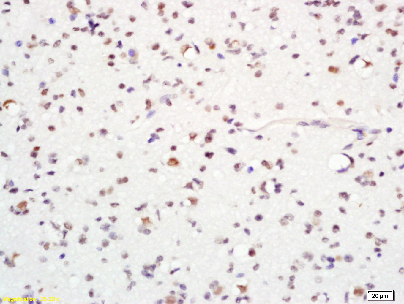 , ,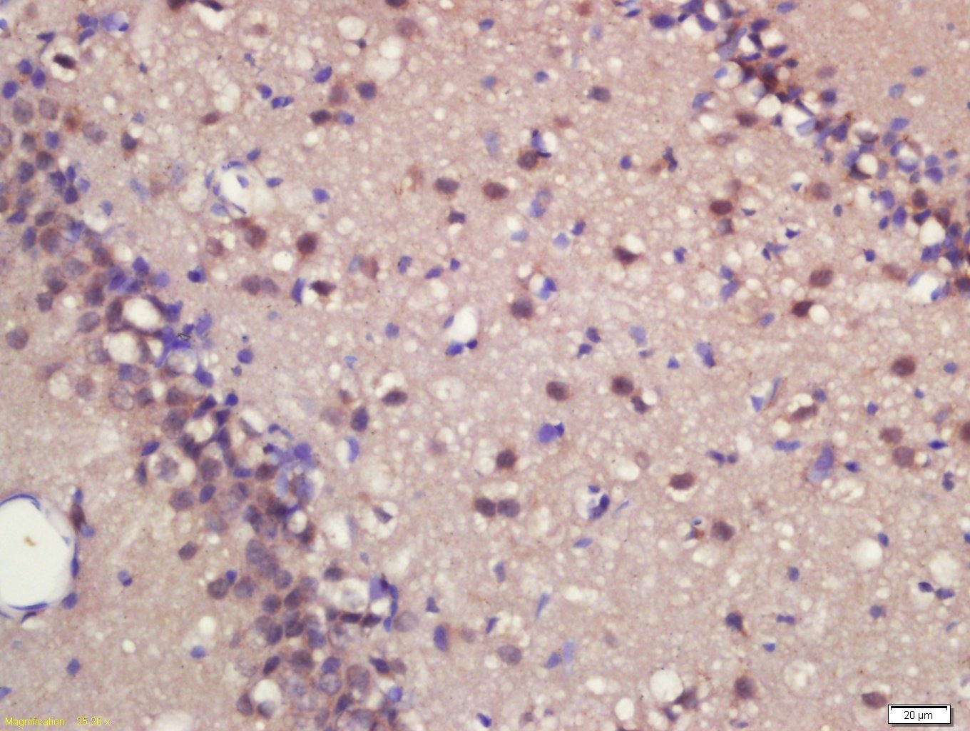 , ,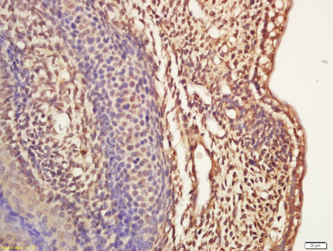 , ,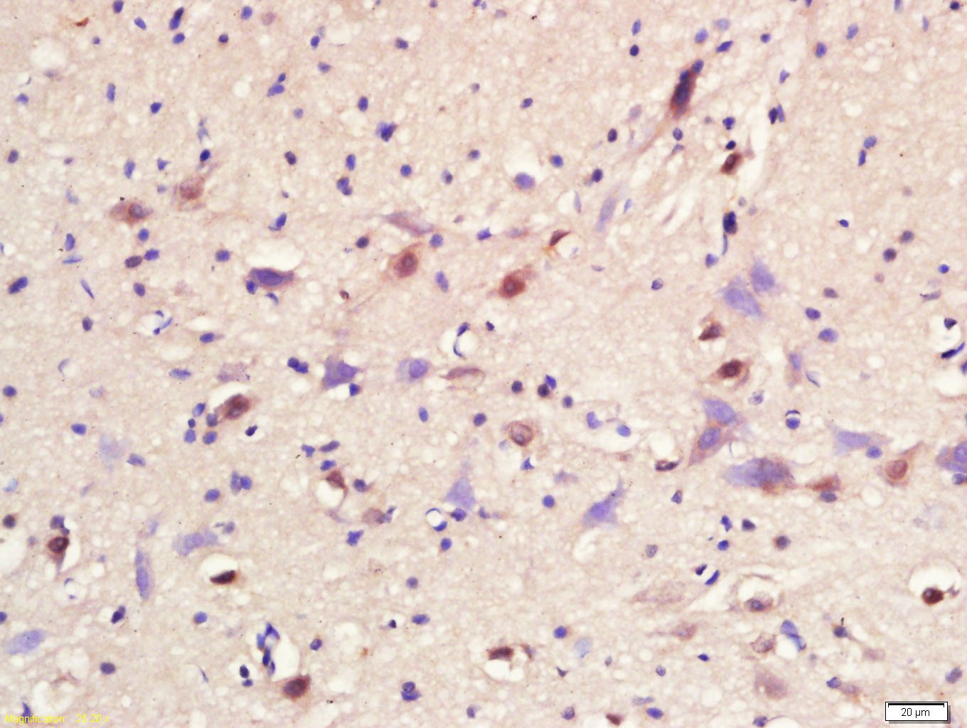 , ,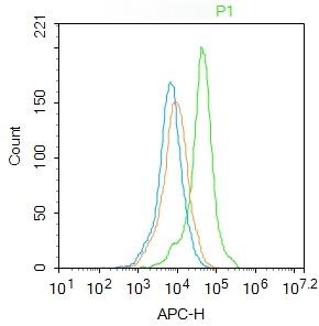 , ,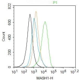 , ,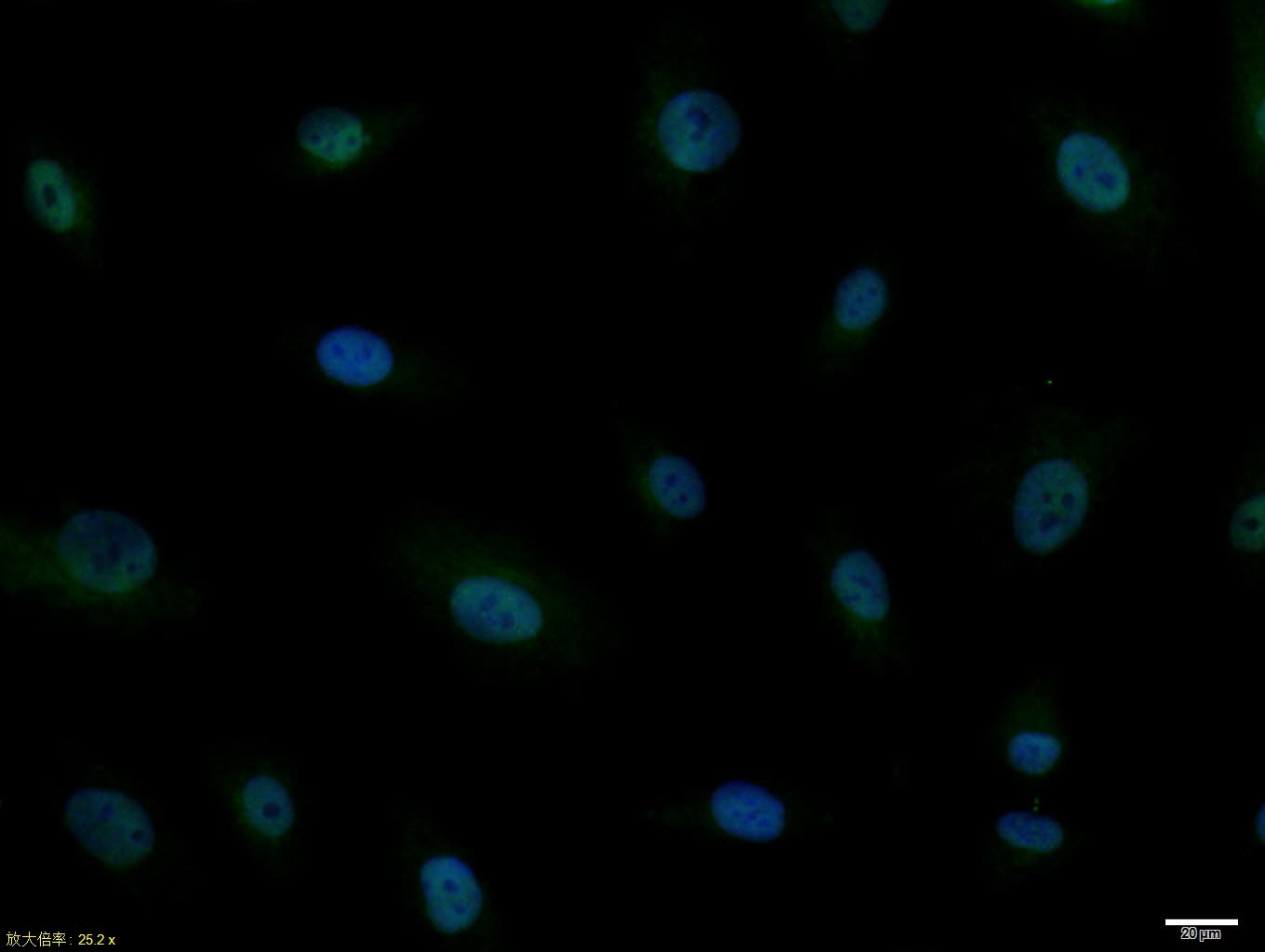 , ,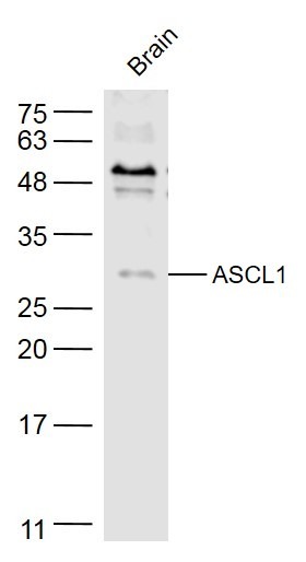 |
| Application | |
| Recommended Dose | FCM=2 μg/Test; ICC/IF=1:100-500; IF=1:100-500; IHC-Fr=1:100-500; IHC-P=1:100-500; WB=1:500-2000 |
| Antibody Type | Polyclonal |
| Host Species | Rabbit |
| Subcellular Localization | Nucleus (Probable). |
| Construction | Polyclonal Antibody |
| Purification | Protein A purified |
| Appearance | Liquid |
| Formulation | 0.01M TBS (pH7.4) with 1% BSA, 0.02% Proclin300 and 50% Glycerol. |
| Concentration | 1 mg/mL |
| Research Background | This gene encodes a member of the basic helix-loop-helix (BHLH) family of transcription factors. The protein activates transcription by binding to the E box (5'-CANNTG-3'). Dimerization with other BHLH proteins is required for efficient DNA binding. This protein plays a role in the neuronal commitment and differentiation and in the generation of olfactory and autonomic neurons. Mutations in this gene may contribute to the congenital central hypoventilation syndrome (CCHS) phenotype in rare cases. [provided by RefSeq, Jul 2008] |
| Immunogen | KLH conjugated synthetic peptide: human ASCL1 |
| Antigen Species | Human |
| Gene Name | ASCL1 |
| Gene ID | |
| Protein Name | Achaete-scute homolog 1 |
| Uniprot ID | |
| Biology Area | HLH,Neurogenesis |
| Function | Transcriptional regulator. May play a role at early stages of development of specific neural lineages in most regions of the CNS, and of several lineages in the PNS. Essential for the generation of olfactory and autonomic neurons. Involved in the initiation of neuronal differentiation. Mediates transcription activation by binding to the E box (5'-CANNTG-3'). |
| Molecular Weight | Theoretical: 26 kDa. Actual: 26 kDa. |
| Stability & Storage | Store at -20°C or -80°C for 12 months. Avoid repeated freeze-thaw cycles. |
| Transport | Shipping with blue ice. |
| Size | Quantity | Unit Price | Amount | Operation |
|---|
 Hello! How can I help you today?
 Copyright © 2015-2026 TargetMol Chemicals Inc. All Rights Reserved.
