
Shopping Cart
Remove All Your shopping cart is currently empty

Synonyms: Aquaporin-7-like, Aquaporin-7, Aquaporin adipose (AQPap), Aquaglyceroporin-7, AQP9, AQP7L, AQP-7, AQP7
Anti-Aquaporin-7/AQP7 Polyclonal Antibody
| Pack Size | Price | USA Stock | Global Stock | Quantity |
|---|---|---|---|---|
| 50 µL | $221 | 7-10 days | 7-10 days | |
| 100 µL | $372 | 7-10 days | 7-10 days | |
| 200 µL | $529 | 7-10 days | 7-10 days |
| Description | Anti-Aquaporin-7/AQP7 Polyclonal Antibody is a Rabbit antibody targeting Aquaporin-7/AQP7. Anti-Aquaporin-7/AQP7 Polyclonal Antibody can be used in FCM,IF,IHC-Fr,IHC-P,WB. |
| Synonyms | Aquaporin-7-like, Aquaporin-7, Aquaporin adipose (AQPap), Aquaglyceroporin-7, AQP9, AQP7L, AQP-7, AQP7 |
| Ig Type | IgG |
| Reactivity | Human,Mouse (predicted:Rat,Chicken,Dog,Pig,Cow) |
| Verified Activity | 1. Blank control: A549. Primary Antibody (green line): Rabbit Anti-AQP7 antibody (TMAB-00141) Dilution: 3 μg/10^6 cells; Isotype Control Antibody (orange line): Rabbit IgG. Secondary Antibody: Goat anti-rabbit IgG-PE Dilution: 3 μg/test. Protocol The cells were incubated in 5% BSA to block non-specific protein-protein interactions for 30 min at at room temperature. Cells stained with Primary Antibody for 30 min at room temperature. The secondary antibody used for 40 min at room temperature. 2. Tissue/cell: Mouse embryos; 4% Paraformaldehyde-fixed and paraffin-embedded; Antigen retrieval: citrate buffer (0.01M, pH6.0), Boiling bathing for 15 min; Block endogenous peroxidase by 3% Hydrogen peroxide for 30 min; Blocking buffer (normal goat serum) at 37°C for 20 min; Incubation: Anti-AQP7 Polyclonal Antibody, Unconjugated (TMAB-00141) 1:200, overnight at 4°C, followed by conjugation to the secondary antibody and DAb staining. 3. Black line: Positive blank control (A549); Negative blank control (HUVEC) Green line: Primary Antibody (Rabbit Anti-AQP7 antibody (TMAB-00141)) Orange line: Isotype Control Antibody (Rabbit IgG). Blue line: Secondary Antibody (Goat anti-rabbit IgG-AF488) A549 (Positive) and HUVEc (Negative control) cells (black) were incubated in 5% BSA blocking buffer for 30 min at room temperature. Cells were then stained with AQP7 Antibody (TMAB-00141) at 1:50 dilution in blocking buffer and incubated for 30 min at room temperature, washed twice with 2% BSA in PBS, followed by secondary antibody (blue) incubation for 40 min at room temperature. Acquisitions of 20,000 events were performed. Cells stained with primary antibody (green), and isotype control (orange). 4. Sample: Lane 1: Adipose (Mouse) Lysate at 40 μg Lane 2: Kidney (Mouse) Lysate at 40 μg Lane 3: Testis (Mouse) Lysate at 40 μg Lane 4: Spleen (Mouse) Lysate at 40 μg Primary: Anti-AQP7 (TMAB-00141) at 1/1000 dilution Secondary: IRDye800CW Goat Anti-Rabbit IgG at 1/20000 dilution Predicted band size: 37/18 kDa Observed band size: 40 kDa 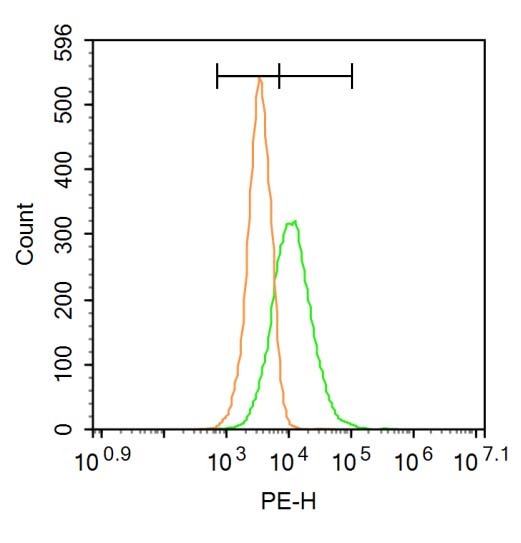 , ,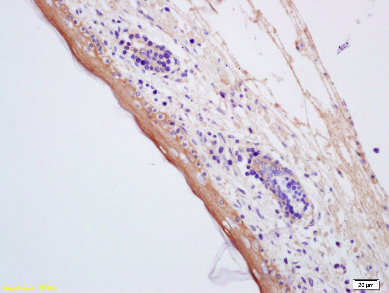 , ,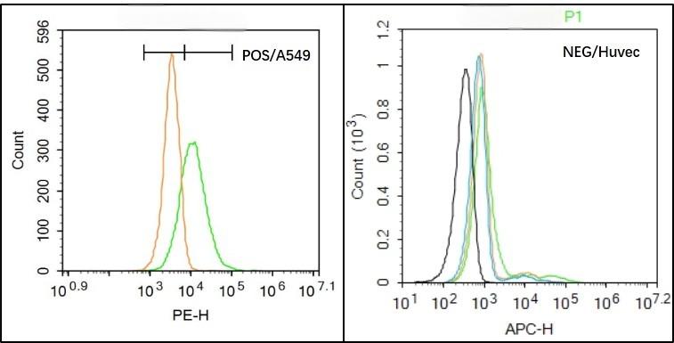 , ,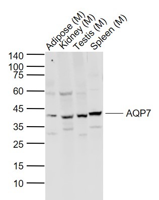 |
| Application | |
| Recommended Dose | WB: 1:500-2000; IHC-P: 1:100-500; IHC-Fr: 1:100-500; IF: 1:100-500; FCM: 2ug/test |
| Antibody Type | Polyclonal |
| Host Species | Rabbit |
| Subcellular Localization | Membrane; Multi-pass membrane protein. |
| Construction | Polyclonal Antibody |
| Purification | Protein A purified |
| Appearance | Liquid |
| Formulation | 0.01M TBS (pH7.4) with 1% BSA, 0.02% Proclin300 and 50% Glycerol. |
| Concentration | 1 mg/mL |
| Research Background | Water is a critical component of all living cells. Interestingly, tissue membranes show a great degree of water permeability. Mammalian red cells, renal proximal tubules, and descending thin limb of Henle are extraordinarily permeable to water. Water crosses hydrophobic plasma membranes either by simple diffusion or through a facilitative transport mechanism mediated by special protein "aquaporin". Over the last decade, genes for several members of aquaporin family have been cloned, expressed, and their distribution studied in many tissues. AQP0 or MIP26 (major intrinsic protein 26kD), and Aquaporin 1 (AQP1, purified from red cells) also called CHIP28 (channel forming integral protein, 28kD; 268aa; gene locus 7p14) has been the foundation of the growing family of aquaporin. The lens specific AQP0 represents up to 80% of total lens membrane protein. Defects in MIP26 are cause of autosomal dominant cataract. The cataract Fraser mutation (CATFR or Shriveled) is a transposon induced splicing error that substitutes a long terminal repeat sequence for the C terminus of MIP. The lens opacity mutation (LOP) is an amino acid substitution that inhibits targeting of MIP to the cell membrane. |
| Immunogen | KLH conjugated synthetic peptide: human AQP7 |
| Antigen Species | Human |
| Gene Name | AQP7 |
| Gene ID | |
| Protein Name | Aquaporin-7 |
| Uniprot ID | |
| Biology Area | Channels,Cancer |
| Function | Forms a channel for water and glycerol (By similarity). |
| Molecular Weight | Theoretical: 37 kDa. Actual: 40 kDa. |
| Stability & Storage | Store at -20°C or -80°C for 12 months. Avoid repeated freeze-thaw cycles. |
| Transport | Shipping with blue ice. |
| Size | Quantity | Unit Price | Amount | Operation |
|---|
 Hello! How can I help you today?
 Copyright © 2015-2026 TargetMol Chemicals Inc. All Rights Reserved.
