
Shopping Cart
Remove All Your shopping cart is currently empty

Synonyms: Tripartite motif-containing protein 63, TRIM63, Striated muscle RING zinc finger protein, SMRZ, RNF28, RING-type E3 ubiquitin transferase TRIM63, RING finger protein 28, Muscle-specific RING finger protein 1 (MuRF-1;MuRF1), MURF1, Iris RING finger protein, IRF, E3 ubiquitin-protein ligase TRIM63
Anti-TRIM63 Polyclonal Antibody
| Pack Size | Price | USA Stock | Global Stock | Quantity |
|---|---|---|---|---|
| 50 µL | $220 | 7-10 days | 7-10 days | |
| 100 µL | $374 | 7-10 days | 7-10 days | |
| 200 µL | $529 | 7-10 days | 7-10 days |
| Description | Anti-TRIM63 Polyclonal Antibody is a Rabbit antibody targeting TRIM63. Anti-TRIM63 Polyclonal Antibody can be used in FCM,IF,IHC-Fr,IHC-P. |
| Synonyms | Tripartite motif-containing protein 63, TRIM63, Striated muscle RING zinc finger protein, SMRZ, RNF28, RING-type E3 ubiquitin transferase TRIM63, RING finger protein 28, Muscle-specific RING finger protein 1 (MuRF-1;MuRF1), MURF1, Iris RING finger protein, IRF, E3 ubiquitin-protein ligase TRIM63 |
| Ig Type | IgG |
| Reactivity | Human,Mouse (predicted:Rat,Pig,Cow,Horse,Rabbit) |
| Verified Activity | 1. Tissue/cell: mouse embryos tissue; 4% Paraformaldehyde-fixed and paraffin-embedded; Antigen retrieval: citrate buffer (0.01M, pH6.0), Boiling bathing for 15 min; Block endogenous peroxidase by 3% Hydrogen peroxide for 30 min; Blocking buffer (normal goat serum) at 37°C for 20 min; Incubation: Anti-MuRF1 Polyclonal Antibody, Unconjugated (TMAB-01897) 1:200, overnight at 4°C, followed by conjugation to the secondary antibody and DAb staining. 2. Blank control: K562. Primary Antibody (green line): Rabbit Anti-MuRF1 antibody (TMAB-01897) Dilution: 2 μg/10^6 cells; Isotype Control Antibody (orange line): Rabbit IgG. Secondary Antibody: Goat anti-rabbit IgG-PE Dilution: 1 μg/test. Protocol The cells were fixed with 4% PFA (10 min at room temperature) and then permeabilized with 90% ice-cold methanol for 20 min at-20°C. The cells were then incubated in 5% BSA to block non-specific protein-protein interactions for 30 min at room temperature. Cells stained with Primary Antibody for 30 min at room temperature. The secondary antibody used for 40 min at room temperature. 3. Paraformaldehyde-fixed, paraffin embedded (human heart); Antigen retrieval by boiling in sodium citrate buffer (pH6.0) for 15 min; Block endogenous peroxidase by 3% hydrogen peroxide for 20 min; Blocking buffer (normal goat serum) at 37°C for 30 min; Incubation with (MuRF1) Polyclonal Antibody, Unconjugated (TMAB-01897) at 1:200 overnight at 4°C, followed by operating according to SP Kit (Rabbit) instructionsand DAB staining. 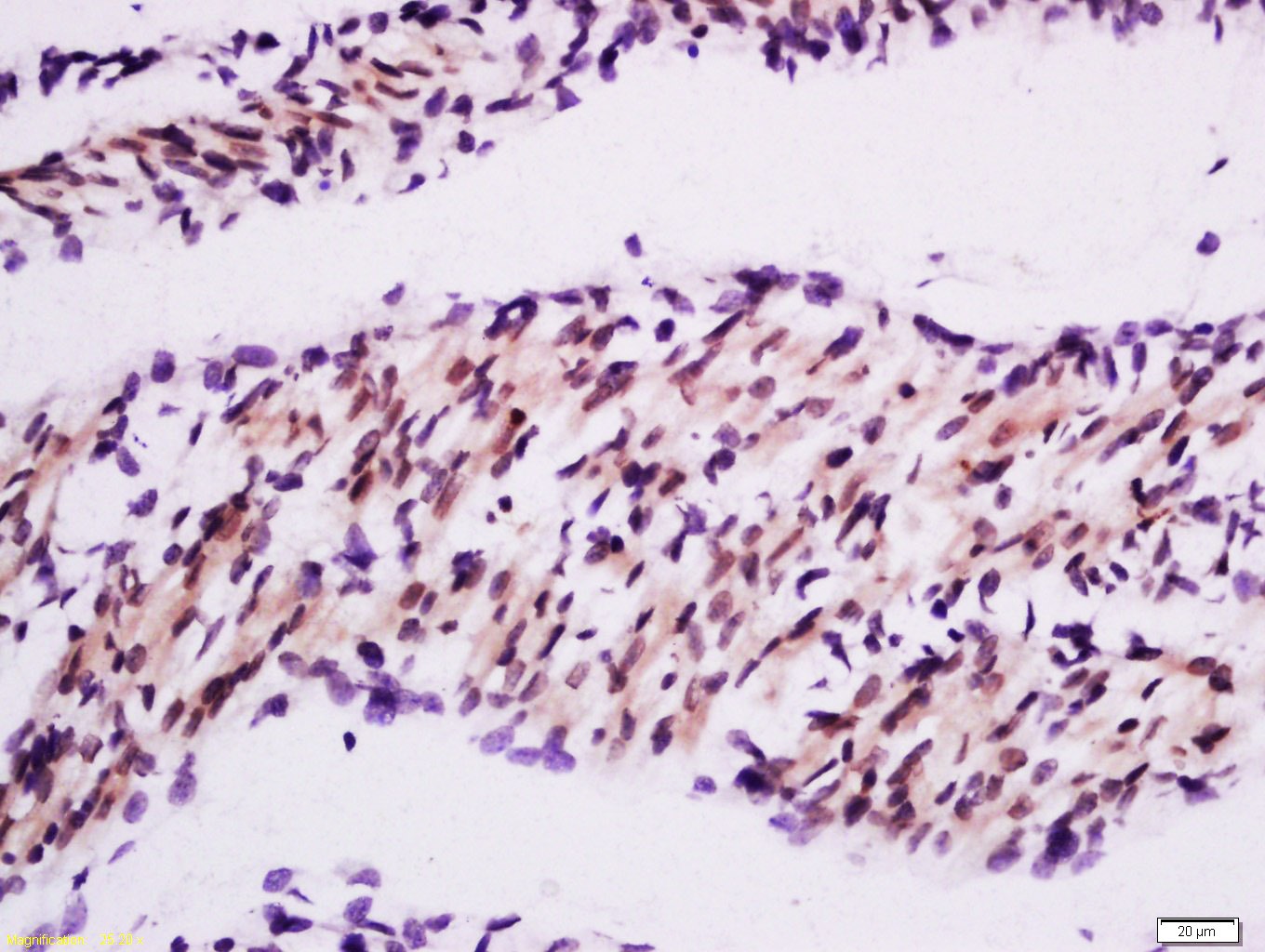 , ,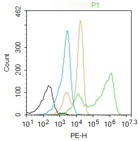 , ,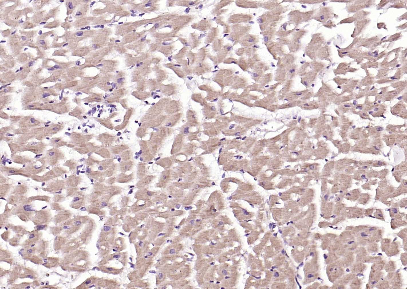 |
| Application | |
| Recommended Dose | IHC-P: 1:100-500; IHC-Fr: 1:100-500; IF: 1:100-500; FCM: 2ug/Test |
| Antibody Type | Polyclonal |
| Host Species | Rabbit |
| Subcellular Localization | Cytoplasm. Nucleus. Cytoplasm, myofibril, sarcomere, M line. Cytoplasm, myofibril, sarcomere, Z line. Note=Colocalizes with TNNI3 in myocytes. Localizes to the M- and Z-lines in skeletal muscle. |
| Tissue Specificity | Muscle specific. Selectively expressed in heart and skeletal muscle. Also expressed in the iris. |
| Construction | Polyclonal Antibody |
| Purification | Protein A purified |
| Appearance | Liquid |
| Formulation | 0.01M TBS (pH7.4) with 1% BSA, 0.02% Proclin300 and 50% Glycerol. |
| Concentration | 1 mg/mL |
| Research Background | This gene encodes a member of the RING zinc finger protein family found in striated muscle and iris. The product of this gene is localized to the Z-line and M-line lattices of myofibrils, where titin's N-terminal and C-terminal regions respectively bind to the sarcomere. In vitro binding studies have shown that this protein also binds directly to titin near the region of titin containing kinase activity. Another member of this protein family binds to microtubules. Since these family members can form heterodimers, this suggests that these proteins may serve as a link between titin kinase and microtubule-dependent signal pathways in muscle. [provided by RefSeq]. |
| Immunogen | KLH conjugated synthetic peptide: human MuRF1 |
| Antigen Species | Human |
| Gene Name | TRIM63 |
| Gene ID | |
| Protein Name | E3 ubiquitin-protein ligase TRIM63 |
| Uniprot ID | |
| Biology Area | Myosins,Troponin,Hypertrophy,RING Finger E3 Ligase,Ubiquitin ligases,E3 Ubiquitin Ligases |
| Function | E3 ubiquitin ligase. Mediates the ubiquitination and subsequent proteasomal degradation of CKM, GMEB1 and HIBADH. Regulates the proteasomal degradation of muscle proteins under amino acid starvation, where muscle protein is catabolized to provide other organs with amino acids. Inhibits de novo skeletal muscle protein synthesis under amino acid starvation. Regulates proteasomal degradation of cardiac troponin I/TNNI3 and probably of other sarcomeric-associated proteins. May play a role in striated muscle atrophy and hypertrophy by regulating an anti-hypertrophic PKC-mediated signaling pathway. May regulate the organization of myofibrils through TTN in muscle cells. |
| Molecular Weight | Theoretical: 39 kDa. |
| Stability & Storage | Store at -20°C or -80°C for 12 months. Avoid repeated freeze-thaw cycles. |
| Transport | Shipping with blue ice. |
| Size | Quantity | Unit Price | Amount | Operation |
|---|
 Hello! How can I help you today?
 Copyright © 2015-2026 TargetMol Chemicals Inc. All Rights Reserved.
