
Shopping Cart
Remove All Your shopping cart is currently empty

Synonyms: VMP1, Vacuole membrane protein 1, Transmembrane protein 49, TMEM 49, TDC1, DKFZP566I133
Anti-TMEM49 Polyclonal Antibody
| Pack Size | Price | USA Stock | Global Stock | Quantity |
|---|---|---|---|---|
| 50 µL | $220 | 7-10 days | 7-10 days | |
| 100 µL | $372 | 7-10 days | 7-10 days | |
| 200 µL | $528 | 7-10 days | 7-10 days |
| Description | Anti-TMEM49 Polyclonal Antibody is a Rabbit antibody targeting TMEM49. Anti-TMEM49 Polyclonal Antibody can be used in IF,IHC-Fr,IHC-P,WB. |
| Synonyms | VMP1, Vacuole membrane protein 1, Transmembrane protein 49, TMEM 49, TDC1, DKFZP566I133 |
| Ig Type | IgG |
| Reactivity | Mouse (predicted:Human,Rat,Pig,Horse,Rabbit) |
| Verified Activity | 1. Sample: Kidney (Mouse) Lysate at 40 μg Primary: Anti-TMEM49 (TMAB-01867) at 1/1000 dilution Secondary: IRDye800CW Goat Anti-Rabbit IgG at 1/20000 dilution Predicted band size: 46 kDa Observed band size: 46 kDa 2. Tissue/cell: mouse pancreas tissue; 4% Paraformaldehyde-fixed and paraffin-embedded; Antigen retrieval: citrate buffer (0.01M, pH6.0), Boiling bathing for 15 min; Block endogenous peroxidase by 3% Hydrogen peroxide for 30 min; Blocking buffer (normal goat serum) at 37°C for 20 min; Incubation: Anti-TMEM49 Polyclonal Antibody, Unconjugated (TMAB-01867) 1:200, overnight at 4°C, followed by conjugation to the secondary antibody and DAb staining. 3. Sample: Lane 1: Mouse Stomach tissue lysates Lane 2: Mouse Cerebrum tissue lysates Primary: Anti-TMEM49 (TMAB-01867) at 1/1000 dilution Secondary: IRDye800CW Goat Anti-Rabbit IgG at 1/20000 dilution Predicted band size: 46 kDa Observed band size: 48 kDa 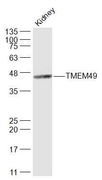 , ,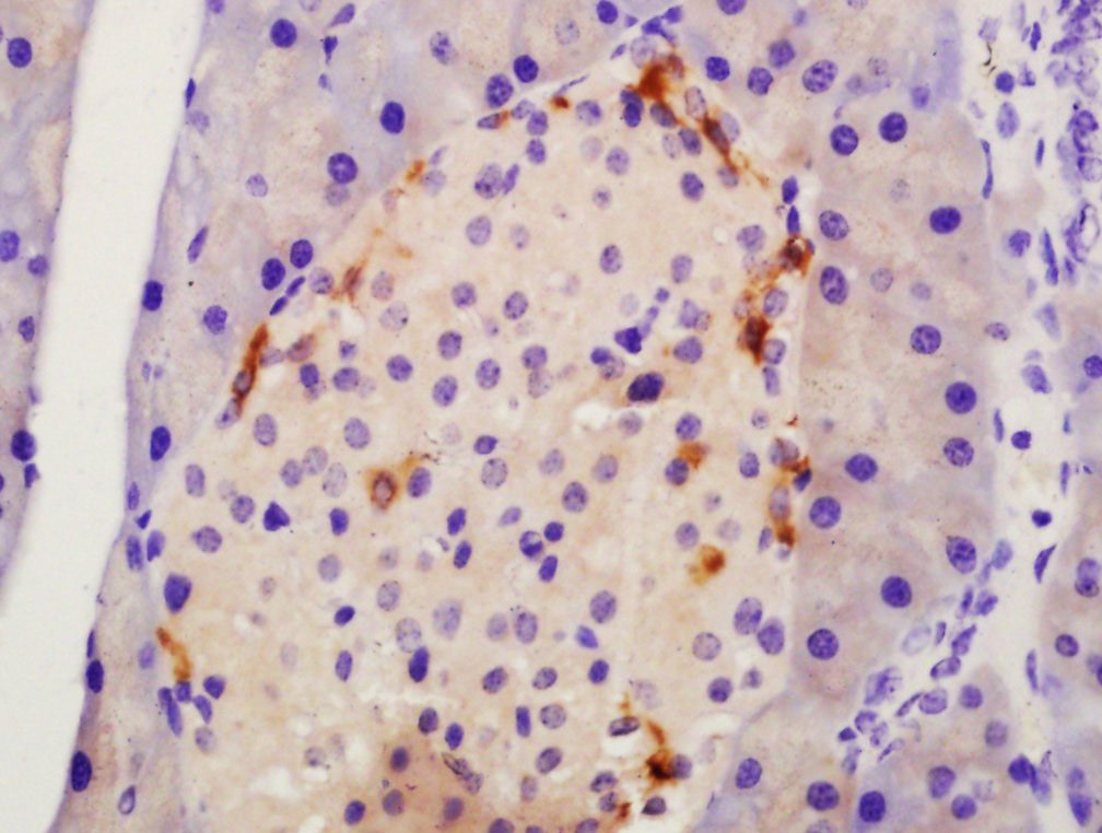 , ,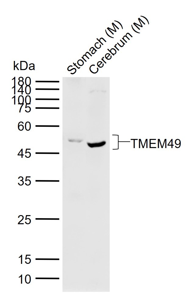 |
| Application | |
| Recommended Dose | WB: 1:500-2000; IHC-P: 1:100-500; IHC-Fr: 1:100-500; IF: 1:100-500 |
| Antibody Type | Polyclonal |
| Host Species | Rabbit |
| Subcellular Localization | Endoplasmic reticulum-Golgi intermediate compartment membrane. Cell membrane. Vacuole membrane. Endoplasmic reticulum. |
| Construction | Polyclonal Antibody |
| Purification | Protein A purified |
| Appearance | Liquid |
| Formulation | 0.01M TBS (pH7.4) with 1% BSA, 0.02% Proclin300 and 50% Glycerol. |
| Concentration | 1 mg/mL |
| Research Background | Vacuole membrane protein 1 (VMP1)/TMEM49 is a transmembrane protein localized to intracellular vacuoles and was discovered as a protein that promotes vacuole formation in acinar cells associated with acute pancreatitis (1). Over-expression of VMP1 promotes vacuole formation and subsequent cell death (1). Subsequent studies have shown that VMP1 expression is induced by starvation and the mTOR inhibitor, rapamycin, and can trigger autophagy (2). VMP1 is targeted, along with LC3, to autophagosome membranes (2). Knockdown of VMP1 can inhibit autophagosome formation (2). VMP1 interacts with Beclin-1, a key autophagy protein that activates the Class III PI3 kinase Vps34, which is regulated by a large network of associated proteins (3). VMP1 functions in the degradation and clearance of zymogen-containing vacuoles during experimental pancreatitis (4). During this process, VMP1 interacts with the ubiquitin protease USP9X, suggesting a possible functional link between the molecular machinery of autophagy and the ubiquitin pathway. Orthologues of VMP1 have been reported in C. elegans (known as EPG-3), Drosophila (known as TANGO-5), and Dictyostelium, and have been shown to play a role in membrane trafficking, orgenelle organization, and autophagy (5-7). |
| Immunogen | KLH conjugated synthetic peptide: human TMEM49 |
| Antigen Species | Human |
| Gene Name | VMP1 |
| Gene ID | |
| Protein Name | Vacuole membrane protein 1 |
| Uniprot ID | |
| Biology Area | Autophagosome,Tumor biomarkers,Autophagosome,Autophagosome |
| Function | Stress-induced protein that, when overexpressed, promotes formation of intracellular vacuoles followed by cell death. May be involved in the cytoplasmic vacuolization of acinar cells during the early stage of acute pancreatitis. Plays a role in the initial stages of the autophagic process through its interaction with BECN1 (By similarity). Involved in cell-cell adhesion. Plays an essential role in formation of cell junctions.Sequence similarities: Belongs to the VMP1 family. |
| Molecular Weight | Theoretical: 46 kDa. Actual: 48 kDa. |
| Stability & Storage | Store at -20°C or -80°C for 12 months. Avoid repeated freeze-thaw cycles. |
| Transport | Shipping with blue ice. |
| Size | Quantity | Unit Price | Amount | Operation |
|---|
 Hello! How can I help you today?
 Copyright © 2015-2026 TargetMol Chemicals Inc. All Rights Reserved.
