
Shopping Cart
Remove All Your shopping cart is currently empty

Synonyms:
Anti-Tissue Factor Polyclonal Antibody
| Pack Size | Price | USA Stock | Global Stock | Quantity |
|---|---|---|---|---|
| 50 µL | $220 | 7-10 days | 7-10 days | |
| 100 µL | $374 | 7-10 days | 7-10 days | |
| 200 µL | $527 | 7-10 days | 7-10 days |
| Description | Anti-Tissue Factor Polyclonal Antibody is a Rabbit antibody targeting Tissue Factor. Anti-Tissue Factor Polyclonal Antibody can be used in IF,IHC-Fr,IHC-P,WB. |
| Ig Type | IgG |
| Reactivity | Human,Mouse,Rat (predicted:Dog,Pig,Cow,Horse,Rabbit,GuineaPig) |
| Verified Activity | 1. Paraformaldehyde-fixed, paraffin embedded (Mouse brain); Antigen retrieval by boiling in sodium citrate buffer (pH6.0) for 15 min; Block endogenous peroxidase by 3% hydrogen peroxide for 20 min; Blocking buffer (normal goat serum) at 37°C for 30 min; Antibody incubation with (Tissue factor) Polyclonal Antibody, Unconjugated (TMAB-01847) at 1:400 overnight at 4°C, followed by operating according to SP Kit (Rabbit) instructionsand DAB staining. 2. Sample: Lane 1: Mouse Lung tissue lysates Lane 2: Mouse Placenta tissue lysates Lane 3: Human A431 cell lysates Lane 4: Human MDA-MB-231 cell lysates Lane 5: Human SiHa cell lysates Primary: Anti-Tissue factor (TMAB-01847) at 1/1000 dilution Secondary: IRDye800CW Goat Anti-Rabbit IgG at 1/20000 dilution Predicted band size: 29 kDa Observed band size: 50 kDa 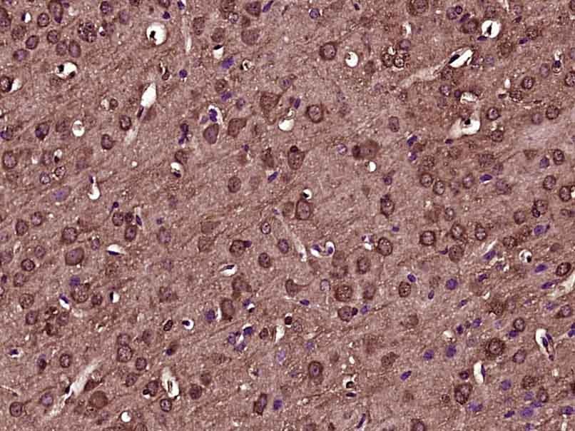 , ,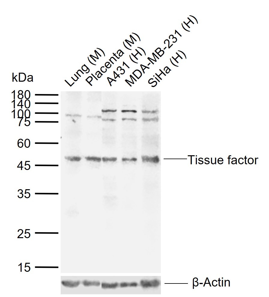 |
| Application | |
| Recommended Dose | WB: 1:500-2000; IHC-P: 1:100-500; IHC-Fr: 1:100-500; IF: 1:100-500 |
| Antibody Type | Polyclonal |
| Host Species | Rabbit |
| Subcellular Localization | Isoform 1: Membrane; Single-pass type I membrane protein. Isoform 2: Secreted. |
| Tissue Specificity | Lung, placenta and pancreas. |
| Construction | Polyclonal Antibody |
| Purification | Protein A purified |
| Appearance | Liquid |
| Formulation | 0.01M TBS (pH7.4) with 1% BSA, 0.02% Proclin300 and 50% Glycerol. |
| Concentration | 1 mg/mL |
| Research Background | This gene encodes coagulation factor III which is a cell surface glycoprotein. This factor enables cells to initiate the blood coagulation cascades, and it functions as the high-affinity receptor for the coagulation factor VII. The resulting complex provides a catalytic event that is responsible for initiation of the coagulation protease cascades by specific limited proteolysis. Unlike the other cofactors of these protease cascades, which circulate as nonfunctional precursors, this factor is a potent initiator that is fully functional when expressed on cell surfaces. There are 3 distinct domains of this factor: extracellular, transmembrane, and cytoplasmic. This protein is the only one in the coagulation pathway for which a congenital deficiency has not been described. Alternate splicing results in multiple transcript variants.[provided by RefSeq, May 2010] |
| Immunogen | KLH conjugated synthetic peptide: human Tissue factor |
| Antigen Species | Human |
| Gene Name | F3 |
| Gene ID | |
| Protein Name | Tissue factor |
| Uniprot ID | |
| Biology Area | Angiogenic Factors,Common,Extrinsic,CD markers ELISA kits |
| Function | Initiates blood coagulation by forming a complex with circulating factor VII or VIIa. The [TF:VIIa] complex activates factors IX or X by specific limited protolysis. TF plays a role in normal hemostasis by initiating the cell-surface assembly and propagation of the coagulation protease cascade. |
| Molecular Weight | Theoretical: 29 kDa. Actual: 50 kDa. |
| Stability & Storage | Store at -20°C or -80°C for 12 months. Avoid repeated freeze-thaw cycles. |
| Transport | Shipping with blue ice. |
| Size | Quantity | Unit Price | Amount | Operation |
|---|
 Hello! How can I help you today?
 Copyright © 2015-2026 TargetMol Chemicals Inc. All Rights Reserved.
