
Shopping Cart
Remove All Your shopping cart is currently empty

Synonyms: Thy-1 cell surface antigen
Anti-Thy1/CD90 Polyclonal Antibody
| Pack Size | Price | USA Stock | Global Stock | Quantity |
|---|---|---|---|---|
| 50 µL | $222 | 7-10 days | 7-10 days | |
| 100 µL | $374 | 7-10 days | 7-10 days | |
| 200 µL | $528 | 7-10 days | 7-10 days |
| Description | Anti-Thy1/CD90 Polyclonal Antibody is a Rabbit antibody targeting Thy1/CD90. Anti-Thy1/CD90 Polyclonal Antibody can be used in FCM,IF,IHC-Fr,WB. |
| Synonyms | Thy-1 cell surface antigen |
| Ig Type | IgG |
| Reactivity | Human,Mouse,Rat (predicted:Sheep,Rabbit,Horse,Pig,Dog) |
| Verified Activity | 1. Sample: Brain (Mouse) Lysate at 40 μg Primary: Anti-CD90 (TMAB-01835) at 1/300 dilution Secondary: IRDye800CW Goat Anti-Rabbit IgG at 1/20000 dilution Predicted band size: 12 kDa Observed band size: 32 kDa 2. Blank control (blue line): U251 (blue). Primary Antibody (green line): Rabbit Anti-CD90 antibody (TMAB-01835) Dilution: 1 μg/10^6 cells; Isotype Control Antibody (orange line): Rabbit IgG. Secondary Antibody (white blue line): Goat anti-rabbit IgG-FITC Dilution: 1 μg/test. Protocol The cells were fixed with 70% ice-cold methanol overnight at 4°C. Cells stained with Primary Antibody for 30 min at room temperature. The cells were then incubated in 1 X PBS/2% BSA/10% goat serum to block non-specific protein-protein interactions followed by the antibody for 15 min at room temperature. The secondary antibody used for 40 min at room temperature. 3. Blank control: U937 (blue). Primary Antibody: Rabbit Anti-CD90 antibody (TMAB-01835), Dilution: 1 μg in 100 μL 1X PBS containing 0.5% BSA; Isotype Control Antibody: Rabbit IgG (orange),used under the same conditions. Secondary Antibody: Goat anti-rabbit IgG-Pe (white blue), Dilution: 1:200 in 1 X PBS containing 0.5% BSA. Protocol The cells were fixed with 2% paraformaldehyde (10 min). Primary antibody (TMAB-01835, 1 μg/1x10^6 cells) were incubated for 30 min on the ice, followed by 1 X PBS containing 0.5% BSA + 10% goat serum (15 min) to block non-specific protein-protein interactions. Then the Goat Anti-rabbit IgG/PE antibody was added into the blocking buffer mentioned above to react with the primary antibody at 1/200 dilution for 30 min on ice. 4. Sample: Brain (Mouse) Lysate at 40 μg Primary: Anti-CD90 (TMAB-01835) at 1/300 dilution Secondary: IRDye800CW Goat Anti-Rabbit IgG at 1/20000 dilution Predicted band size: 12 kDa Observed band size: 32 kDa 5. Blank control: Mouse brain. Primary Antibody (green line): Rabbit Anti-CD90 antibody (TMAB-01835) Dilution: 1 μg/10^6 cells; Isotype Control Antibody (orange line): Rabbit IgG. Secondary Antibody: Goat anti-rabbit IgG-AF488 Dilution: 1 μg/test. Protocol The cells were incubated in 5% BSA to block non-specific protein-protein interactions for 30 min at room temperature. Cells stained with Primary Antibody for 30 min at room temperature. The secondary antibody used for 40 min at room temperature. 6. Blank control: Mouse brain. Primary Antibody (green line): Rabbit Anti-CD90 antibody (TMAB-01835) Dilution: 1 μg/10^6 cells; Isotype Control Antibody (orange line): Rabbit IgG. Secondary Antibody: Goat anti-rabbit IgG-AF488 Dilution: 1 μg/test. Protocol The cells were incubated in 5% BSA to block non-specific protein-protein interactions for 30 min at room temperature. Cells stained with Primary Antibody for 30 min at room temperature. The secondary antibody used for 40 min at room temperature. 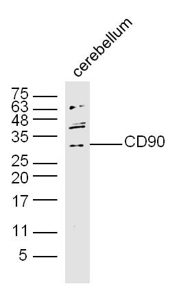 , ,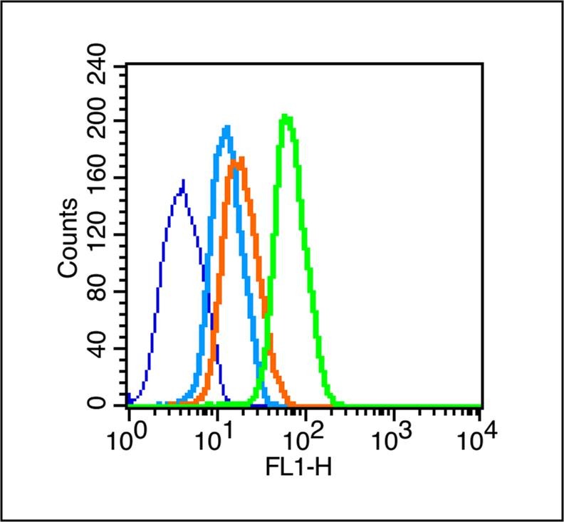 , ,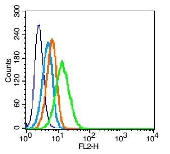 , ,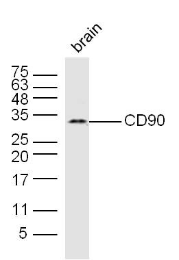 , ,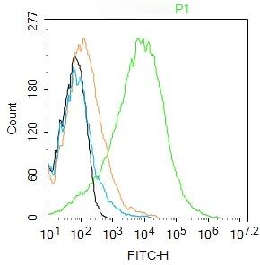 , ,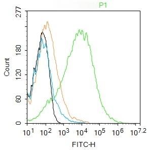 |
| Application | |
| Recommended Dose | WB: 1:500-2000; IHC-Fr: 1:100-500; IF: 1:100-500; FCM: 1μg/Test |
| Antibody Type | Polyclonal |
| Host Species | Rabbit |
| Subcellular Localization | Cell membrane; Lipid-anchor, GPI-anchor. |
| Tissue Specificity | Abundant in lymphoid tissues. |
| Construction | Polyclonal Antibody |
| Purification | Protein A purified |
| Appearance | Liquid |
| Formulation | 0.01M TBS (pH7.4) with 1% BSA, 0.02% Proclin300 and 50% Glycerol. |
| Concentration | 1 mg/mL |
| Research Background | Thy-1 or CD90 (Cluster of Differentiation 90) is a 25–37 kDa heavily N-glycosylated, glycophosphatidylinositol (GPI) anchored conserved cell surface protein with a single V-like immunoglobulin domain, originally discovered as a thymocyte antigen. Thy-1can be used as a marker for a variety of stem cells and for the axonal processes of mature neurons. Structural study of Thy-1 lead to the foundation of the Immunoglobulin superfamily, of which it is the smallest member, and led to some of the initial biochemical description and characterization of a vertebrate GPI anchor and also the first demonstration of tissue specific differential glycosylation. |
| Immunogen | KLH conjugated synthetic peptide: rat Thy-1 |
| Antigen Species | Rat |
| Gene Name | THY1 |
| Gene ID | |
| Protein Name | Thy-1 membrane glycoprotein |
| Uniprot ID | |
| Biology Area | CD,Synapse marker,B Lymphocytic Lineage,T Lymphocytic Lineage,Monocytic Lineage |
| Function | May play a role in cell-cell or cell-ligand interactions during synaptogenesis and other events in the brain. |
| Molecular Weight | Theoretical: 12 kDa. Actual: 32 kDa. |
| Stability & Storage | Store at -20°C or -80°C for 12 months. Avoid repeated freeze-thaw cycles. |
| Transport | Shipping with blue ice. |
| Size | Quantity | Unit Price | Amount | Operation |
|---|
 Hello! How can I help you today?
 Copyright © 2015-2026 TargetMol Chemicals Inc. All Rights Reserved.
