
Shopping Cart
Remove All Your shopping cart is currently empty

Synonyms: SMAD, mothers against DPP homolog 7(Drosophila), SMAD, mothers against DPP homolog 7, SMAD family member 7, SMAD 7, SMAD, SMA-AND MAD-RELATED PROTEIN 7, Mothers against DPP homolog 8, Mothers against DPP homolog 7, Mothers against decapentaplegic homolog 8, Mothers against decapentaplegic homolog 7, Mothers Against Decapentaplegic Drosophila Homolog of 7, MADH8, MADH 8, MADH 7, MAD(mothers against decapentaplegic Drosophila) homolog 7, MAD mothers against decapentaplegic homolog 7, Mad homolog 7, MAD
Anti-SMAD7 Polyclonal Antibody
| Pack Size | Price | USA Stock | Global Stock | Quantity |
|---|---|---|---|---|
| 50 µL | $220 | 7-10 days | 7-10 days | |
| 100 µL | $374 | 7-10 days | 7-10 days | |
| 200 µL | $529 | 7-10 days | 7-10 days |
| Description | Anti-SMAD7 Polyclonal Antibody is a Rabbit antibody targeting SMAD7. Anti-SMAD7 Polyclonal Antibody can be used in FCM, ICC/IF, IF, IHC-Fr, IHC-P, WB. |
| Synonyms | SMAD, mothers against DPP homolog 7(Drosophila), SMAD, mothers against DPP homolog 7, SMAD family member 7, SMAD 7, SMAD, SMA-AND MAD-RELATED PROTEIN 7, Mothers against DPP homolog 8, Mothers against DPP homolog 7, Mothers against decapentaplegic homolog 8, Mothers against decapentaplegic homolog 7, Mothers Against Decapentaplegic Drosophila Homolog of 7, MADH8, MADH 8, MADH 7, MAD(mothers against decapentaplegic Drosophila) homolog 7, MAD mothers against decapentaplegic homolog 7, Mad homolog 7, MAD |
| Ig Type | IgG |
| Reactivity | Human,Mouse,Rat (predicted:Pig,Cow) |
| Verified Activity | 1. Sample: Lung (Mouse) Lysate at 30 μg Placenta (Mouse) Lysate at 30 μg Large intestine (Mouse) Lysate at 30 μg Primary: Anti-MADH7/Smad7 (TMAB-01742) at 1/300 dilution Secondary: IRDye800CW Goat Anti-Rabbit IgG at 1/20000 dilution Predicted band size: 46 kDa Observed band size: 50 kDa 2. Paraformaldehyde-fixed, paraffin embedded (rat lung); Antigen retrieval by boiling in sodium citrate buffer (pH6.0) for 15 min; Block endogenous peroxidase by 3% hydrogen peroxide for 20 min; Blocking buffer (normal goat serum) at 37°C for 30 min; Antibody incubation with (Smad7) Polyclonal Antibody, Unconjugated (TMAB-01742) at 1:600 overnight at 4°C, followed by a conjugated secondary for 20 min and DAB staining. 3. Blank control: SH-SY5Y. Primary Antibody (green line): Rabbit Anti-MADH7/Smad7 antibody (TMAB-01742) Dilution: 1 μg/10^6 cells; Isotype Control Antibody (orange line): Rabbit IgG. Secondary Antibody: Goat anti-rabbit IgG-AF647 Dilution: 1 μg/test. Protocol The cells were fixed with 4% PFA (10 min at room temperature) and then permeabilized with 90% ice-cold methanol for 20 min at-20°C. The cells were then incubated in 5% BSA to block non-specific protein-protein interactions for 30 min at room temperature. Cells stained with Primary Antibody for 30 min at room temperature. The secondary antibody used for 40 min at room temperature. 4. Sample: Lane 1: Cerebrum (Mouse) Lysate at 40 μg Lane 2: Cerebrum (Rat) Lysate at 40 μg Lane 3: Stomach (Mouse) Lysate at 40 μg Lane 4: Lung (Mouse) Lysate at 40 μg Lane 5: Kidney (Mouse) Lysate at 40 μg Primary: Anti-MADH7/Smad7 (TMAB-01742) at 1/1000 dilution Secondary: IRDye800CW Goat Anti-Rabbit IgG at 1/20000 dilution Predicted band size: 50 kDa Observed band size: 50 kDa 5. Paraformaldehyde-fixed, paraffin embedded (rat stomach); Antigen retrieval by boiling in sodium citrate buffer (pH6.0) for 15 min; Block endogenous peroxidase by 3% hydrogen peroxide for 20 min; Blocking buffer (normal goat serum) at 37°C for 30 min; Antibody incubation with (MADH7) Polyclonal Antibody, Unconjugated (TMAB-01742) at 1:200 overnight at 4°C, followed by operating according to SP Kit (Rabbit) instructionsand DAB staining. 6. U-2OS cell; 4% Paraformaldehyde-fixed; Triton X-100 at room temperature for 20 min; Blocking buffer (normal goat serum) at 37°C for 20 min; Antibody incubation with (MADH7/Smad7) polyclonal Antibody, Unconjugated (TMAB-01742) 1:100, 90 minutes at 37°C; followed by a conjugated Goat Anti-Rabbit IgG antibody at 37°C for 90 minutes, DAPI (blue) was used to stain the cell nucleus. 7. Sample: Lane 1: Stomach (Mouse) Lysate at 40 μg Lane 2: Spleen (Mouse) Lysate at 40 μg Lane 3: Lung (Mouse) Lysate at 40 μg Primary: Anti-MADH7/Smad7 (TMAB-01742) at 1/1000 dilution Secondary: IRDye800CW Goat Anti-Rabbit IgG at 1/20000 dilution Predicted band size: 50 kDa Observed band size: 50 kDa 8. Paraformaldehyde-fixed, paraffin embedded (rat kidney); Antigen retrieval by boiling in sodium citrate buffer (pH6.0) for 15 min; Block endogenous peroxidase by 3% hydrogen peroxide for 20 min; Blocking buffer (normal goat serum) at 37°C for 30 min; Antibody incubation with (MADH7) Polyclonal Antibody, Unconjugated (TMAB-01742) at 1:200 overnight at 4°C, followed by operating according to SP Kit (Rabbit) instructionsand DAB staining. 9. Paraformaldehyde-fixed, paraffin embedded (mouse kidney); Antigen retrieval by boiling in sodium citrate buffer (pH6.0) for 15 min; Block endogenous peroxidase by 3% hydrogen peroxide for 20 min; Blocking buffer (normal goat serum) at 37°C for 30 min; Antibody incubation with (MADH7) Polyclonal Antibody, Unconjugated (TMAB-01742) at 1:200 overnight at 4°C, followed by operating according to SP Kit (Rabbit) instructionsand DAB staining. 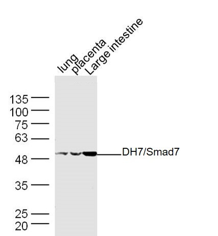 , ,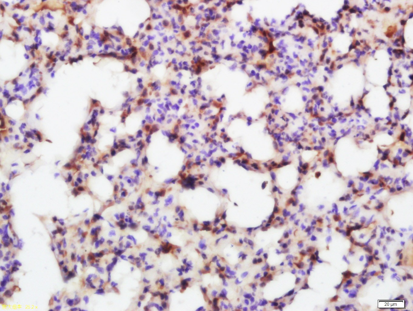 , ,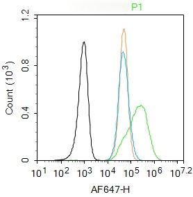 , ,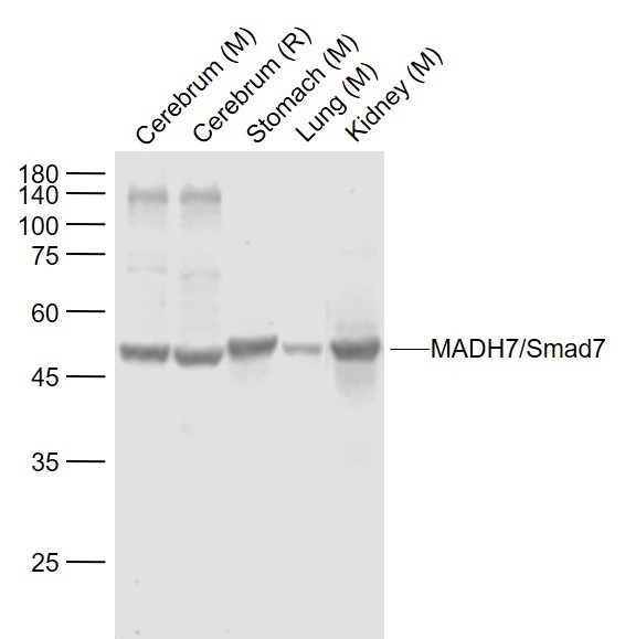 , ,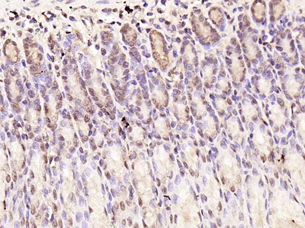 , ,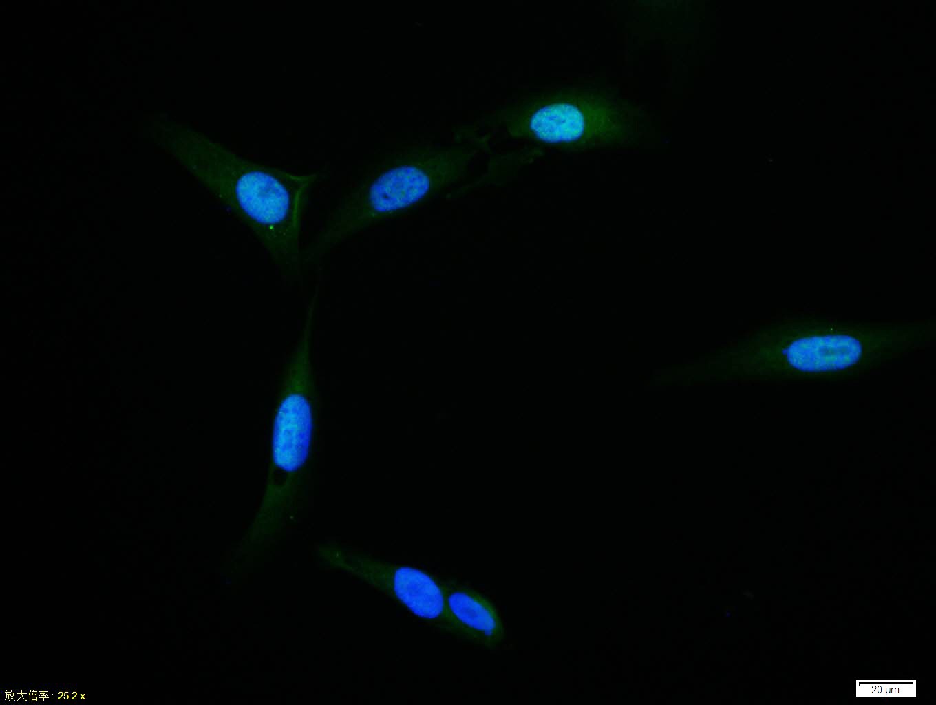 , ,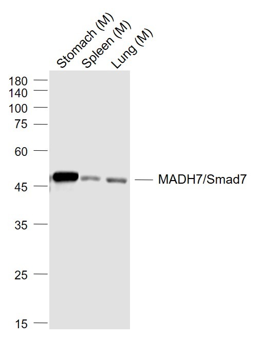 , ,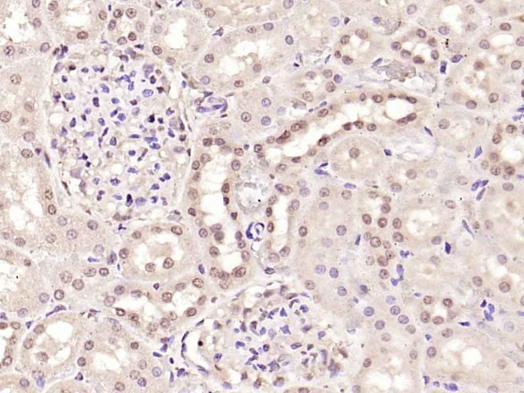 , ,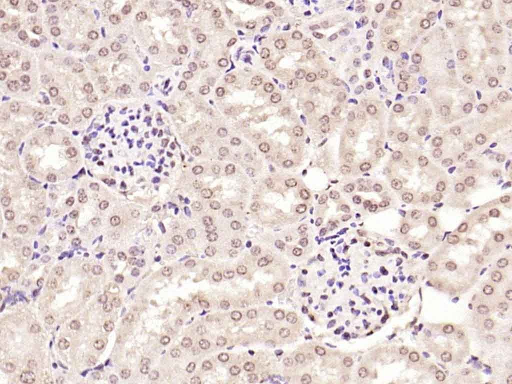 |
| Application | |
| Recommended Dose | FCM=1 μg/Test; ICC/IF=1:100-500; IF=1:100-500; IHC-Fr=1:100-500; IHC-P=1:100-500; WB=1:500-2000 |
| Antibody Type | Polyclonal |
| Host Species | Rabbit |
| Subcellular Localization | Nucleus. Cytoplasm. Note=Interaction with NEDD4L or RNF111 or induces translocation from the nucleus to the cytoplasm. TGF-beta stimulates its translocation from the nucleus to the cytoplasm. PDPK1 inhibits its translocation from the nucleus to the cytoplasm in response to TGF-beta. |
| Tissue Specificity | Ubiquitous with higher expression in the lung and vascular endothelium. |
| Construction | Polyclonal Antibody |
| Purification | Protein A purified |
| Appearance | Liquid |
| Formulation | 0.01M TBS (pH7.4) with 1% BSA, 0.02% Proclin300 and 50% Glycerol. |
| Concentration | 1 mg/mL |
| Research Background | The protein encoded by this gene is a nuclear protein that binds the E3 ubiquitin ligase SMURF2. Upon binding, this complex translocates to the cytoplasm, where it interacts with TGF-beta receptor type-1 (TGFBR1), leading to the degradation of both the encoded protein and TGFBR1. Expression of this gene is induced by TGFBR1. Variations in this gene are a cause of susceptibility to colorectal cancer type 3 (CRCS3). Several transcript variants encoding different isoforms have been found for this gene. [provided by RefSeq, Jun 2010] |
| Immunogen | KLH conjugated synthetic peptide: human Smad7 |
| Antigen Species | Human |
| Gene Name | SMAD7 |
| Gene ID | |
| Protein Name | Mothers against decapentaplegic homolog 7 |
| Uniprot ID | |
| Biology Area | SMADs,SMADs |
| Function | Antagonist of signaling by TGF-beta (transforming growth factor) type 1 receptor superfamily members; has been shown to inhibit TGF-beta (Transforming growth factor) and activin signaling by associating with their receptors thus preventing SMAD2 access. Functions as an adapter to recruit SMURF2 to the TGF-beta receptor complex. Also acts by recruiting the PPP1R15A-PP1 complex to TGFBR1, which promotes its dephosphorylation. Positively regulates PDPK1 kinase activity by stimulating its dissociation from the 14-3-3 protein YWHAQ which acts as a negative regulator. |
| Molecular Weight | Theoretical: 46 kDa. Actual: 50 kDa. |
| Stability & Storage | Store at -20°C or -80°C for 12 months. Avoid repeated freeze-thaw cycles. |
| Transport | Shipping with blue ice. |
| Size | Quantity | Unit Price | Amount | Operation |
|---|
 Hello! How can I help you today?
 Copyright © 2015-2026 TargetMol Chemicals Inc. All Rights Reserved.
