
Shopping Cart
Remove All Your shopping cart is currently empty

Synonyms: retinoblastoma binding protein 4, RBAP48, mRbAp48
Anti-RBBP4 Antibody
(7R532)
| Pack Size | Price | USA Stock | Global Stock | Quantity |
|---|---|---|---|---|
| 50 µL | $298 | 7-10 days | 7-10 days | |
| 100 µL | $496 | 7-10 days | 7-10 days |
| Description | Anti-RBBP4 Antibody (7R532) is a Rabbit antibody targeting RBBP4. Anti-RBBP4 Antibody (7R532) can be used in FCM,ICC,IHC,IP,WB. |
| Synonyms | retinoblastoma binding protein 4, RBAP48, mRbAp48 |
| Ig Type | IgG |
| Clone | 7R532 |
| Reactivity | Human,Mouse,Rat |
| Verified Activity | 1. Western blot analysis of RbAP48 on different lysates using anti-RbAP48 antibody at 1/500 dilution. Positive control: Lane 1: NIH-3T3, Lane 2: Hela, Lane 3: Rat brain, Lane 4: Mouse testis. 2. Immunohistochemical analysis of paraffin-embedded rat brain tissue using anti-RbAP48 antibody. Counter stained with hematoxylin. 3. Immunohistochemical analysis of paraffin-embedded human tonsil tissue using anti-RbAP48 antibody. Counter stained with hematoxylin. 4. Immunohistochemical analysis of paraffin-embedded mouse testis tissue using anti-RbAP48 antibody. Counter stained with hematoxylin. 5. Immunohistochemical analysis of paraffin-embedded human lung cancer tissue using anti-RbAP48 antibody. Counter stained with hematoxylin. 6. Immunohistochemical analysis of paraffin-embedded human liver tissue using anti-RbAP48 antibody. Counter stained with hematoxylin. 7. ICC staining RbAP48 in Hela cells (green). The nuclear counter stain is DAPI (blue). Cells were fixed in paraformaldehyde, permeabilised with 0.25% Triton X100/PBS. 8. ICC staining RbAP48 in MCF-7 cells (green). The nuclear counter stain is DAPI (blue). Cells were fixed in paraformaldehyde, permeabilised with 0.25% Triton X100/PBS. 9. ICC staining RbAP48 in SH-SY5Y cells (green). The nuclear counter stain is DAPI (blue). Cells were fixed in paraformaldehyde, permeabilised with 0.25% Triton X100/PBS. 10. Flow cytometric analysis of Hela cells with RbAP48 antibody at 1/50 dilution (red) compared with an unlabelled control (cells without incubation with primary antibody; black). Alexa Fluor 488-conjugated goat anti rabbit IgG was used as the secondary antibody. 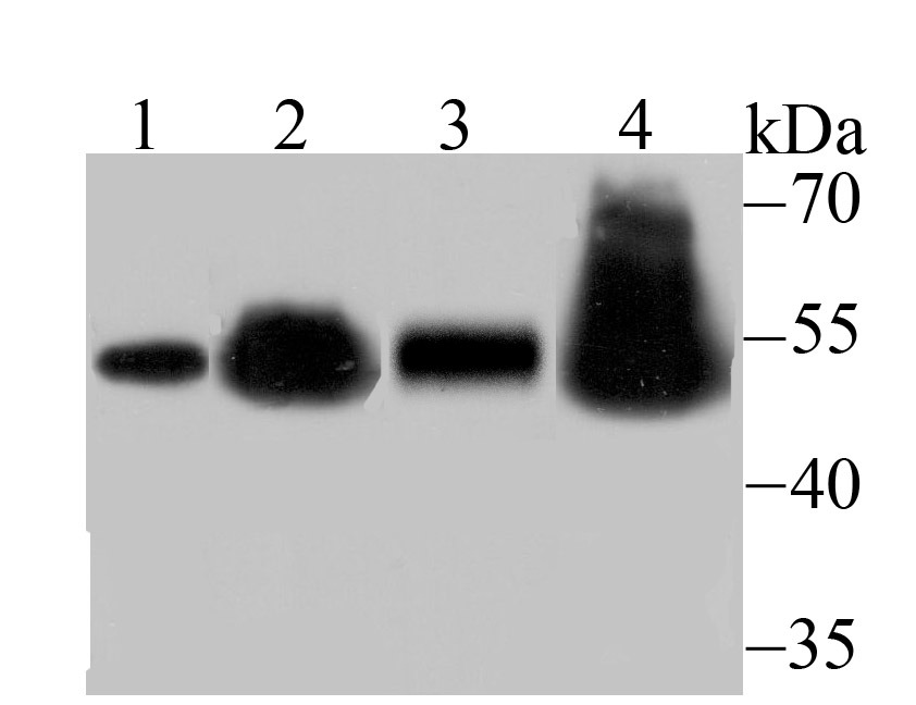 , ,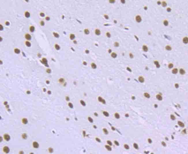 , ,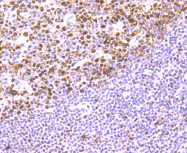 , ,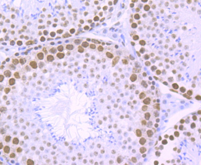 , ,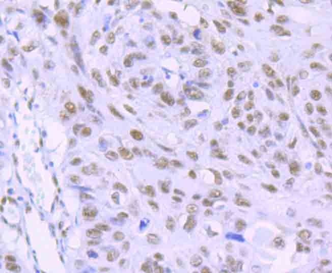 , ,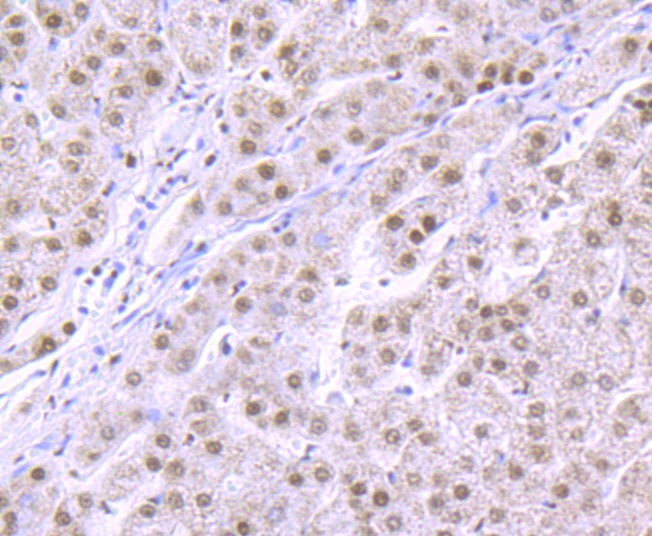 , ,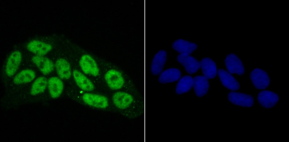 , ,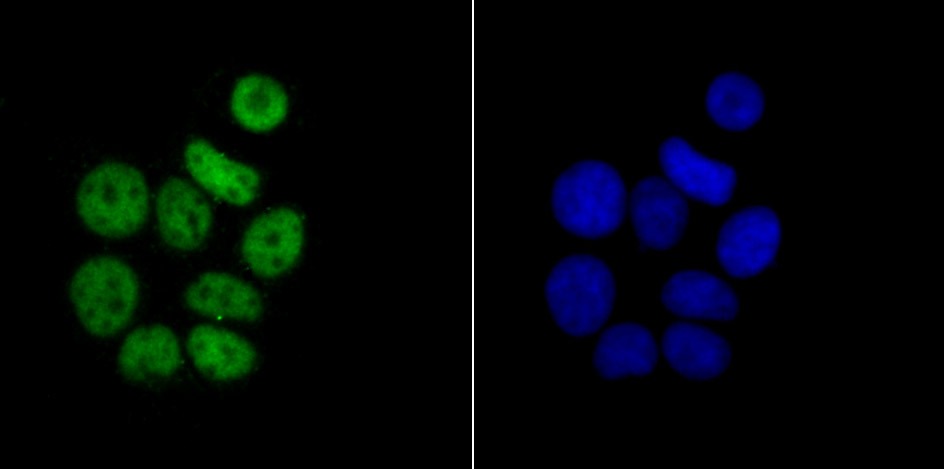 , ,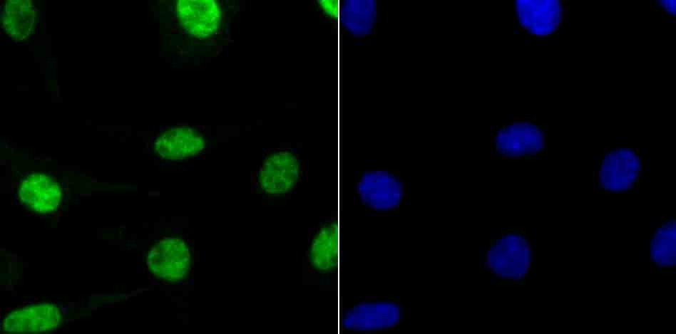 , ,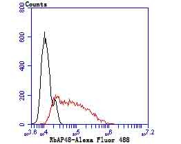 |
| Application | |
| Recommended Dose | WB: 1:500-2000; IHC: 1:50-200; ICC: 1:50-200; IP:1:10-50; FCM: 1:50-100 |
| Antibody Type | Monoclonal |
| Host Species | Rabbit |
| Construction | Recombinant Antibody |
| Purification | ProA affinity purified |
| Appearance | Liquid |
| Formulation | 1*TBS (pH7.4), 1%BSA, 40%Glycerol. Preservative: 0.05% Sodium Azide. |
| Research Background | In the intact cell, DNA is closely associated with histones and other nuclear proteins to form chromatin.The remodeling of chromatin is believed to be a critical component of transcriptional regulation, and a major source of this remodeling is brought about by the acetylation of nucleosomal histones. Acetylation of lysine residues in the amino-terminal tail domain of histone results in an allosteric change in the nucleosomal conformation, and an increased accessiblity of DNA to transcription factors. Mammalian HDAC1 (also designated HD1), HDAC2 (also designated RPD3) and HDAC3, all of which are related to the yeast transcriptional regulator Rpd3p, have been identified as histone deacetylases. The retinoblastoma binding proteins RbAp46 and RbAp48 have been identified as histone binding proteins, and they are components of the histone deacetylase complex. |
| Conjucates | Unconjugated |
| Immunogen | Recombinant Protein |
| Uniprot ID |
| Molecular Weight | Theoretical: 48 kDa. |
| Stability & Storage | Store at -20°C or -80°C for 12 months. Avoid repeated freeze-thaw cycles. |
| Transport | Shipping with blue ice. |
| Size | Quantity | Unit Price | Amount | Operation |
|---|
 Hello! How can I help you today?
 Copyright © 2015-2026 TargetMol Chemicals Inc. All Rights Reserved.
