
Shopping Cart
Remove All Your shopping cart is currently empty

Synonyms: PSMA 1, PSC2, PSC 2, PSA1_HUMAN, Protein P30 33K, Proteasome subunit nu, Proteasome subunit alpha type-1, Proteasome subunit alpha type I, Proteasome subunit alpha type 1, Proteasome nu chain, Proteasome component C2, Proteasome alpha 1 subunit, Proteasome (prosome macropain) subunit alpha type 1, PROS-30, PROS30, PROS 30, NU, Multicatalytic endopeptidase complex subunit C2, MGC23915, MGC22853, MGC21459, MGC1667, MGC14751, MGC14575, MGC14542, Macropain subunit nu, Macropain subunit C2, HC2, HC 2, 30 kDa prosomal protein
Anti-PSMA1 Antibody
(8C777)
| Pack Size | Price | USA Stock | Global Stock | Quantity |
|---|---|---|---|---|
| 50 µL | $298 | 7-10 days | 7-10 days | |
| 100 µL | $497 | 7-10 days | 7-10 days |
| Description | Anti-PSMA1 Antibody (8C777) is a Rabbit antibody targeting PSMA1. Anti-PSMA1 Antibody (8C777) can be used in FCM,ICC/IF,IHC,IP,WB. |
| Synonyms | PSMA 1, PSC2, PSC 2, PSA1_HUMAN, Protein P30 33K, Proteasome subunit nu, Proteasome subunit alpha type-1, Proteasome subunit alpha type I, Proteasome subunit alpha type 1, Proteasome nu chain, Proteasome component C2, Proteasome alpha 1 subunit, Proteasome (prosome macropain) subunit alpha type 1, PROS-30, PROS30, PROS 30, NU, Multicatalytic endopeptidase complex subunit C2, MGC23915, MGC22853, MGC21459, MGC1667, MGC14751, MGC14575, MGC14542, Macropain subunit nu, Macropain subunit C2, HC2, HC 2, 30 kDa prosomal protein |
| Ig Type | IgG |
| Clone | 8C777 |
| Reactivity | Human,Mouse,Rat |
| Verified Activity | 1. Western blot analysis of PSMA1 on different cell lysates using anti-PSMA1 antibody at 1/500 dilution. Positive control: Lane 1: Jurkat, Lane 2: Hela, Lane 3: HepG2, Lane 4: 293. 2. Western blot analysis of PSMA1 on different lysates using anti-PSMA1 antibody at 1/500 dilution. Positive control: Lane 1: PC-12, Lane 2: Mouse spleen tissue, Lane 3: Rat spleen tissue. 3. Immunohistochemical analysis of paraffin-embedded human liver cancer tissue using anti-PSMA1 antibody. Counter stained with hematoxylin. 4. Immunohistochemical analysis of paraffin-embedded rat testis tissue using anti-PSMA1 antibody. Counter stained with hematoxylin. 5. Immunohistochemical analysis of paraffin-embedded mouse cerebellum tissue using anti-PSMA1 antibody. Counter stained with hematoxylin. 6. Immunohistochemical analysis of paraffin-embedded human pancreas tissue using anti-PSMA1 antibody. Counter stained with hematoxylin. 7. ICC staining PSMA1 in HepG2 cells (green). The nuclear counter stain is DAPI (blue). Cells were fixed in paraformaldehyde, permeabilised with 0.25% Triton X100/PBS. 8. ICC staining PSMA1 in MCF-7 cells (green). The nuclear counter stain is DAPI (blue). Cells were fixed in paraformaldehyde, permeabilised with 0.25% Triton X100/PBS. 9. ICC staining PSMA1 in PC-3M cells (green). The nuclear counter stain is DAPI (blue). Cells were fixed in paraformaldehyde, permeabilised with 0.25% Triton X100/PBS. 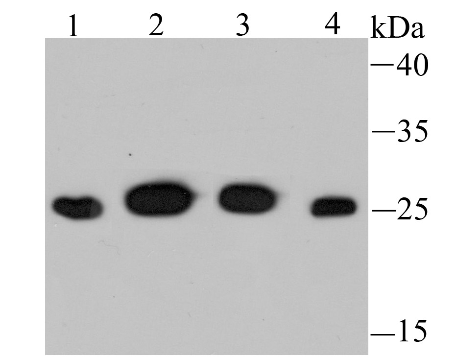 , ,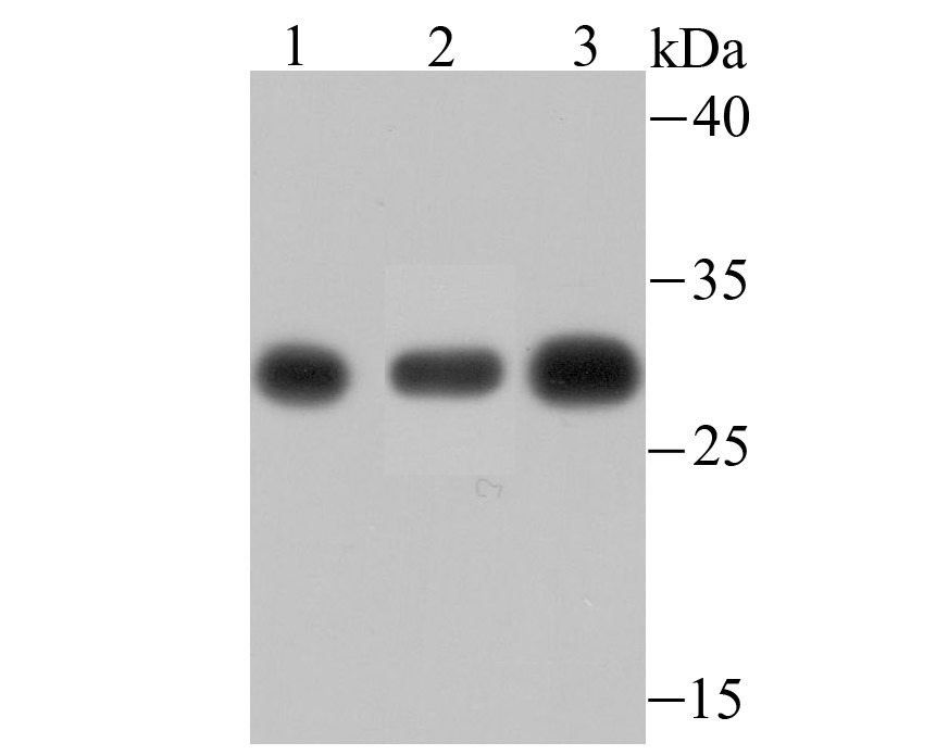 , ,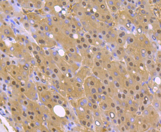 , ,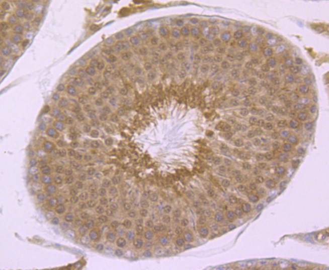 , ,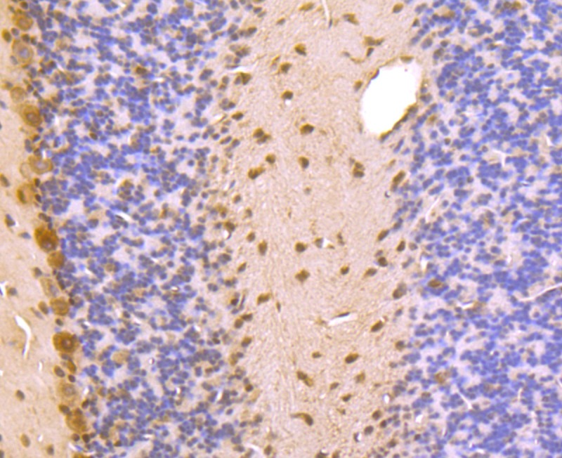 , ,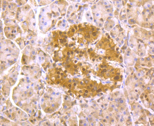 , ,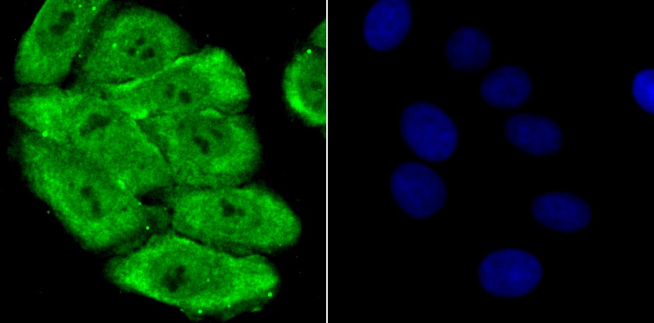 , ,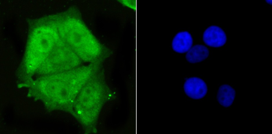 , ,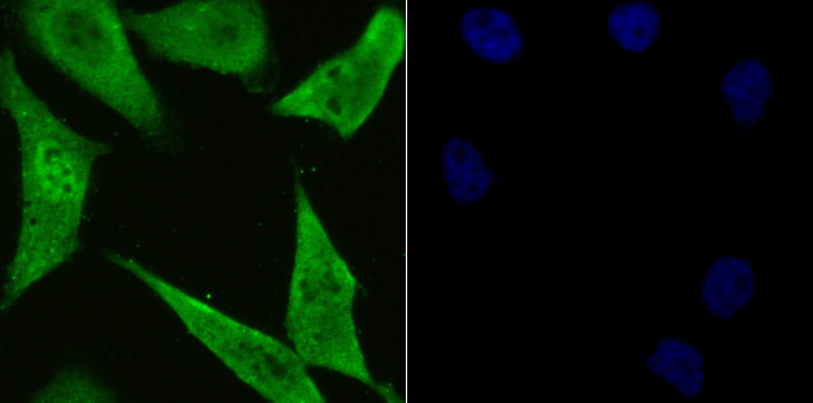 |
| Application | |
| Recommended Dose | WB: 1:500-2000; IHC: 1:50-200; ICC/IF: 1:200-1000; FCM: 1:50-100 |
| Antibody Type | Monoclonal |
| Host Species | Rabbit |
| Construction | Recombinant Antibody |
| Purification | ProA affinity purified |
| Appearance | Liquid |
| Formulation | 1*TBS (pH7.4), 1%BSA, 40%Glycerol. Preservative: 0.05% Sodium Azide. |
| Research Background | The proteasome is a multicatalytic proteinase complex which is characterized by its ability to cleave peptides with Arg, Phe, Tyr, Leu, and Glu adjacent to the leaving group at neutral or slightly basic pH. The proteasome has an ATP-dependent proteolytic activity. Mediates the lipopolysaccharide-induced signal transduction in the macrophage proteasome. Might be involved in the inflammatory response of macrophages during the interaction with C.albicans heat-inactivated cells. |
| Conjucates | Unconjugated |
| Immunogen | Recombinant Protein |
| Uniprot ID |
| Molecular Weight | Theoretical: 30 kDa. |
| Stability & Storage | Store at -20°C or -80°C for 12 months. Avoid repeated freeze-thaw cycles. |
| Transport | Shipping with blue ice. |
| Size | Quantity | Unit Price | Amount | Operation |
|---|
 Hello! How can I help you today?
 Copyright © 2015-2026 TargetMol Chemicals Inc. All Rights Reserved.
