
Shopping Cart
Remove All Your shopping cart is currently empty

Synonyms: PPAR-α, PPARα, PPAR-alpha, PPARalpha, Ppar, peroxisome proliferator-activated receptor α, peroxisome proliferator-activated receptor alpha, Nr1c1, AW742785, 4933429D07Rik
Anti-PPAR alpha/PPARA Polyclonal Antibody 2
| Pack Size | Price | USA Stock | Global Stock | Quantity |
|---|---|---|---|---|
| 50 µL | $222 | 7-10 days | 7-10 days | |
| 100 µL | $374 | 7-10 days | 7-10 days | |
| 200 µL | $529 | 7-10 days | 7-10 days |
| Description | Anti-PPAR alpha/PPARA Polyclonal Antibody 2 is a Rabbit antibody targeting PPAR alpha/PPARA. Anti-PPAR alpha/PPARA Polyclonal Antibody 2 can be used in FCM,IF,IHC-Fr,IHC-P,WB. |
| Synonyms | PPAR-α, PPARα, PPAR-alpha, PPARalpha, Ppar, peroxisome proliferator-activated receptor α, peroxisome proliferator-activated receptor alpha, Nr1c1, AW742785, 4933429D07Rik |
| Ig Type | IgG |
| Reactivity | Human,Mouse,Rat (predicted:Chicken,Pig,Cow,Horse,Rabbit) |
| Verified Activity | 1. Sample: Lane1: Heart (Mouse) Lysate at 30 μg Lane2: Liver (Mouse) Cell Lysate at 30 μg Primary: Anti-PPAR alpha (TMAB-01565) at 1:300 dilution; Secondary: HRP conjugated Goat-Anti-rabbit Igg (secondary antibody) at 1: 5000dilution; Predicted band size: 51 kDa Observed band size: 51 kDa 2. Rat splenocytes stained with Anti-PPAR alpha Polyclonal Antibody, PE-CY5 Conjugated (TMAB-01565-PE-Cy5) at 1:50. 3. Sample: Heart (Mouse) Lysate at 40 μg Primary: Anti-PPAP alpha (TMAB-01565) at 1/300 dilution Secondary: IRDye800CW Goat Anti-Rabbit IgG at 1/10000 dilution Predicted band size: 51 kDa Observed band size: 51 kDa 4. Blank control: HepG2. Primary Antibody (green line): Rabbit Anti-PPAR alpha antibody (TMAB-01565) Dilution: 1 μg/10^6 cells; Isotype Control Antibody (orange line): Rabbit IgG. Secondary Antibody: Goat anti-rabbit IgG-AF647 Dilution: 1 μg/test. Protocol The cells were fixed with 4% PFA (10 min at room temperature) and then permeabilized with 90% ice-cold methanol for 20 min at-20°C. The cells were then incubated in 5% BSA to block non-specific protein-protein interactions for 30 min at room temperature. Cells stained with Primary Antibody for 30 min at room temperature. The secondary antibody used for 40 min at room temperature. 5. Paraformaldehyde-fixed, paraffin embedded (rat brain); Antigen retrieval by boiling in sodium citrate buffer (pH6.0) for 15 min; Block endogenous peroxidase by 3% hydrogen peroxide for 20 min; Blocking buffer (normal goat serum) at 37°C for 30 min; Antibody incubation with (PPAR alpha) Polyclonal Antibody, Unconjugated (TMAB-01565) at 1:200 overnight at 4°C, followed by operating according to SP Kit (Rabbit) instructionsand DAB staining. 6. Blank control: Jurkat. Primary Antibody (green line): Rabbit Anti-PPAR alpha antibody (TMAB-01565) Dilution: 1 μg/10^6 cells; Isotype Control Antibody (orange line): Rabbit IgG. Secondary Antibody: Goat anti-rabbit IgG-FITC Dilution: 1 μg/test. Protocol The cells were fixed with 4% PFA (10 min at room temperature) and then permeabilized with 90% ice-cold methanol for 20 min at-20°C. The cells were then incubated in 5% BSA to block non-specific protein-protein interactions for 30 min at room temperature. Cells stained with Primary Antibody for 30 min at room temperature. The secondary antibody used for 40 min at room temperature. 7. Sample: Heart (Mouse) Lysate at 40 μg Heart (Rat) Lysate at 40 μg Liver (Mouse) Lysate at 40 μg Urinary bladder (Mouse) Lysate at 40 μg Kidney (Mouse) Lysate at 40 μg Primary: Anti-PPAR alpha (TMAB-01565) at 1/1000 dilution Secondary: IRDye800CW Goat Anti-Rabbit IgG at 1/20000 dilution Predicted band size: 52/19 kDa Observed band size: 52 kDa 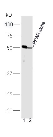 , ,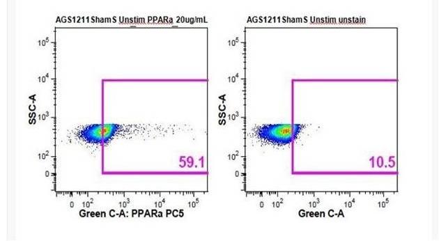 , ,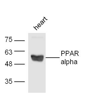 , ,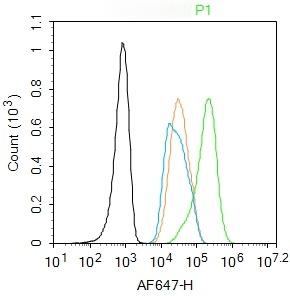 , ,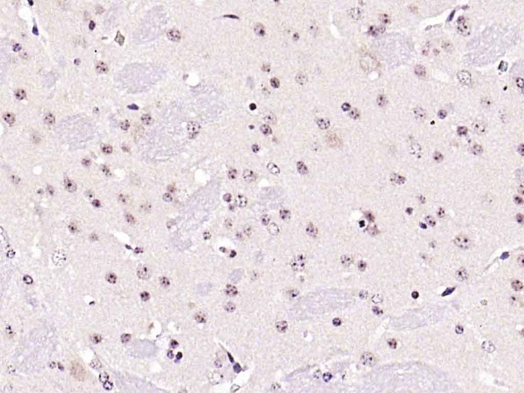 , ,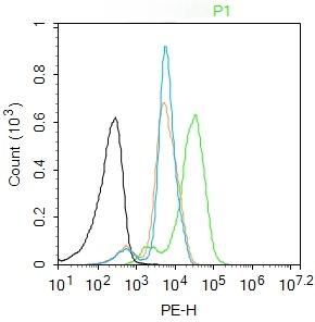 , ,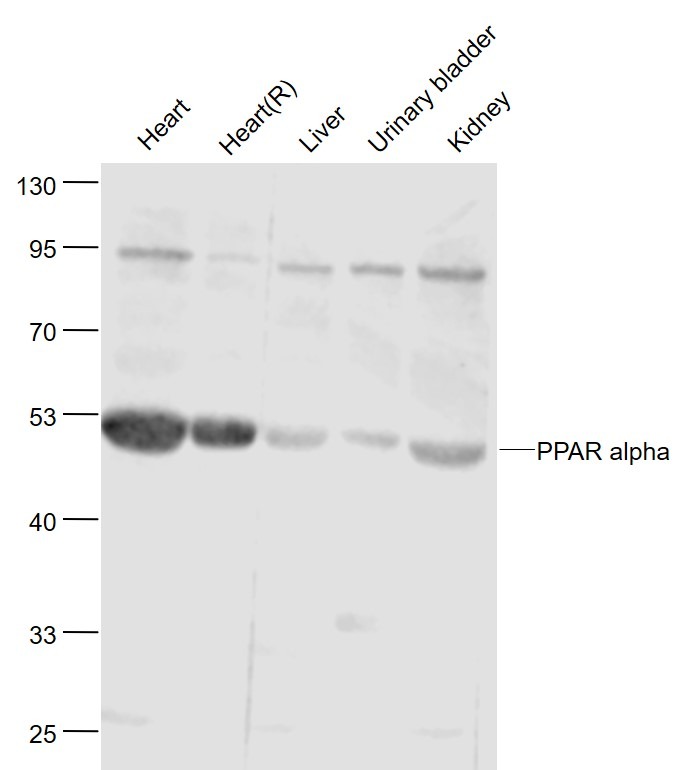 |
| Application | |
| Recommended Dose | WB: 1:500-2000; IHC-P: 1:100-500; IHC-Fr: 1:100-500; IF: 1:100-500; FCM: 1μg /test |
| Antibody Type | Polyclonal |
| Host Species | Rabbit |
| Subcellular Localization | Nucleus. |
| Tissue Specificity | Skeletal muscle, liver, heart and kidney. |
| Construction | Polyclonal Antibody |
| Purification | Protein A purified |
| Appearance | Liquid |
| Formulation | 0.01M TBS (pH7.4) with 1% BSA, 0.02% Proclin300 and 50% Glycerol. |
| Concentration | 1 mg/mL |
| Research Background | Peroxisome proliferators are nongenotoxic carcinogens which are purported to exert their effect on cells through their interaction with members of the nuclear hormone receptor family, termed Peroxisome Proliferator Activated Receptors (PPARs). Nuclear hormone receptors are ligand dependent intracellular proteins that stimulate transcription of specific genes by binding to specific DNA sequences following activation by the appropriate ligand. Studies indicate that PPARs are activated by peroxisome proliferators such as clofibric acid, nafenopin, and WY-14,643, as well as by some fatty acids. It has also been shown that PPARs can induce transcription of acyl coenzyme A oxidase and cytochrome P450 A6 (CYP450 A6) through interaction with specific response elements. PPAR alpha is activated by free fatty acids including linoleic, arachidonic, and oleic acids. Induction of peroxisomes by this mechanism leads to a reduction in blood triglyceride levels. PPAR alpha is expressed mainly in skeletal muscle, heart, liver, and kidney and is thought to regulate many genes involved in the beta-oxidation of fatty acids. Activation of rat liver PPAR alpha has been shown to suppress hepatocyte apoptosis. PPAR alpha, like several other nuclear hormone receptors, heterodimerizes with retinoic X receptor (RXR) alpha to form a transcriptionally competent complex. |
| Immunogen | KLH conjugated synthetic peptide: human PPAR alpha |
| Antigen Species | Human |
| Gene Name | Ppara |
| Gene ID | |
| Protein Name | Peroxisome proliferator-activated receptor alpha |
| Uniprot ID | |
| Biology Area | Metabolism of lipids and lipoproteins,Response to hypoxia,Nuclear hormone receptors,Metabolism,Zinc Finger,Fatty acids,Lipid metabolism,Mitochondrial transcription,Hypoxia,Mitochondrial Biogenesis,Fatty acid oxidation,Obesity |
| Function | Ligand-activated transcription factor. Key regulator of lipid metabolism. Activated by the endogenous ligand 1-palmitoyl-2-oleoyl-sn-glycerol-3-phosphocholine (16:0/18:1-GPC). Activated by oleylethanolamide, a naturally occurring lipid that regulates satiety (By similarity). Receptor for peroxisome proliferators such as hypolipidemic drugs and fatty acids. Regulates the peroxisomal beta-oxidation pathway of fatty acids. Functions as transcription activator for the ACOX1 and P450 genes. Transactivation activity requires heterodimerization with RXRA and is antagonized by NR2C2. |
| Molecular Weight | Theoretical: 51 kDa. Actual: 51 kDa. |
| Stability & Storage | Store at -20°C or -80°C for 12 months. Avoid repeated freeze-thaw cycles. |
| Transport | Shipping with blue ice. |
| Size | Quantity | Unit Price | Amount | Operation |
|---|
 Hello! How can I help you today?
 Copyright © 2015-2026 TargetMol Chemicals Inc. All Rights Reserved.
