
Shopping Cart
Remove All Your shopping cart is currently empty

Synonyms: Programmed cell death 6-interacting protein, PDCD6-interacting protein, KIAA1375, Hp95, ALIX, ALG-2-interacting protein X, ALG-2-interacting protein 1, AIP1
Anti-PDCD6IP Polyclonal Antibody
| Pack Size | Price | USA Stock | Global Stock | Quantity |
|---|---|---|---|---|
| 50 µL | $220 | 7-10 days | 7-10 days | |
| 100 µL | $372 | 7-10 days | 7-10 days | |
| 200 µL | $529 | 7-10 days | 7-10 days |
| Description | Anti-PDCD6IP Polyclonal Antibody is a Rabbit antibody targeting PDCD6IP. Anti-PDCD6IP Polyclonal Antibody can be used in IF, IHC-Fr, IHC-P, WB. |
| Synonyms | Programmed cell death 6-interacting protein, PDCD6-interacting protein, KIAA1375, Hp95, ALIX, ALG-2-interacting protein X, ALG-2-interacting protein 1, AIP1 |
| Ig Type | IgG |
| Reactivity | Human,Mouse (predicted:Rat) |
| Verified Activity | 1. Tissue/cell: human lung carcinoma; 4% Paraformaldehyde-fixed and paraffin-embedded; Antigen retrieval: citrate buffer (0.01M, pH6.0), Boiling bathing for 15 min; Block endogenous peroxidase by 3% Hydrogen peroxide for 30 min; Blocking buffer (normal goat serum) at 37°C for 20 min; Incubation: Anti-IL-10 Polyclonal Antibody, Unconjugated 1:200, overnight at 4°C, followed by conjugation to the secondary antibody and DAb staining. 2. Sample: Lane 1: Testis (Mouse) Lysate at 40 μg Lane 2: Cerebrum (Mouse) Lysate at 40 μg Lane 3: Testis (Rat) Lysate at 40 μg Lane 4: Cerebrum (Rat) Lysate at 40 μg Lane 5: Jurkat (Human) Cell Lysate at 30 μg Primary: Anti-ALIX (TMAB-01348) at 1/1000 dilution Secondary: IRDye800CW Goat Anti-Rabbit IgG at 1/20000 dilution Predicted band size: 95 kDa Observed band size: 100 kDa 3. 25 μg total protein per Lane of various lysates probed with PDCD6IP polyclonal antibody, unconjugated (TMAB-01348) at 1:1000 dilution and 4°C overnight incubation. Followed by conjugated secondary antibody incubation at RT for 60 min. 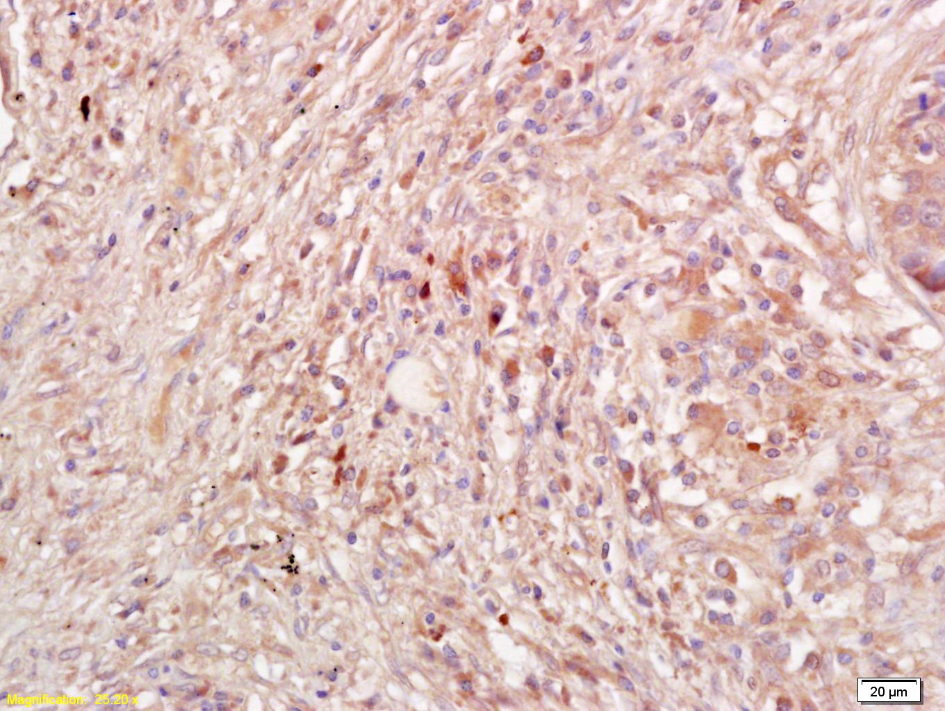 , ,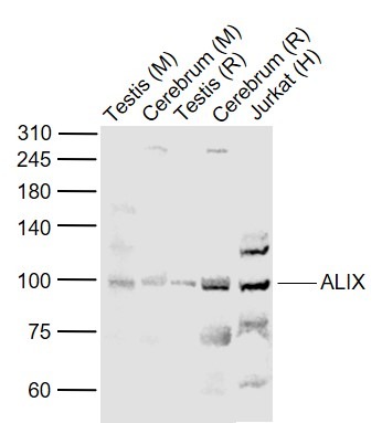 , ,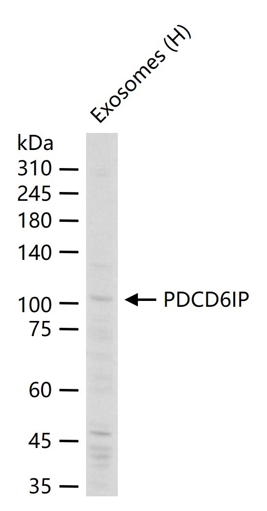 |
| Application | |
| Recommended Dose | WB: 1:500-2000; IHC-P: 1:100-500; IHC-Fr: 1:100-500; IF: 1:100-500 |
| Antibody Type | Polyclonal |
| Host Species | Rabbit |
| Subcellular Localization | Cytoplasm, cytosol. Melanosome. Cytoplasm, cytoskeleton, centrosome. Note=Identified by mass spectrometry in melanosome fractions from stage I to stage IV. Colocalized with CEP55 in the midbody during cytokinesis. Colocalized with CEP55 at centrosomes of non-dividing cells. |
| Construction | Polyclonal Antibody |
| Purification | Protein A purified |
| Appearance | Liquid |
| Formulation | 0.01M TBS (pH7.4) with 1% BSA, 0.02% Proclin300 and 50% Glycerol. |
| Concentration | 1 mg/mL |
| Research Background | This gene encodes a protein that functions within the ESCRT pathway in the abscission stage of cytokinesis, in intralumenal endosomal vesicle formation, and in enveloped virus budding. Studies using mouse cells have shown that overexpression of this protein can block apoptosis. In addition, the product of this gene binds to the product of the PDCD6 gene, a protein required for apoptosis, in a calcium-dependent manner. This gene product also binds to endophilins, proteins that regulate membrane shape during endocytosis. Overexpression of this gene product and endophilins results in cytoplasmic vacuolization, which may be partly responsible for the protection against cell death. Several alternatively spliced transcript variants encoding different isoforms have been found for this gene. Related pseudogenes have been identified on chromosome 15. [provided by RefSeq, Jan 2012] |
| Immunogen | KLH conjugated synthetic peptide: human PDCD6IP |
| Antigen Species | Human |
| Gene Name | PDCD6IP |
| Gene ID | |
| Protein Name | Programmed cell death 6-interacting protein |
| Uniprot ID | |
| Biology Area | Death Ligands,Host Virus Interaction,Regulation |
| Function | Class E VPS protein involved in concentration and sorting of cargo proteins of the multivesicular body (MVB) for incorporation into intralumenal vesicles (ILVs) that are generated by invagination and scission from the limiting membrane of the endosome. Binds to the phospholipid lysobisphosphatidic acid (LBPA) which is abundant in MVBs internal membranes. The MVB pathway appears to require the sequential function of ESCRT-O, -I,-II and -III complexes. The ESCRT machinery also functions in topologically equivalent membrane fission events, such as the terminal stages of cytokinesis and enveloped virus budding (HIV-1 and other lentiviruses). Appears to be an adapter for a subset of ESCRT-III proteins, such as CHMP4, to function at distinct membranes. Required for completion of cytokinesis. Involved in HIV-1 virus budding. Can replace TSG101 it its role of supporting HIV-1 release; this function implies the interaction with CHMP4B. May play a role in the regulation of both apoptosis and cell proliferation. |
| Molecular Weight | Theoretical: 95 kDa. Actual: 100 kDa. |
| Stability & Storage | Store at -20°C or -80°C for 12 months. Avoid repeated freeze-thaw cycles. |
| Transport | Shipping with blue ice. |
| Size | Quantity | Unit Price | Amount | Operation |
|---|
 Hello! How can I help you today?
 Copyright © 2015-2026 TargetMol Chemicals Inc. All Rights Reserved.
