
Shopping Cart
Remove All Your shopping cart is currently empty


Anti-PD-L1 Polyclonal Antibody 4 is a Rabbit antibody targeting PD-L1. Anti-PD-L1 Polyclonal Antibody 4 can be used in FCM, WB.
| Pack Size | Price | USA Warehouse | Global Warehouse | Quantity |
|---|---|---|---|---|
| 50 μL | $220 | 7-10 days | 7-10 days | |
| 100 μL | $372 | 7-10 days | 7-10 days | |
| 200 μL | $529 | 7-10 days | 7-10 days |
| Description | Anti-PD-L1 Polyclonal Antibody 4 is a Rabbit antibody targeting PD-L1. Anti-PD-L1 Polyclonal Antibody 4 can be used in FCM, WB. |
| Synonyms | Pdl1, Pdcd1lg1, Pdcd1l1, CD274 molecule, B7h1, A530045L16Rik |
| Ig Type | IgG |
| Reactivity | Mouse (predicted:Human,Rat) |
| Verified Activity | 1. Sample: Eye (Mouse) Lysate at 40 μg LympHnode (Mouse) Lysate at 40 μg Primary: Anti-PD-L1 (TMAB-01362) at 1/1000 dilution Secondary: IRDye800CW Goat Anti-Rabbit IgG at 1/20000 dilution Predicted band size: 48 kDa Observed band size: 50 kDa 2. Blank control (black line): RAW264.7. Primary Antibody (green line): Rabbit Anti-PD-L1 antibody (TMAB-01362) Dilution: 1 μg/Test; Secondary Antibody (white blue line): Goat anti-rabbit IgG-AF488 Dilution: 0.5 μg/Test. Isotype control (orange line): Normal Rabbit IgG Protocol The cells were fixed with 4% PFA (10 min at room temperature) and then permeabilized with 90% ice-cold methanol for 20 min at-20°C, The cells were then incubated in 5% BSA to block non-specific protein-protein interactions for 30 min at room temperature. Cells stained with Primary Antibody for 30 min at room temperature. The secondary antibody used for 40 min at room temperature. 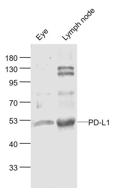 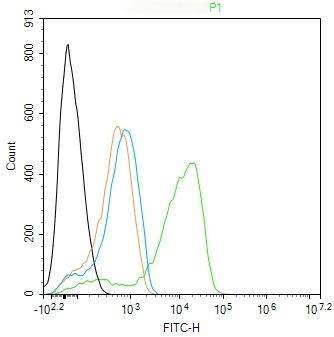 |
| Application | |
| Recommended Dose | WB: 1:500-2000; FCM: 1ug/Test |
| Antibody Type | Polyclonal |
| Host Species | Rabbit |
| Subcellular Localization | Isoform 1: Cell membrane; Single-pass type I membrane protein. Isoform 2: Endomembrane system; Single-pass type I membrane protein. |
| Tissue Specificity | Highly expressed in the heart, skeletal muscle, placenta and lung. Weakly expressed in the thymus, spleen, kidney and liver. Expressed on activated T- and B-cells, dendritic cells, keratinocytes and monocytes. |
| Construction | Polyclonal Antibody |
| Purification | Protein A purified |
| Appearance | Liquid |
| Formulation | 0.01M TBS (pH7.4) with 1% BSA, 0.02% Proclin300 and 50% Glycerol. |
| Concentration | 1 mg/mL |
| Research Background | This gene encodes an immune inhibitory receptor ligand that is expressed by hematopoietic and non-hematopoietic cells, such as T cells and B cells and various types of tumor cells. The encoded protein is a type I transmembrane protein that has immunoglobulin V-like and C-like domains. Interaction of this ligand with its receptor inhibits T-cell activation and cytokine production. During infection or inflammation of normal tissue, this interaction is important for preventing autoimmunity by maintaining homeostasis of the immune response. In tumor microenvironments, this interaction provides an immune escape for tumor cells through cytotoxic T-cell inactivation. Expression of this gene in tumor cells is considered to be prognostic in many types of human malignancies, including colon cancer and renal cell carcinoma. Alternative splicing results in multiple transcript variants. [provided by RefSeq, Sep 2015] |
| Immunogen | KLH conjugated synthetic peptide: mouse PD-L1 |
| Antigen Species | Mouse |
| Gene Name | CD274 |
| Gene ID | |
| Protein Name | Programmed cell death 1 ligand 1 |
| Uniprot ID | |
| Biology Area | Tumor antigens,Tumor-associated antigens,Non-lineage,Autoimmune,pd-l1 |
| Function | Involved in the costimulatory signal, essential for T-cell proliferation and production of IL10 and IFNG, in an IL2-dependent and a PDCD1-independent manner. Interaction with PDCD1 inhibits T-cell proliferation and cytokine production. |
| Molecular Weight | Theoretical: 32 kDa. |
| Stability & Storage | Store at -20°C or -80°C for 12 months. Avoid repeated freeze-thaw cycles. |
| Transport | Shipping with blue ice. |

| Size | Quantity | Unit Price | Amount | Operation |
|---|
 Hello! How can I help you today?
 Copyright © 2015-2026 TargetMol Chemicals Inc. All Rights Reserved.
