
Shopping Cart
Remove All Your shopping cart is currently empty

Synonyms: Testicular receptor 3, ST-59, Orphan nuclear receptor TR3, Orphan nuclear receptor HMR, Nuclear receptor subfamily 4immunitygroup A member 1, Nuclear hormone receptor NUR/77 (Nur77), NR4A1, NAK1, HMR, GFRP1, Early response protein NAK1
Anti-NR4A1 Antibody
(7O127)
| Pack Size | Price | USA Stock | Global Stock | Quantity |
|---|---|---|---|---|
| 50 µL | $296 | 7-10 days | 7-10 days | |
| 100 µL | $498 | 7-10 days | 7-10 days |
| Description | Anti-NR4A1 Antibody (7O127) is a Rabbit antibody targeting NR4A1. Anti-NR4A1 Antibody (7O127) can be used in ICC/IF,IHC,WB. |
| Synonyms | Testicular receptor 3, ST-59, Orphan nuclear receptor TR3, Orphan nuclear receptor HMR, Nuclear receptor subfamily 4immunitygroup A member 1, Nuclear hormone receptor NUR/77 (Nur77), NR4A1, NAK1, HMR, GFRP1, Early response protein NAK1 |
| Ig Type | IgG |
| Clone | 7O127 |
| Reactivity | Human,Mouse,Rat |
| Verified Activity | 1. Western blot analysis of NUR77 on rat brain cells lysates using anti-NUR77 antibody at 1/500 dilution. 2. ICC staining of NUR77 in Hela cells (green). 4% PFA fixed cells 20 minutes, washed with PBS. Cells were probed with the primary antibody (49510, 1/50) overnight at 4℃ washed with PBS. CoraLite488 Goat anti-Rabbit lgG was used as the secondary antibody at 1/100 dilution. The nuclear counter stain is DAPI (blue). 3. ICC staining of NUR77 in 3T3-L1 cells (green). 4% PFA fixed cells 20 minutes, washed with PBS. Cells were probed with the primary antibody (49510, 1/50) overnight at 4℃ washed with PBS. CoraLite488 Goat anti-Rabbit lgG was used as the secondary antibody at 1/100 dilution. The nuclear counter stain is DAPI (blue). 4. Immunohistochemical analysis of paraffin-embedded human liver tissue with Rabbit anti-NUR77 antibody (ET1703-97) at 1/1,000 dilution. The section was pre-treated using heat mediated antigen retrieval with sodium citrate buffer (pH 6.0) for 2 minutes. The tissues were blocked in 1% BSA for 20 minutes at room temperature, washed with ddH2O and PBS, and then probed with the primary antibody at 1/1,000 dilution for 1 hour at room temperature. The detection was performed using an HRP conjugated compact polymer system. DAB was used as the chromogen. Tissues were counterstained with hematoxylin and mounted with DPX. 5. Immunohistochemical analysis of paraffin-embedded human colon carcinoma tissue with Rabbit anti-NUR77 antibody at 1/1,000 dilution. The section was pre-treated using heat mediated antigen retrieval with sodium citrate buffer (pH 6.0) for 2 minutes. The tissues were blocked in 1% BSA for 20 minutes at room temperature, washed with ddH2O and PBS, and then probed with the primary antibody at 1/1,000 dilution for 1 hour at room temperature. The detection was performed using an HRP conjugated compact polymer system. DAB was used as the chromogen. Tissues were counterstained with hematoxylin and mounted with DPX. 6. Immunohistochemical analysis of paraffin-embedded human breast tissue using anti-NUR77 antibody. The section was pre-treated using heat mediated antigen retrieval with Tris-EDTA buffer (pH 8.0-8.4) for 20 minutes.The tissues were blocked in 5% BSA for 30 minutes at room temperature, washed with ddH2O and PBS, and then probed with the primary antibody (1/50) for 30 minutes at room temperature. The detection was performed using an HRP conjugated compact polymer system. DAB was used as the chromogen. Tissues were counterstained with hematoxylin and mounted with DPX. 7. Immunohistochemical analysis of paraffin-embedded human breast tissue using anti-NUR77 antibody. The section was pre-treated using heat mediated antigen retrieval with Tris-EDTA buffer (pH 8.0-8.4) for 20 minutes.The tissues were blocked in 5% BSA for 30 minutes at room temperature, washed with ddH2O and PBS, and then probed with the primary antibody (1/50) for 30 minutes at room temperature. The detection was performed using an HRP conjugated compact polymer system. DAB was used as the chromogen. Tissues were counterstained with hematoxylin and mounted with DPX. 8. Immunohistochemical analysis of paraffin-embedded mouse ovarian tissue using anti-NUR77 antibody. The section was pre-treated using heat mediated antigen retrieval with Tris-EDTA buffer (pH 8.0-8.4) for 20 minutes.The tissues were blocked in 5% BSA for 30 minutes at room temperature, washed with ddH2O and PBS, and then probed with the primary antibody (1/50) for 30 minutes at room temperature. The detection was performed using an HRP conjugated compact polymer system. DAB was used as the chromogen. Tissues were counterstained with hematoxylin and mounted with DPX. 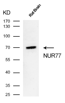 , ,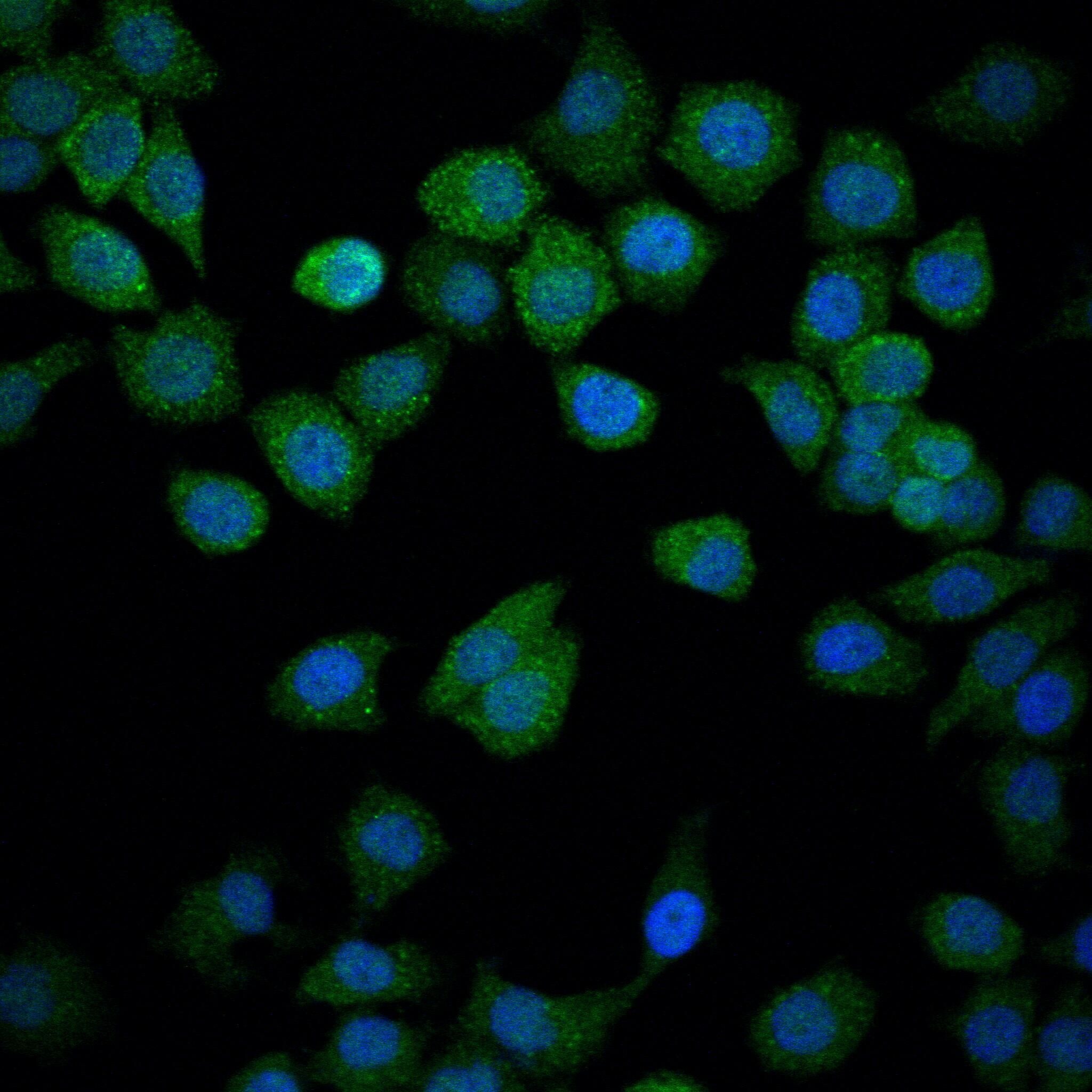 , ,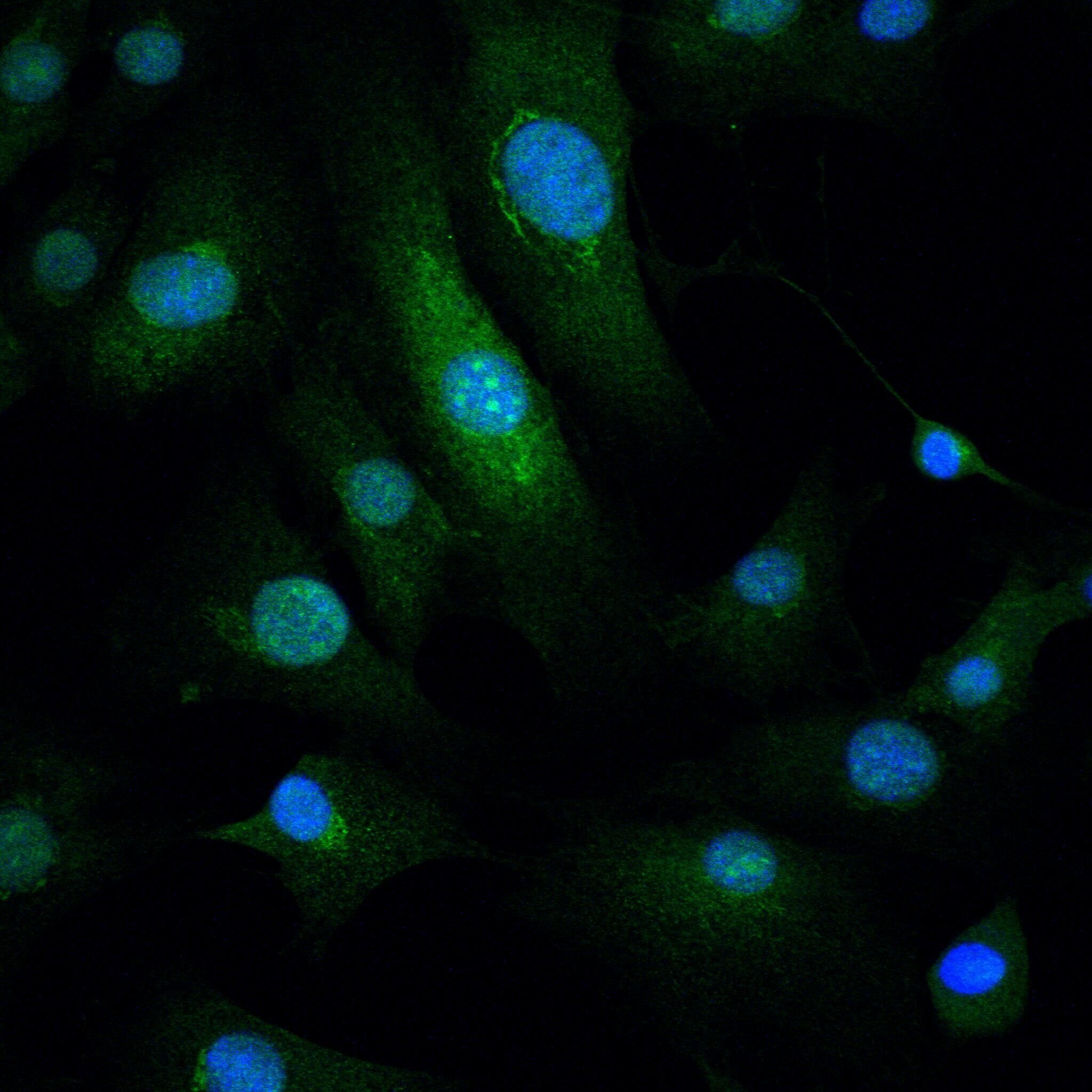 , ,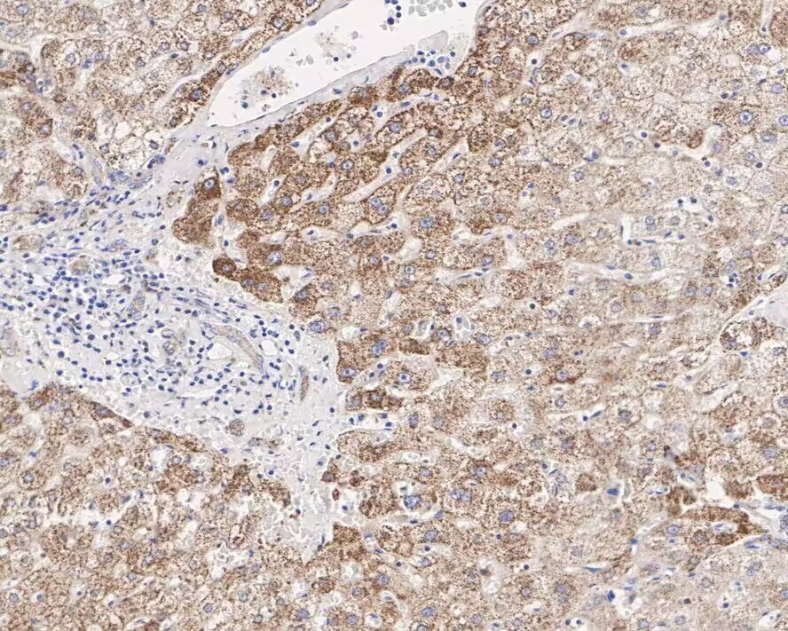 , ,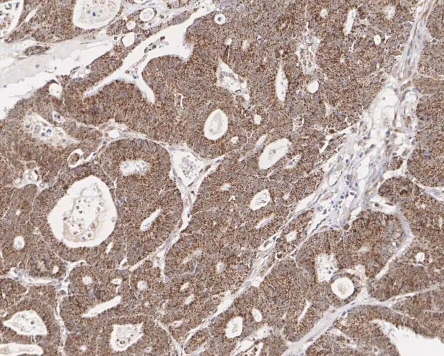 , ,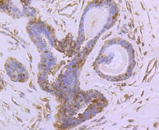 , ,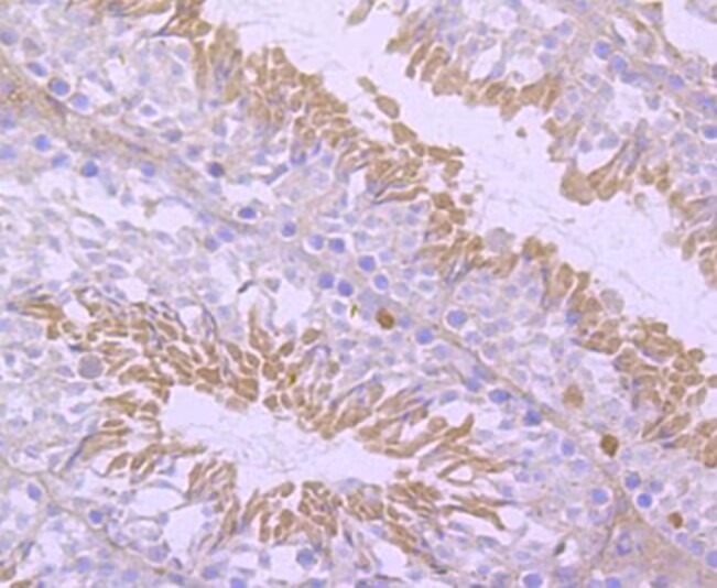 , ,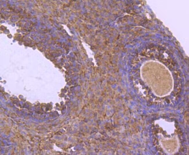 |
| Application | |
| Recommended Dose | WB: 1:500-1000; IHC: 1:50-200; ICC/IF: 1:50-200 |
| Antibody Type | Monoclonal |
| Host Species | Rabbit |
| Construction | Recombinant Antibody |
| Purification | ProA affinity purified |
| Appearance | Liquid |
| Formulation | 1*TBS (pH7.4), 0.05% BSA, 40% Glycerol. Preservative: 0.05% Sodium Azide. |
| Research Background | Nurr1 (Nur-related factor 1) and Nur77 (also designated NGFI-B) encode orphan nuclear receptors which may comprise an additional subfamily within the nuclear receptor superfamily. The rat and human homologs of mouse Nurr1 are designated RNR1 and NOT, respectively. Both Nurr1 and Nur77 are growth factor inducible immediate early response genes. Induction of both Nurr1 and Nur77 is seen after membrane depolarization while only Nur77 induction is seen with NGF stimulation. JunD acts as a mediator for Nur77. An increase in Nur77 expression is seen in activated T cells during G0 to G1 transition and throughout the G1 phase. In addition to its function as an immediate early gene, Nur77 may play a role in TCR-mediated apoptosis. Cyclosporin A, a potent immunosuppressant, has been shown to inhibit the ability of Nur77 to bind DNA. A dominant negative form of Nur77 can protect T cell hybridomas from activation-induced apoptosis. However, the absolute requirement of Nur77 for TCR-mediated apoptosis is still under debate. |
| Conjucates | Unconjugated |
| Immunogen | Synthetic peptide within Human NUR77 aa 10-49 / 598 |
| Antigen Species | Human |
| Uniprot ID |
| Molecular Weight | Theoretical: 64 kDa. |
| Stability & Storage | Store at -20°C or -80°C for 12 months. Avoid repeated freeze-thaw cycles. |
| Transport | Shipping with blue ice. |
| Size | Quantity | Unit Price | Amount | Operation |
|---|
 Hello! How can I help you today?
 Copyright © 2015-2026 TargetMol Chemicals Inc. All Rights Reserved.
