
Shopping Cart
Remove All Your shopping cart is currently empty

Synonyms: SH2/SH3 Adaptor Protein NCK-α, SH2/SH3 Adaptor Protein NCK-Alpha, Nck-1, NCK1, NCK adaptor Protein 1, NCK, Cytoplasmic Protein NCK1
Anti-NCK1 Antibody
(6Y736)
| Pack Size | Price | USA Stock | Global Stock | Quantity |
|---|---|---|---|---|
| 50 µL | $297 | 7-10 days | 7-10 days | |
| 100 µL | $498 | 7-10 days | 7-10 days |
| Description | Anti-NCK1 Antibody (6Y736) is a Rabbit antibody targeting NCK1. Anti-NCK1 Antibody (6Y736) can be used in FCM,ICC,IF,IHC,IP,WB. |
| Synonyms | SH2/SH3 Adaptor Protein NCK-α, SH2/SH3 Adaptor Protein NCK-Alpha, Nck-1, NCK1, NCK adaptor Protein 1, NCK, Cytoplasmic Protein NCK1 |
| Ig Type | IgG |
| Clone | 6Y736 |
| Reactivity | Human,Mouse,Rat |
| Verified Activity | 1. Western blot analysis of NCK1 on different lysates using anti-NCK1 antibody at 1/500 dilution. Positive control: Lane 1: Hela, Lane 1: NIH-3T3, Lane 2: Rat kidney tissue, Lane 3: Mouse testis tissue. 2. Immunohistochemical analysis of paraffin-embedded mouse testis tissue using anti-NCK1 antibody. Counter stained with hematoxylin. 3. Immunohistochemical analysis of paraffin-embedded rat testis tissue using anti-NCK1 antibody. Counter stained with hematoxylin. 4. Immunohistochemical analysis of paraffin-embedded human tonsil tissue using anti-NCK1 antibody. Counter stained with hematoxylin. 5. Immunohistochemical analysis of paraffin-embedded human kidney tissue using anti-NCK1 antibody. Counter stained with hematoxylin. 6. ICC staining NCK1 in LOVO cells (green). The nuclear counter stain is DAPI (blue). Cells were fixed in paraformaldehyde, permeabilised with 0.25% Triton X100/PBS. 7. ICC staining NCK1 in A431 cells (green). The nuclear counter stain is DAPI (blue). Cells were fixed in paraformaldehyde, permeabilised with 0.25% Triton X100/PBS. 8. ICC staining NCK1 in HUVEC cells (green). The nuclear counter stain is DAPI (blue). Cells were fixed in paraformaldehyde, permeabilised with 0.25% Triton X100/PBS. 9. Flow cytometric analysis of Jurkat cells with NCK1 antibody at 1/50 dilution (red) compared with an unlabelled control (cells without incubation with primary antibody; black). Alexa Fluor 488-conjugated goat anti rabbit IgG was used as the secondary antibody. 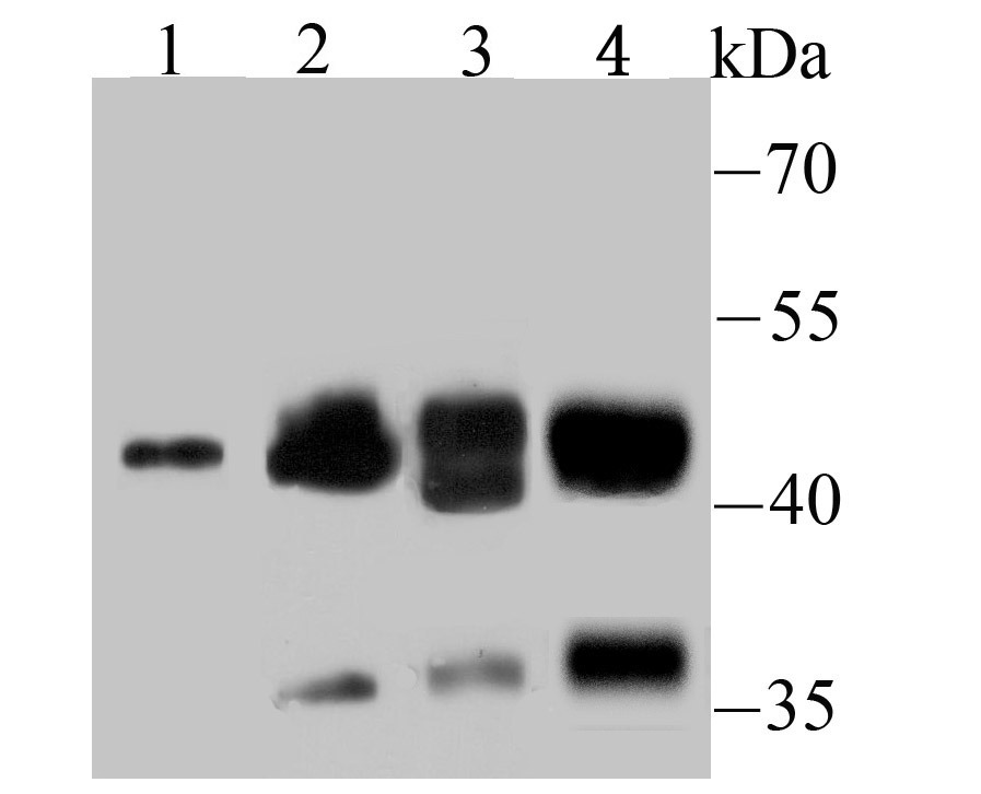 , ,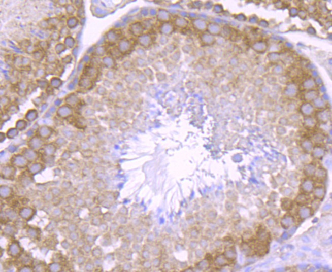 , ,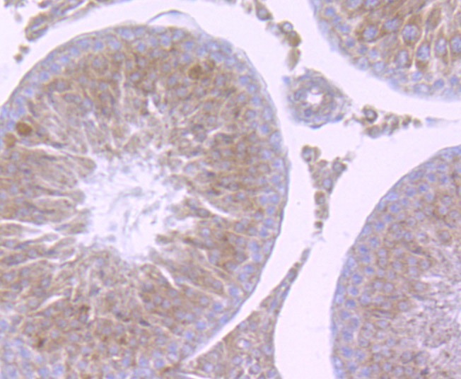 , ,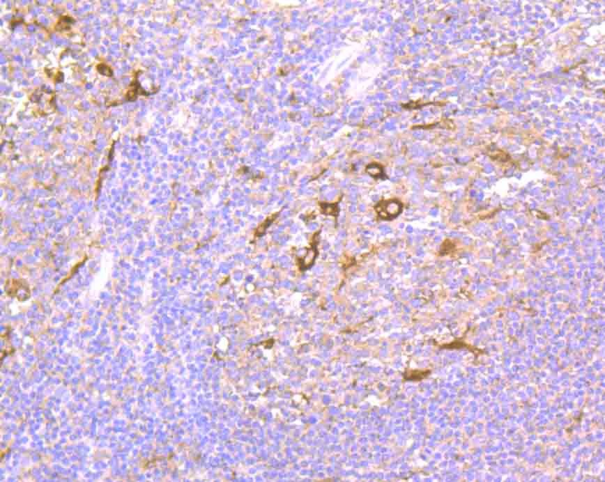 , ,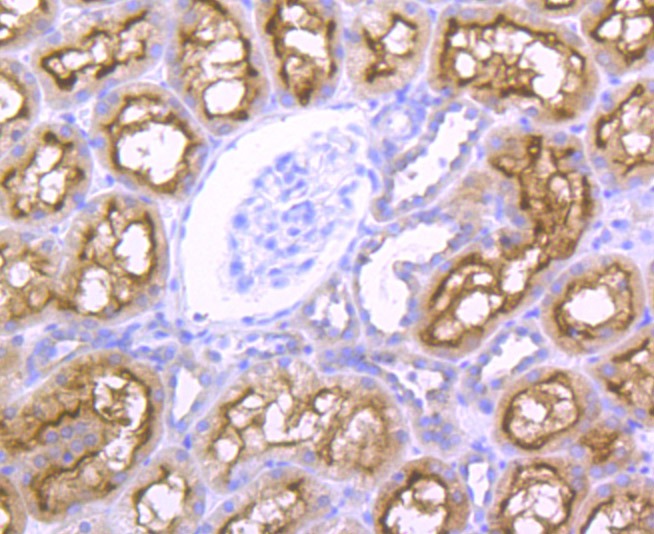 , ,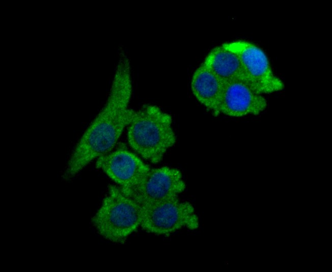 , ,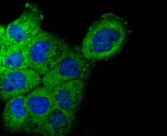 , ,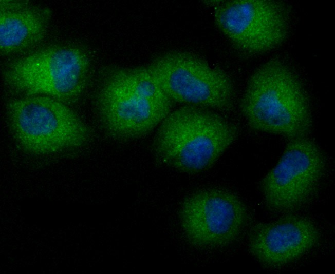 , ,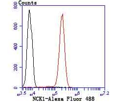 |
| Application | |
| Recommended Dose | WB: 1:500-2000; IHC: 1:50-200; ICC: 1:50-200; IP: 1:10-50; FCM: 1:50-100 |
| Antibody Type | Monoclonal |
| Host Species | Rabbit |
| Construction | Recombinant Antibody |
| Purification | ProA affinity purified |
| Appearance | Liquid |
| Formulation | 1*TBS (pH7.4), 1%BSA, 40%Glycerol. Preservative: 0.05% Sodium Azide. |
| Research Background | The NCK family of SH2/SH3 adaptor proteins consists of two members, NCK1 (NCKα) and NCK2 (NCKβ), which couple tyrosine kinase signaling, including the EGF and PDGF receptor-pathways, to downstream signaling proteins. Specifically, overexpression of Nck1 in NIH/3T3 cells decreases DNA synthesis stimulated by EGF. Furthermore, the SH2 domain of NCK2 inhibits EGF- and PDGF-induced DNA synthesis. The SH3 domain of NCK binds a proline-rich domain on PAK, a known actin cytoskeleton regulator. The NCK protein thus mediates the interaction between PAK and RAC. The NCK2 protein binds human PDGFR-b (Tyr 1009); overexpression of Nck2 inhibits PDGF-induced membrane ruffling and lamellipod formation. Various growth factor receptors, cell surface antigens and adhesion molecules phosphorylate mammalian NCK1 and NCK2. The human NCK1 and NCK2 genes map to chromosomes 3q22.3 and 2q12.2, respectively. |
| Conjucates | Unconjugated |
| Immunogen | Recombinant Protein |
| Uniprot ID |
| Molecular Weight | Theoretical: 43/35 kDa. |
| Stability & Storage | Store at -20°C or -80°C for 12 months. Avoid repeated freeze-thaw cycles. |
| Transport | Shipping with blue ice. |
| Size | Quantity | Unit Price | Amount | Operation |
|---|
 Hello! How can I help you today?
 Copyright © 2015-2026 TargetMol Chemicals Inc. All Rights Reserved.
