
Shopping Cart
Remove All Your shopping cart is currently empty

Synonyms:
| Pack Size | Price | USA Stock | Global Stock | Quantity |
|---|---|---|---|---|
| 50 µL | $208 | 7-10 days | 7-10 days | |
| 100 µL | $349 | 7-10 days | 7-10 days |
| Description | Anti-NANOG Antibody (1M448) is a Mouse antibody targeting NANOG. Anti-NANOG Antibody (1M448) can be used in ELISA,FCM,ICC,IF,WB. |
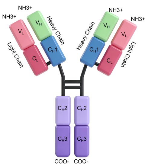
| Ig Type | IgG1 |
| Clone | 1M448 |
| Reactivity | Human, Mouse, Rat |
| Verified Activity | 1. Western Blot -Positive WB detected in: Rat brain tissue, Mouse brain tissue -All lanes: NANOG antibody at 1:500 -Secondary: Goat polyclonal to Mouse IgG at 1/10000 dilution -Predicted band size: 35, 33 kDa -Observed band size: 46, 42 kDa 2. Western Blot -Positive WB detected in: MCF-7 whole cell lysate, Ntera-2 whole cell lysate, A549 whole cell lysate -All lanes: NANOG antibody at 1:500 -Secondary: Goat polyclonal to Mouse IgG at 1/10000 dilution -Predicted band size: 35, 33 kDa -Observed band size: 46, 40 kDa 3. Immunocytochemistry analysis of TMAH-00803 diluted at 1:100 and staining in Hela cells performed on a Leica BondTM system. The cells were fixed in 4% formaldehyde, permeabilized using 0.2% Triton X-100 and blocked with 10% normal goat serum 30min at RT. Then primary antibody (1% BSA) was incubated at 4°C overnight. The primary is detected by a biotinylated secondary antibody and visualized using an HRP conjugated SP system. 4. Immunocytochemistry analysis of TMAH-00803 diluted at 1:100 and staining in Ntera-2 cells performed on a Leica BondTM system. The cells were fixed in 4% formaldehyde, permeabilized using 0.2% Triton X-100 and blocked with 10% normal goat serum 30min at RT. Then primary antibody (1% BSA) was incubated at 4°C overnight. The primary is detected by a biotinylated secondary antibody and visualized using an HRP conjugated SP system. 5. Immunofluorescence staining of Hela cells with TMAH-00803 at 1:100, counter-stained with DAPI. The cells were blocked in 10% normal Goat Serum and then incubated with the primary antibody overnight at 4°C. The secondary antibody was Alexa Fluor 488-congugated AffiniPure Goat Anti-Mouse IgG(H+L). 6. Immunofluorescence staining of Ntera-2 cells with TMAH-00803 at 1:100, counter-stained with DAPI. The cells were blocked in 10% normal Goat Serum and then incubated with the primary antibody overnight at 4°C. The secondary antibody was Alexa Fluor 488-congugated AffiniPure Goat Anti-Mouse IgG(H+L). 7. Overlay histogram showing Hela cells stained with TMAH-00803 (red line) at 1:250. The cells were incubated in 1x PBS /10% normal goat serum to block non-specific protein-protein interactions followed by primary antibody for 1 h at 4°C. The secondary antibody used was FITC goat anti-mouse IgG(H+L) at 1/200 dilution for 1 h at 4°C. Isotype control antibody (green line) was used under the same conditions. Acquisition of >10,000 events was performed. 8. Overlay histogram showing MCF-7 cells stained with TMAH-00803 (red line) at 1:250. The cells were incubated in 1x PBS /10% normal goat serum to block non-specific protein-protein interactions followed by primary antibody for 1 h at 4°C. The secondary antibody used was FITC goat anti-mouse IgG(H+L) at 1/200 dilution for 1 h at 4°C. Isotype control antibody (green line) was used under the same conditions. Acquisition of >10,000 events was performed. 9. Overlay histogram showing Ntera-2 cells stained with TMAH-00803 (red line) at 1:250. The cells were incubated in 1x PBS /10% normal goat serum to block non-specific protein-protein interactions followed by primary antibody for 1 h at 4°C. The secondary antibody used was FITC goat anti-mouse IgG(H+L) at 1/200 dilution for 1 h at 4°C. Isotype control antibody (green line) was used under the same conditions. Acquisition of >10,000 events was performed. |
| Application | |
| Antibody Type | Monoclonal |
| Host Species | Mouse |
| Subcellular Localization | Nucleus. |
| Tissue Specificity | Expressed in testicular carcinoma and derived germ cell tumors (at protein level). Expressed in fetal gonads, ovary and testis. Also expressed in ovary teratocarcinoma cell line and testicular embryonic carcinoma. Not expressed in many somatic organs and |
| Construction | Hybridoma Monoclonal Antibody |
| Purification | Protein G purified |
| Appearance | Liquid |
| Formulation | Preservative: 0.03% Proclin 300. Constituents: 50% Glycerol, 0.01M PBS, PH 7.4. |
| Purity | >95% |
| Research Background | Transcription regulator involved in inner cell mass and embryonic stem (ES) cells proliferation and self-renewal. Imposes pluripotency on ES cells and prevents their differentiation towards extraembryonic endoderm and trophectoderm lineages. Blocks bone morphogenetic protein-induced mesoderm differentiation of ES cells by physically interacting with SMAD1 and interfering with the recruitment of coactivators to the active SMAD transcriptional complexes. Acts as a transcriptional activator or repressor. Binds optimally to the DNA consensus sequence 5'-TAAT[GT][GT]-3' or 5'-[CG][GA][CG]C[GC]ATTAN[GC]-3'. Binds to the POU5F1/OCT4 promoter. Able to autorepress its expression in differentiating (ES) cells: binds to its own promoter following interaction with ZNF281/ZFP281, leading to recruitment of the NuRD complex and subsequent repression of expression. When overexpressed, promotes cells to enter into S phase and proliferation. |
| Conjucates | Unconjugated |
| Immunogen | Recombinant Protein: Human Homeobox Protein NANOG Protein (1-305AA) |
| Antigen Species | Human |
| Gene Name | NANOG |
| Gene ID | |
| Uniprot ID | |
| Biology Area | Stem Cells |
| Stability & Storage | Store at -20°C or -80°C for 12 months. Avoid repeated freeze-thaw cycles. |
| Transport | Shipping with blue ice. |
| Size | Quantity | Unit Price | Amount | Operation |
|---|
 Hello! How can I help you today?
 Copyright © 2015-2026 TargetMol Chemicals Inc. All Rights Reserved.
