
Shopping Cart
Remove All Your shopping cart is currently empty

Synonyms: TK25, TK14, K-SAM, KGFR, JWS, fibroblast growth factor receptor 2, FGFR2 α(IIIb), ECT1, CFD1, CEK3, CD332, BFR-1, BEK, BBDS
Anti-FGFR2 Polyclonal Antibody
| Pack Size | Price | USA Stock | Global Stock | Quantity |
|---|---|---|---|---|
| 50 µL | $222 | 7-10 days | 7-10 days | |
| 100 µL | $373 | 7-10 days | 7-10 days | |
| 200 µL | $527 | 7-10 days | 7-10 days |
| Description | Anti-FGFR2 Polyclonal Antibody is a Rabbit antibody targeting FGFR2. Anti-FGFR2 Polyclonal Antibody can be used in FCM,ICC/IF,IF,IHC-Fr,IHC-P,WB. |
| Synonyms | TK25, TK14, K-SAM, KGFR, JWS, fibroblast growth factor receptor 2, FGFR2 α(IIIb), ECT1, CFD1, CEK3, CD332, BFR-1, BEK, BBDS |
| Ig Type | IgG |
| Reactivity | Human,Mouse (predicted:Rat) |
| Verified Activity | 1. Image was kindly submitted by Dr. Uthaman from Yale University. L6 cells were transfected with FGFR2, and stained with RABBIT ANTI-FGFR2 POLYCLONAL ANTIBODY, conjugated (TMAB-00675-FITC) at 1:100 dilution 2. Paraformaldehyde-fixed, paraffin embedded (human stomach cancer); Antigen retrieval by boiling in sodium citrate buffer (pH6.0) for 15 min; Block endogenous peroxidase by 3% hydrogen peroxide for 20 min; Blocking buffer (normal goat serum) at 37°C for 30 min; Antibody incubation with (FGFR2) Polyclonal Antibody, Unconjugated (TMAB-00675) at 1:400 overnight at 4°C, followed by a conjugated secondary for 20 min and DAB staining. 3. Paraformaldehyde-fixed, paraffin embedded (human stomach cancer); Antigen retrieval by boiling in sodium citrate buffer (pH6.0) for 15 min; Block endogenous peroxidase by 3% hydrogen peroxide for 20 min; Blocking buffer (normal goat serum) at 37°C for 30 min; Antibody incubation with (FGFR2) Polyclonal Antibody, Unconjugated (TMAB-00675) at 1:400 overnight at 4°C, followed by a conjugated secondary for 20 min and DAB staining. 4. Paraformaldehyde-fixed, paraffin embedded (mouse brain tissue); Antigen retrieval by boiling in sodium citrate buffer (pH6.0) for 15 min; Block endogenous peroxidase by 3% hydrogen peroxide for 20 min; Blocking buffer (normal goat serum) at 37°C for 30 min; Antibody incubation with (FGFR2) Polyclonal Antibody, Unconjugated (TMAB-00675) at 1:400 overnight at 4°C, followed by a conjugated secondary for 20 min and DAB staining. 5. Tissue/cell: MCF7; 4% Paraformaldehyde-fixed; Triton X-100 at room temperature for 20 min; Blocking buffer (normal goat serum) at 37°C for 20 min; Antibody incubation with (FGFR2) Polyclonal Antibody, Unconjugated (TMAB-00675) 1:200, 90 minutes at 37°C; followed by a conjugated Goat Anti-Rabbit IgG antibody at 37°C for 90 minutes, DAPI (blue) was used to stain the cell nucleus. 6. Sample: Lane 1: Mouse Spinal cord tissue lysates Lane 2: Mouse NIH/3T3 cell lysates Lane 3: Human 293T cell lysates Lane 4: Human A431 cell lysates Lane 5: Human HeLa cell lysates Primary: Anti-FGFR2 (TMAB-00675) at 1/1000 dilution Secondary: IRDye800CW Goat Anti-Rabbit IgG at 1/20000 dilution Predicted band size: 89 kDa Observed band size: 142 kDa 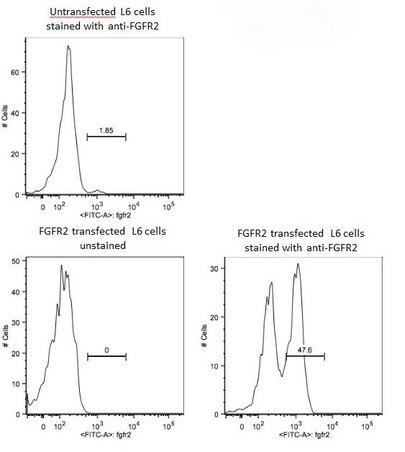 , ,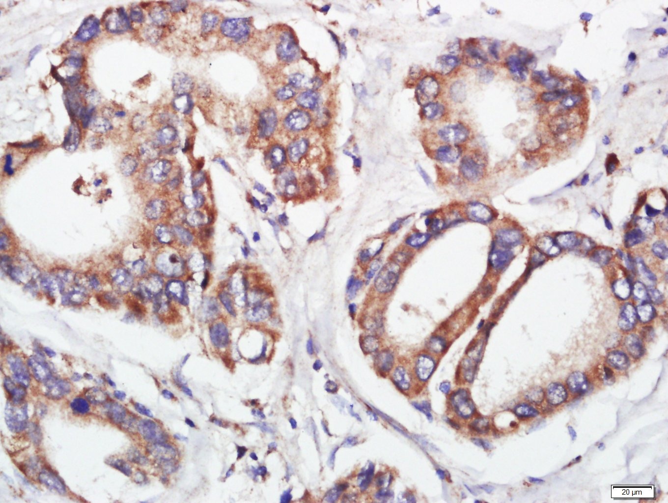 , ,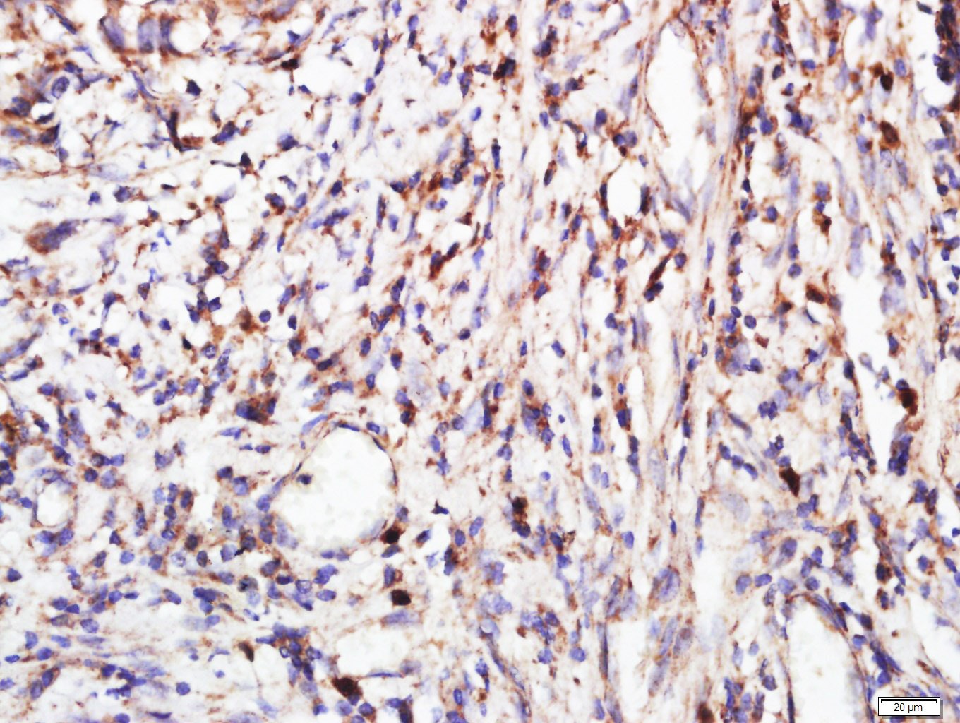 , ,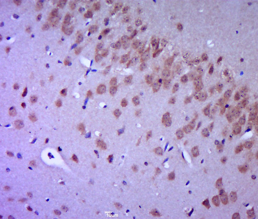 , ,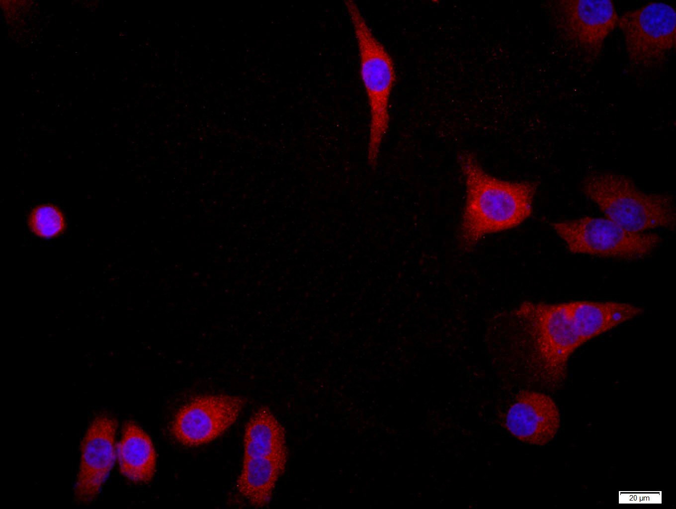 , ,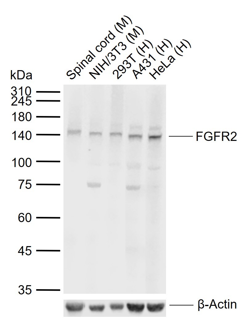 |
| Application | |
| Recommended Dose | WB: 1:500-2000; IHC-P: 1:100-500; IHC-Fr: 1:100-500; ICC/IF: 1:100-500; IF: 1:100-500; FCM: 1μg/Test |
| Antibody Type | Polyclonal |
| Host Species | Rabbit |
| Subcellular Localization | Cell membrane; Single-pass type I membrane protein. Golgi apparatus. Cytoplasmic vesicle. Note=Detected on osteoblast plasma membrane lipid rafts. After ligand binding, the activated receptor is rapidly internalized and degraded.Isoform 1: Cell membrane; Single-pass type I membrane protein. Note=After ligand binding, the activated receptor is rapidly internalized and degraded.Isoform 3: Cell membrane; Single-pass type I membrane protein. Note=After ligand binding, the activated receptor is rapidly internalized and degraded. |
| Construction | Polyclonal Antibody |
| Purification | Protein A purified |
| Appearance | Liquid |
| Formulation | 0.01M TBS (pH7.4) with 1% BSA, 0.02% Proclin300 and 50% Glycerol. |
| Concentration | 1 mg/mL |
| Research Background | bs-0675P is one synthetic peptide derived from human FGFR2. Fibroblast growth factors (FGFs) are members of a large family of structurally related polypeptides that are potent physiological regulators of growth and differentiation for a wide variety of cells of mesodermal, ectodermal and endodermal origin. Four genes encoding for high affinity cell surface FGF receptors (FGFRs) have been identified: FGFR1, FGFR2, FGFR3 and FGFR4. FGFRs are members of the tyrosine kinase family of growth factor receptors. FGFR2 is highly expressed in developing human tissues including the brain, choroids plexus, lung etc. Alternative names: Bacteria expressed kinase; BEK; BFR 1; BFR1; CD 332; CD332; CD332 antigen; CEK 3; CEK3; CFD 1; CFD1; Craniofacial dysostosis 1; Crouzon syndrome; ECT 1; ECT1; FGFR 2; Fibroblast growth factor receptor 2; Hydroxyaryl protein kinase; Jackson Weiss syndrome; JWS; K SAM; K sam protein; Keratinocyte growth factor receptor 2; Keratinocyte growth factor receptor; KGFR; KSAM; Pfeiffer syndrome; Protein tyrosine kinase receptor like 14; TK14; TK25; Tyrosylprotein kinase. |
| Immunogen | KLH conjugated synthetic peptide: human FGFR2 |
| Antigen Species | Human |
| Gene Name | FGFR2 |
| Gene ID | |
| Protein Name | Fibroblast growth factor receptor 2 |
| Uniprot ID | |
| Biology Area | FGF,Receptor tyrosine kinases,FGF Receptors,Ectoderm,Neurogenesis,FGF,Receptor Tyrosine Kinases,Ectoderm |
| Function | Tyrosine-protein kinase that acts as cell-surface receptor for fibroblast growth factors and plays an essential role in the regulation of cell proliferation, differentiation, migration and apoptosis, and in the regulation of embryonic development. Required for normal embryonic patterning, trophoblast function, limb bud development, lung morphogenesis, osteogenesis and skin development. Plays an essential role in the regulation of osteoblast differentiation, proliferation and apoptosis, and is required for normal skeleton development. Promotes cell proliferation in keratinocytes and immature osteoblasts, but promotes apoptosis in differentiated osteoblasts. Phosphorylates PLCG1, FRS2 and PAK4. Ligand binding leads to the activation of several signaling cascades. Activation of PLCG1 leads to the production of the cellular signaling molecules diacylglycerol and inositol 1,4,5-trisphosphate. Phosphorylation of FRS2 triggers recruitment of GRB2, GAB1, PIK3R1 and SOS1, and mediates activation of RAS, MAPK1/ERK2, MAPK3/ERK1 and the MAP kinase signaling pathway, as well as of the AKT1 signaling pathway. FGFR2 signaling is down-regulated by ubiquitination, internalization and degradation. Mutations that lead to constitutive kinase activation or impair normal FGFR2 maturation, internalization and degradation lead to aberrant signaling. Over-expressed FGFR2 promotes activation of STAT1. |
| Molecular Weight | Theoretical: 89 kDa. Actual: 142 kDa. |
| Stability & Storage | Store at -20°C or -80°C for 12 months. Avoid repeated freeze-thaw cycles. |
| Transport | Shipping with blue ice. |
| Size | Quantity | Unit Price | Amount | Operation |
|---|
 Hello! How can I help you today?
 Copyright © 2015-2026 TargetMol Chemicals Inc. All Rights Reserved.
