
Shopping Cart
Remove All Your shopping cart is currently empty

Synonyms: Pyruvate dehydrogenase complex component E2 (PDC-E2;PDCE2), mitochondrial, M2 antigen complex 70 kDa subunit, DLTA, DLAT, Dihydrolipoyllysine-residue acetyltransferase component of pyruvate dehydrogenase complex, mitochondrial, Dihydrolipoamide acetyltransferase component of pyruvate dehydrogenase complex, 70 kDa mitochondrial autoantigen of primary biliary cirrhosis (PBC)
Anti-DLAT Polyclonal Antibody
| Pack Size | Price | USA Stock | Global Stock | Quantity |
|---|---|---|---|---|
| 50 µL | $222 | 7-10 days | 7-10 days | |
| 100 µL | $374 | 7-10 days | 7-10 days | |
| 200 µL | $528 | 7-10 days | 7-10 days |
| Description | Anti-DLAT Polyclonal Antibody is a Rabbit antibody targeting DLAT. Anti-DLAT Polyclonal Antibody can be used in IF, IHC-Fr, IHC-P, WB. |
| Synonyms | Pyruvate dehydrogenase complex component E2 (PDC-E2;PDCE2), mitochondrial, M2 antigen complex 70 kDa subunit, DLTA, DLAT, Dihydrolipoyllysine-residue acetyltransferase component of pyruvate dehydrogenase complex, mitochondrial, Dihydrolipoamide acetyltransferase component of pyruvate dehydrogenase complex, 70 kDa mitochondrial autoantigen of primary biliary cirrhosis (PBC) |
| Ig Type | IgG |
| Reactivity | Human,Mouse,Rat (predicted:Dog,Pig,Cow,Horse,Rabbit,Zebrafish,Sheep,GuineaPig,Cat) |
| Verified Activity | 1. Paraformaldehyde-fixed, paraffin embedded (rat stomach tissue); Antigen retrieval by boiling in sodium citrate buffer (pH6.0) for 15 min; Block endogenous peroxidase by 3% hydrogen peroxide for 20 min; Blocking buffer (normal goat serum) at 37°C for 30 min; Antibody incubation with (PDCE2) Polyclonal Antibody, Unconjugated (TMAB-00550) at 1:400 overnight at 4°C, followed by operating according to SP Kit (Rabbit) instructionsand DAB staining. 2. Sample: Lane 1: Mouse Cerebrum tissue lysates Lane 2: Mouse Heart tissue lysates Lane 3: Mouse Muscle tissue lysates Lane 4: Rat Cerebrum tissue lysates Lane 5: Rat Muscle tissue lysates Lane 6: Human PANC-1 cell lysates Primary: Anti-PDCE2 (TMAB-00550) at 1/1000 dilution Secondary: IRDye800CW Goat Anti-Rabbit IgG at 1/20000 dilution Predicted band size: 63 kDa Observed band size: 63 kDa 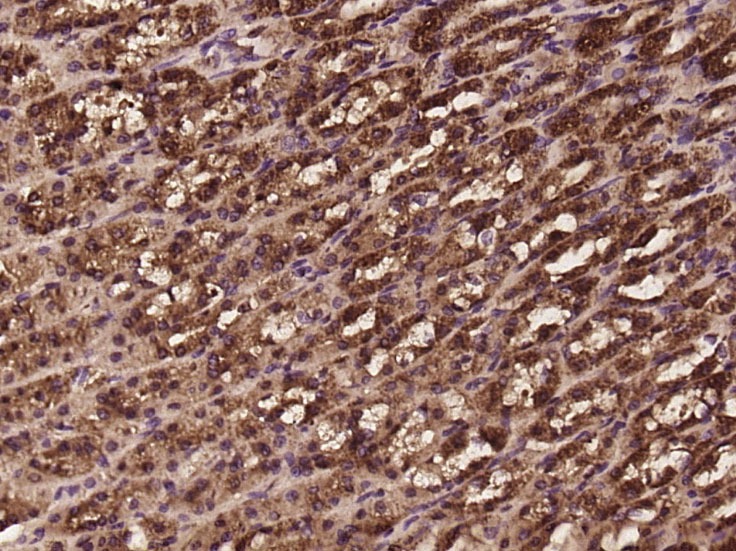 , ,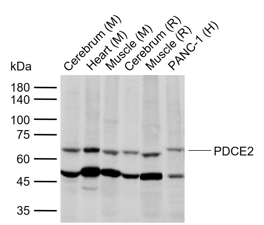 |
| Application | |
| Recommended Dose | WB: 1:500-2000; IHC-P: 1:100-500; IHC-Fr: 1:100-500; IF: 1:100-500 |
| Antibody Type | Polyclonal |
| Host Species | Rabbit |
| Subcellular Localization | Mitochondrion matrix. |
| Construction | Polyclonal Antibody |
| Purification | Protein A purified |
| Appearance | Liquid |
| Formulation | 0.01M TBS (pH7.4) with 1% BSA, 0.02% Proclin300 and 50% Glycerol. |
| Concentration | 1 mg/mL |
| Research Background | This gene encodes component E2 of the multi-enzyme pyruvate dehydrogenase complex (PDC). PDC resides in the inner mitochondrial membrane and catalyzes the conversion of pyruvate to acetyl coenzyme A. The protein product of this gene, dihydrolipoamide acetyltransferase, accepts acetyl groups formed by the oxidative decarboxylation of pyruvate and transfers them to coenzyme A. Dihydrolipoamide acetyltransferase is the antigen for antimitochondrial antibodies. These autoantibodies are present in nearly 95% of patients with the autoimmune liver disease primary biliary cirrhosis (PBC). In PBC, activated T lymphocytes attack and destroy epithelial cells in the bile duct where this protein is abnormally distributed and overexpressed. PBC enventually leads to cirrhosis and liver failure. Mutations in this gene are also a cause of pyruvate dehydrogenase E2 deficiency which causes primary lactic acidosis in infancy and early childhood.[provided by RefSeq, Oct 2009] |
| Immunogen | KLH conjugated synthetic peptide: human PDCE2 |
| Antigen Species | Human |
| Gene Name | DLAT |
| Gene ID | |
| Protein Name | Dihydrolipoyllysine-residue acetyltransferase component of pyruvate dehydrogenase complex, mitochondrial |
| Uniprot ID | |
| Biology Area | Metabolism of carbohydrates,Carbohydrate metabolism,Energy Metabolism,Mitochondrial markers,Cancer,Energy Metabolism,Mitochondrial |
| Function | The pyruvate dehydrogenase complex catalyzes the overall conversion of pyruvate to acetyl-CoA and CO(2). It contains multiple copies of three enzymatic components: pyruvate dehydrogenase (E1), dihydrolipoamide acetyltransferase (E2) and lipoamide dehydrogenase (E3). |
| Molecular Weight | Theoretical: 63 kDa. Actual: 63 kDa. |
| Stability & Storage | Store at -20°C or -80°C for 12 months. Avoid repeated freeze-thaw cycles. |
| Transport | Shipping with blue ice. |
| Size | Quantity | Unit Price | Amount | Operation |
|---|
 Hello! How can I help you today?
 Copyright © 2015-2026 TargetMol Chemicals Inc. All Rights Reserved.
