
Shopping Cart
Remove All Your shopping cart is currently empty

Synonyms: Cytochrome C, CYCS, CYC
Anti-CYCS Antibody
(9A456)
| Pack Size | Price | USA Stock | Global Stock | Quantity |
|---|---|---|---|---|
| 50 µL | $296 | 7-10 days | 7-10 days | |
| 100 µL | $497 | 7-10 days | 7-10 days |
| Description | Anti-CYCS Antibody (9A456) is a Rabbit antibody targeting CYCS. Anti-CYCS Antibody (9A456) can be used in ICC,IHC,IP,WB. |
| Synonyms | Cytochrome C, CYCS, CYC |
| Ig Type | IgG |
| Clone | 9A456 |
| Reactivity | Human,Mouse,Rat |
| Verified Activity | 1. Western blot analysis of Cytochrome C on different lysates using anti-Cytochrome C antibody at 1/1,000 dilution. Positive control: Lane 1: Mouse kidney, Lane 2: Rat kidney. 2. Immunohistochemical analysis of paraffin-embedded human liver tissue using anti-Cytochrome C antibody. Counter stained with hematoxylin. 3. Immunohistochemical analysis of paraffin-embedded human kidney tissue using anti-Cytochrome C antibody. Counter stained with hematoxylin. 4. Immunohistochemical analysis of paraffin-embedded mouse colon tissue using anti-Cytochrome C antibody. Counter stained with hematoxylin. 5. Immunohistochemical analysis of paraffin-embedded mouse skin tissue using anti-Cytochrome C antibody. Counter stained with hematoxylin. 6. Immunohistochemical analysis of paraffin-embedded mouse kidney tissue using anti-Cytochrome C antibody. Counter stained with hematoxylin. 7. ICC staining Cytochrome C in Hela cells (green). The nuclear counter stain is DAPI (blue). Cells were fixed in paraformaldehyde, permeabilised with 0.25% Triton X100/PBS. 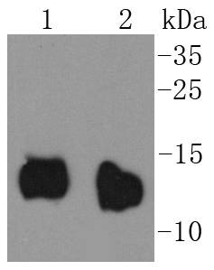 , ,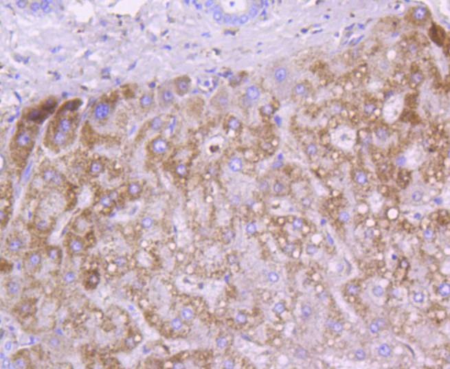 , ,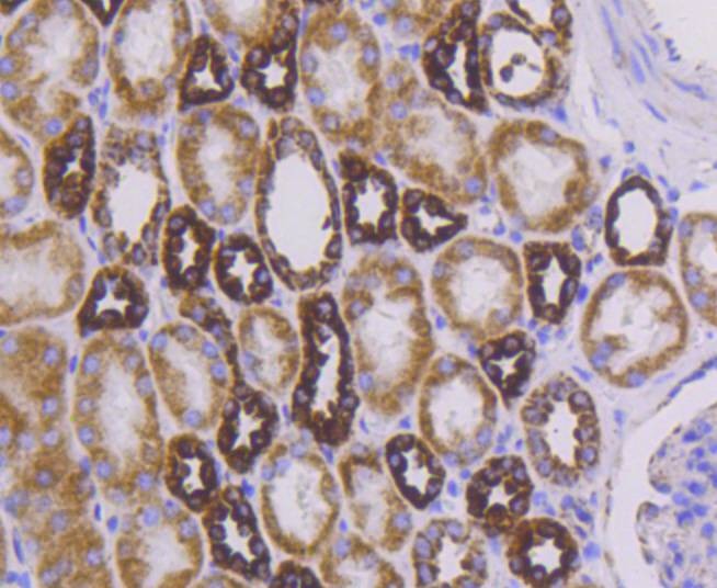 , ,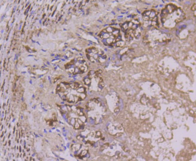 , ,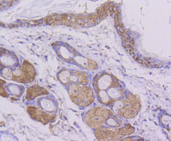 , ,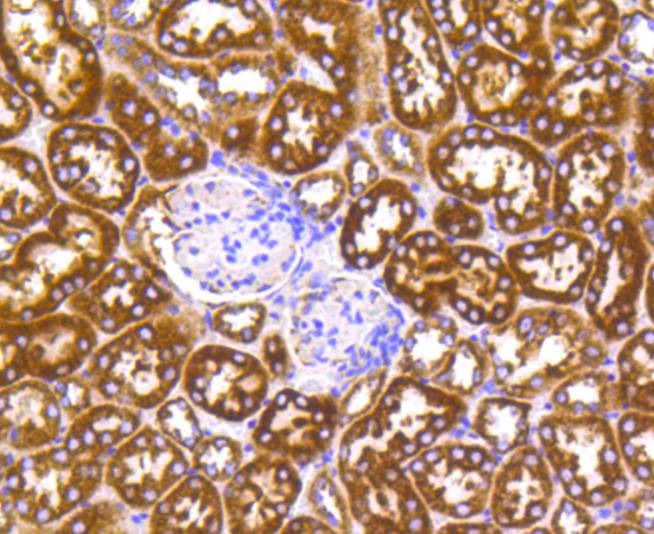 , ,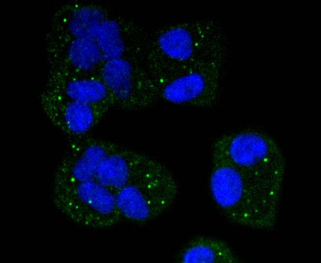 |
| Application | |
| Recommended Dose | WB: 1:1000-5000; IHC: 1:50-200; ICC: 1:50-200 |
| Antibody Type | Monoclonal |
| Host Species | Rabbit |
| Construction | Recombinant Antibody |
| Purification | ProA affinity purified |
| Appearance | Liquid |
| Formulation | 1*TBS (pH7.4), 1%BSA, 40%Glycerol. Preservative: 0.05% Sodium Azide. |
| Research Background | Cytochrome c is a well characterized mobile electron transport protein that is essential to energy conversion in all aerobic organisms. In mammalian cells, this highly conserved protein is normally localized to the mitochondrial intermembrane space. More recent studies have identifed cytosolic cytochrome c as a factor necessary for activation of apoptosis. During apoptosis, cytochrome c is translocated from the mitochondrial membrane to the cytosol, where it is required for activation of caspase-3 (CPP32). Overexpression of Bcl-2 has been shown to prevent the translocation of cytochrome c, thereby blocking the apoptotic process. Overexpression of Bax has been shown to induce the release of cytochrome c and to induce cell death. The release of cytochrome c from the mitochondria is thought to trigger an apoptotic cascade, whereby Apaf-1 binds to Apaf-3 (caspase-9) in a cytochrome c-dependent manner, leading to caspase-9 cleavage of caspase-3. |
| Conjucates | Unconjugated |
| Immunogen | Recombinant Protein |
| Uniprot ID |
| Molecular Weight | Theoretical: 12 kDa. |
| Stability & Storage | Store at -20°C or -80°C for 12 months. Avoid repeated freeze-thaw cycles. |
| Transport | Shipping with blue ice. |
| Size | Quantity | Unit Price | Amount | Operation |
|---|
 Hello! How can I help you today?
 Copyright © 2015-2026 TargetMol Chemicals Inc. All Rights Reserved.
