
Shopping Cart
Remove All Your shopping cart is currently empty

Synonyms: PSK-J3, cyclin-dependent kinase 4, CMM3
Anti-CDK4 Polyclonal Antibody
| Pack Size | Price | USA Stock | Global Stock | Quantity |
|---|---|---|---|---|
| 50 µL | $221 | 7-10 days | 7-10 days | |
| 100 µL | $372 | 7-10 days | 7-10 days | |
| 200 µL | $528 | 7-10 days | 7-10 days |
| Description | Anti-CDK4 Polyclonal Antibody is a Rabbit antibody targeting CDK4. Anti-CDK4 Polyclonal Antibody can be used in FCM, ICC/IF, IF, IHC-Fr, IHC-P, WB. |
| Synonyms | PSK-J3, cyclin-dependent kinase 4, CMM3 |
| Ig Type | IgG |
| Reactivity | Human,Mouse,Rat (predicted:Pig,Cow) |
| Verified Activity | 1. Sample: Spleen (Mouse) Lysate at 40 μg Primary: Anti-CDK4 (TMAB-00405) at 1/300 dilution Secondary: IRDye800CW Goat Anti-Rabbit IgG at 1/20000 dilution Predicted band size: 34 kDa Observed band size: 34 kDa 2. Sample: Brain (Rat) Lysate at 30 μg Colon carcinoma (Human) lysate at 30 μg Primary: Anti-CDK4 (TMAB-00405) at 1/200 dilution Secondary: HRP conjugated Goat-Anti-rabbit IgG (secondary antibody) at 1/3000 dilution Predicted band size: 33 kDa Observed band size: 33 kDa 3. Sample: Cerebrum (Mouse) Lysate at 40 μg Primary: Anti-CDK4 (TMAB-00405) at 1/300 dilution Secondary: IRDye800CW Goat Anti-Rabbit IgG at 1/20000 dilution Predicted band size: 34 kDa Observed band size: 34 kDa 4. Paraformaldehyde-fixed, paraffin embedded (human gastric carcinoma); Antigen retrieval by boiling in sodium citrate buffer (pH6.0) for 15 min; Block endogenous peroxidase by 3% hydrogen peroxide for 20 min; Blocking buffer (normal goat serum) at 37°C for 30 min; Antibody incubation with (CDK4) Polyclonal Antibody, Unconjugated (TMAB-00405) at 1:200 overnight at 4°C, followed by operating according to SP Kit (Rabbit) instructionsand DAB staining. 5. Tissue/cell: Hela cell; 4% Paraformaldehyde-fixed; Triton X-100 at room temperature for 20 min; Blocking buffer (normal goat serum) at 37°C for 20 min; Antibody incubation with (CDK4) polyclonal Antibody, Unconjugated (TMAB-00405) 1:100, 90 minutes at 37°C; followed by a FITC conjugated Goat Anti-Rabbit IgG antibody at 37°C for 90 minutes, DAPI (blue) was used to stain the cell nucleus. 6. Paraformaldehyde-fixed, paraffin embedded (rat brain); Antigen retrieval by boiling in sodium citrate buffer (pH6.0) for 15 min; Block endogenous peroxidase by 3% hydrogen peroxide for 20 min; Blocking buffer (normal goat serum) at 37°C for 30 min; Antibody incubation with (CDK4) Polyclonal Antibody, Unconjugated (TMAB-00405) at 1:200 overnight at 4°C, followed by operating according to SP Kit (Rabbit) instructionsand DAB staining. 7. Blank control (black line): HepG2 (black) (The cells were fixed with 2% paraformaldehyde (10 min), then permeabilized with PBST for 30 min on room temperature) Primary Antibody (Red line): Rabbit Anti-CDK4 antibody (TMAB-00405); Dilution: 1 μg/10^6 cells; Isotype Control Antibody (green line): Rabbit IgG. Secondary Antibody (white blue line): Goat anti-rabbit IgG-FITC;Dilution: 1 μg/test. 8. Paraformaldehyde-fixed, paraffin embedded (rat kidney tissue); Antigen retrieval by boiling in sodium citrate buffer (pH6.0) for 15 min; Block endogenous peroxidase by 3% hydrogen peroxide for 20 min; Blocking buffer (normal goat serum) at 37°C for 30 min; Antibody incubation with (CDK4) Polyclonal Antibody, Unconjugated (TMAB-00405) at 1:400 overnight at 4°C, followed by operating according to SP Kit (Rabbit) instructionsand DAB staining. 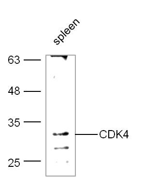 , ,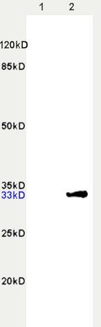 , ,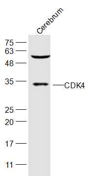 , ,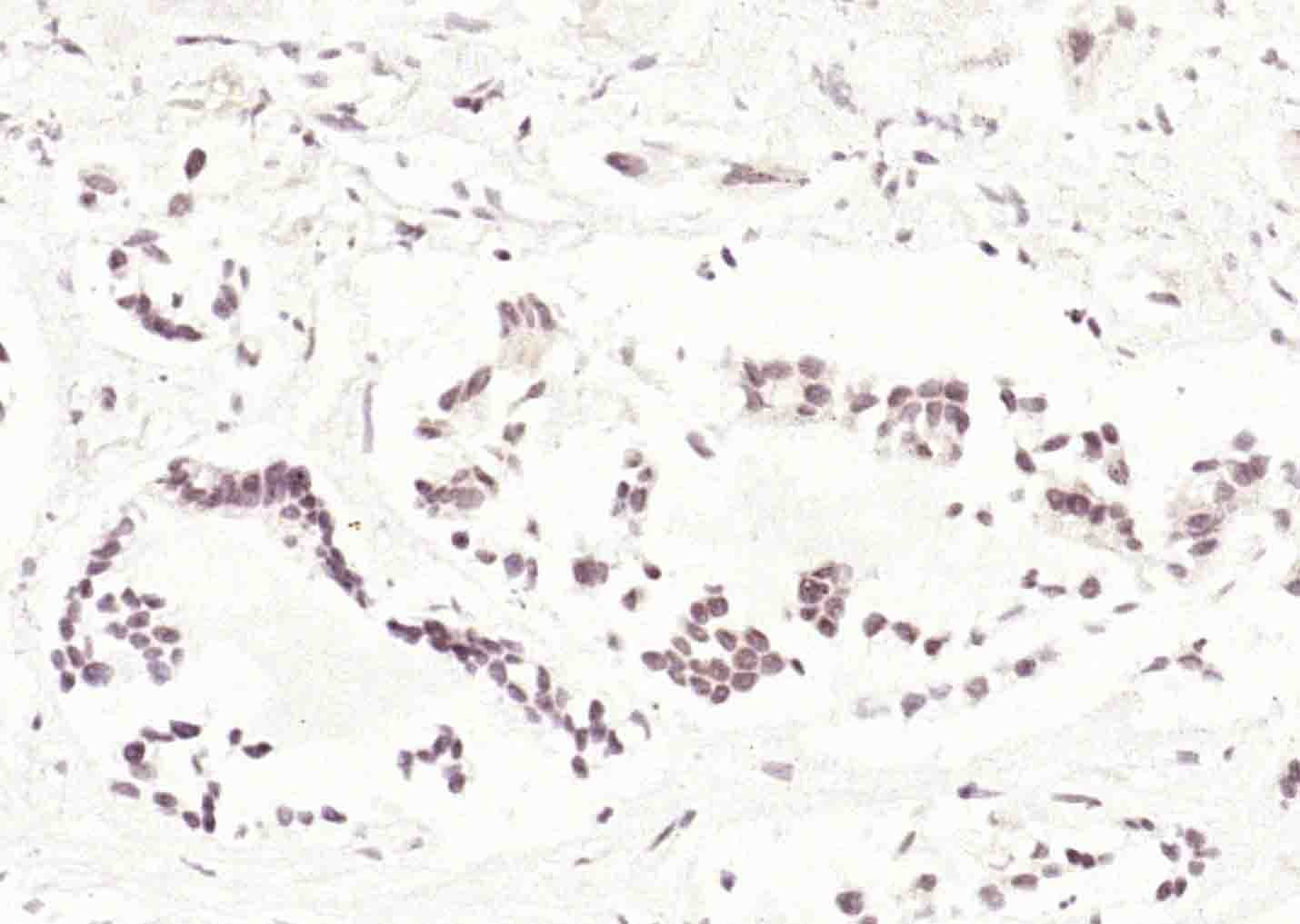 , ,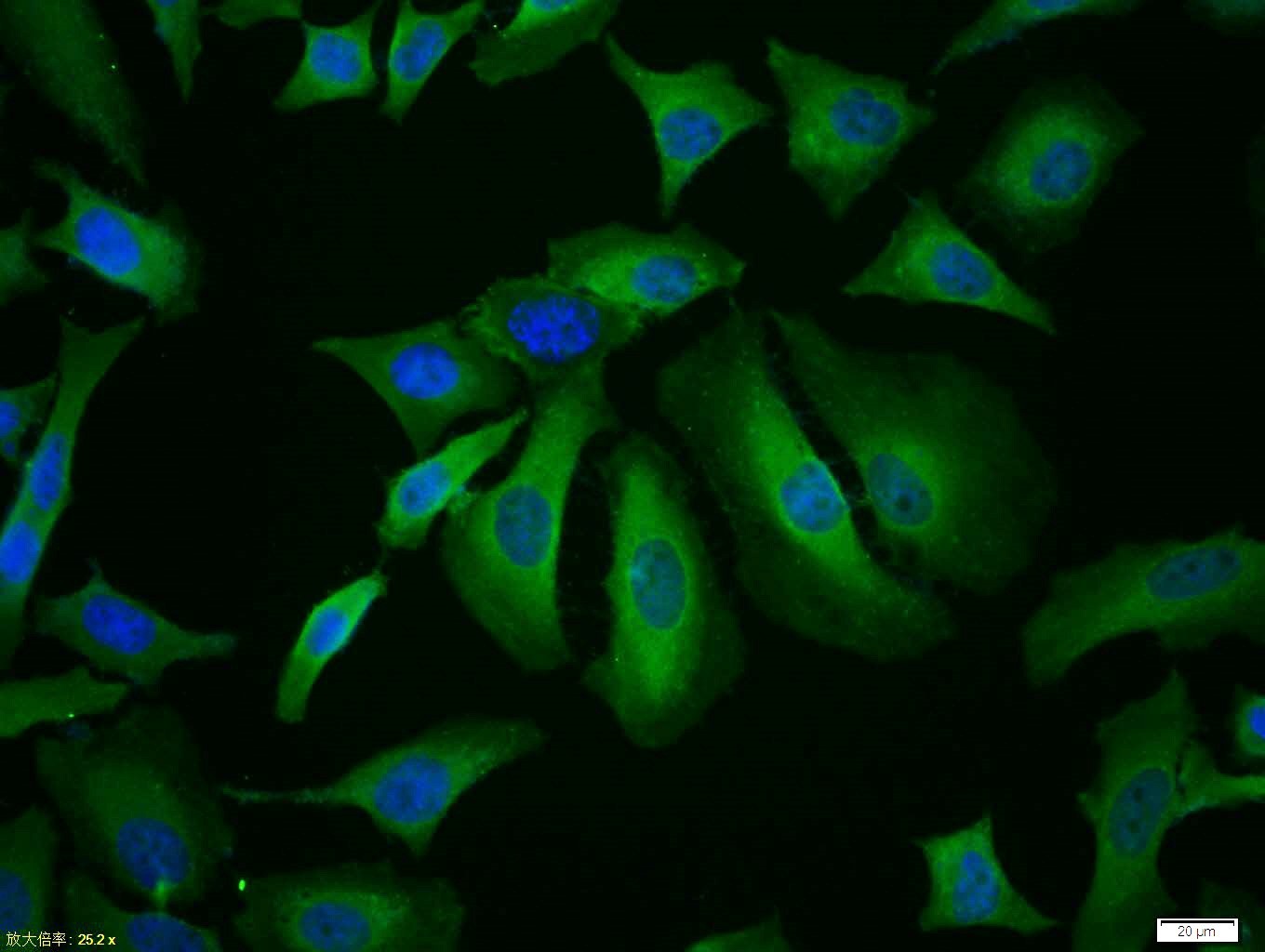 , ,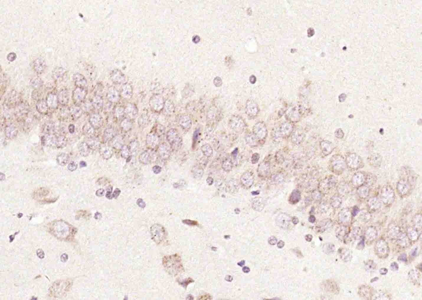 , ,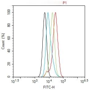 , ,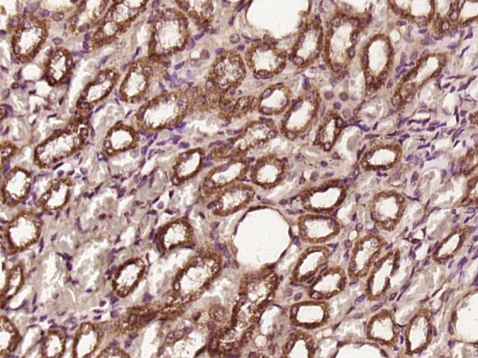 |
| Application | |
| Recommended Dose | FCM=1 μg/Test; ICC/IF=1:100-500; IF=1:100-500; IHC-Fr=1:100-500; IHC-P=1:100-500; WB=1:500-2000 |
| Antibody Type | Polyclonal |
| Host Species | Rabbit |
| Subcellular Localization | Cytoplasm. Nucleus. Membrane. Cytoplasmic when non-complexed. Forms a cyclin D-CDK4 complex in the cytoplasm as cells progress through G(1) phase. The complex accumulates on the nuclear membrane and enters the nucleus on transition from G(1) to S phase. Also present in nucleoli and heterochromatin lumps. Colocalizes with RB1 after release into the nucleus. |
| Construction | Polyclonal Antibody |
| Purification | Protein A purified |
| Appearance | Liquid |
| Formulation | 0.01M TBS (pH7.4) with 1% BSA, 0.02% Proclin300 and 50% Glycerol. |
| Concentration | 1 mg/mL |
| Research Background | The protein encoded by this gene is a member of the Ser/Thr protein kinase family. This protein is highly similar to the gene products of S. cerevisiae cdc28 and S. pombe cdc2. It is a catalytic subunit of the protein kinase complex that is important for cell cycle G1 phase progression. The activity of this kinase is restricted to the G1-S phase, which is controlled by the regulatory subunits D-type cyclins and CDK inhibitor p16(INK4a). This kinase was shown to be responsible for the phosphorylation of retinoblastoma gene product (Rb). Mutations in this gene as well as in its related proteins including D-type cyclins, p16(INK4a) and Rb were all found to be associated with tumorigenesis of a variety of cancers. Multiple polyadenylation sites of this gene have been reported. [provided by RefSeq, Jul 2008] |
| Immunogen | KLH conjugated synthetic peptide: human CDK4 |
| Antigen Species | Human |
| Gene Name | CDK4 |
| Gene ID | |
| Protein Name | Cyclin-dependent kinase 4 Cyclin-dependent kinase 4 |
| Uniprot ID | |
| Biology Area | Cdk4,Cdks,Cdks,Cdks |
| Function | Ser/Thr-kinase component of cyclin D-CDK4 (DC) complexes that phosphorylate and inhibit members of the retinoblastoma (RB) protein family including RB1 and regulate the cell-cycle during G(1)/S transition. Phosphorylation of RB1 allows dissociation of the transcription factor E2F from the RB/E2F complexes and the subsequent transcription of E2F target genes which are responsible for the progression through the G(1) phase. Hypophosphorylates RB1 in early G(1) phase. Cyclin D-CDK4 complexes are major integrators of various mitogenenic and antimitogenic signals. Also phosphorylates SMAD3 in a cell-cycle-dependent manner and represses its transcriptional activity. Component of the ternary complex, cyclin D/CDK4/CDKN1B, required for nuclear translocation and activity of the cyclin D-CDK4 complex. |
| Molecular Weight | Theoretical: 34 kDa. Actual: 34 kDa. |
| Stability & Storage | Store at -20°C or -80°C for 12 months. Avoid repeated freeze-thaw cycles. |
| Transport | Shipping with blue ice. |
| Size | Quantity | Unit Price | Amount | Operation |
|---|
 Hello! How can I help you today?
 Copyright © 2015-2026 TargetMol Chemicals Inc. All Rights Reserved.
