
Shopping Cart
Remove All Your shopping cart is currently empty

Synonyms: MIP3 beta Receptor, MIP 3 beta receptor, MGC108519, Lymphocyte Specific G Protein Coupled Peptide Receptor, EVI1, EVI 1, Epstein Barr virus induced gene 1, Epstein Barr virus induced G protein coupled receptor, EBV Induced G Protein Coupled Receptor 1, Ebi1h, EBI1, EBI 1, Chemokine receptor 7-like protein, Chemokine C C receptor 7, Chemokine C C motif receptor 7, CDW197, CD197 antigen, CD197, CD 197, CCR7, CCR 7, CCCKR7, CC CKR 7, CC chemokine receptor type 7, CC chemokine receptor 7, C C CKR 7, C C chemokine receptor type 7, BLR2, BLR 2
Anti-CCR7 Polyclonal Antibody
| Pack Size | Price | USA Stock | Global Stock | Quantity |
|---|---|---|---|---|
| 50 µL | $220 | 7-10 days | 7-10 days | |
| 100 µL | $374 | 7-10 days | 7-10 days | |
| 200 µL | $529 | 7-10 days | 7-10 days |
| Description | Anti-CCR7 Polyclonal Antibody is a Rabbit antibody targeting CCR7. Anti-CCR7 Polyclonal Antibody can be used in FCM, ICC/IF, WB. |
| Synonyms | MIP3 beta Receptor, MIP 3 beta receptor, MGC108519, Lymphocyte Specific G Protein Coupled Peptide Receptor, EVI1, EVI 1, Epstein Barr virus induced gene 1, Epstein Barr virus induced G protein coupled receptor, EBV Induced G Protein Coupled Receptor 1, Ebi1h, EBI1, EBI 1, Chemokine receptor 7-like protein, Chemokine C C receptor 7, Chemokine C C motif receptor 7, CDW197, CD197 antigen, CD197, CD 197, CCR7, CCR 7, CCCKR7, CC CKR 7, CC chemokine receptor type 7, CC chemokine receptor 7, C C CKR 7, C C chemokine receptor type 7, BLR2, BLR 2 |
| Ig Type | IgG |
| Reactivity | Human,Mouse,Rat (predicted:Dog) |
| Verified Activity | 1. Blank control (Black line): Mouse spleen (Black). Primary Antibody (green line): Rabbit Anti-CD4 antibody (TMAB-00326-PE) Dilution: 3 μg/10^6 cells; Isotype Control Antibody (orange line): Rabbit IgG-PE. Secondary Antibody (white blue line): Goat anti-rabbit IgG-PE Dilution: 1 μg/test. Protocol The cells were fixed with 4% PFA (10 min at room temperature) and then permeabilized with 90% ice-cold methanol for 20 min at room temperature. The cells were then incubated in 5% BSA to block non-specific protein-protein interactions for 30 min at room temperature. Cells stained with Primary Antibody for 30 min at room temperature. The secondary antibody used for 40 min at room temperature. 2. Tissue/cell: human laryngocarcinoma; 4% Paraformaldehyde-fixed and paraffin-embedded; Antigen retrieval: citrate buffer (0.01M, pH6.0), Boiling bathing for 15 min; Block endogenous peroxidase by 3% Hydrogen peroxide for 30 min; Blocking buffer (normal goat serum) at 37°C for 20 min; Incubation: Anti-CCR7 Polyclonal Antibody, Unconjugated (TMAB-00326) 1:200, overnight at 4°C, followed by conjugation to the secondary antibody and DAb staining. 3. Tissue/cell: human gastric tissue;4% Paraformaldehyde-fixed and paraffin-embedded; Antigen retrieval: citrate buffer (0.01M, pH6.0), Boiling bathing for 15 min; Blocking buffer (normal goat serum) at 37°C for 20 min; Incubation: Anti-CCR7 Polyclonal Antibody, Unconjugated (TMAB-00326) 1:200, overnight at 4°C; The secondary antibody was Goat Anti-Rabbit IgG, FITC conjugated used at 1:200 dilution for 40 minutes at 37°C. DAPI (5 μg/ml,blue) was used to stain the cell nucleus. 4. Blank control: Raji (blue). Primary Antibody: Rabbit Anti-CCR7 antibody (TMAB-00326), Dilution: 1 μg in 100 μL 1X PBS containing 0.5% BSA; Isotype Control Antibody: Rabbit Igg (orange),used under the same conditions); Secondary Antibody: Goat anti-rabbit IgG-Pe (white blue), Dilution: 1:200 in 1 X PBS containing 0.5% BSA. Protocol The cells were fixed with 2% paraformaldehyde (10 min). Primary antibody (TMAB-00326, 1 μg/1x10^6 cells) were incubated for 30 min on the ice, followed by 1 X PBS containing 0.5% BSA + 10% goat serum (15 min) to block non-specific protein-protein interactions. Then the Goat Anti-rabbit IgG/PE antibody was added into the blocking buffer mentioned above to react with the primary antibody at 1/200 dilution for 30 min on ice. 5. Hela cell; 4% Paraformaldehyde-fixed; Triton X-100 at room temperature for 20 min; Blocking buffer (normal goat serum) at 37°C for 20 min; Antibody incubation with (CCR7) polyclonal Antibody, Unconjugated (TMAB-00326) 1:100, 90 minutes at 37°C; followed by a conjugated Goat Anti-Rabbit IgG antibody at 37°C for 90 minutes, DAPI (blue) was used to stain the cell nucleus. 6. Hela cell; 4% Paraformaldehyde-fixed; Triton X-100 at room temperature for 20 min; Blocking buffer (normal goat serum) at 37°C for 20 min; Antibody incubation with (CCR7) polyclonal Antibody, Unconjugated (TMAB-00326) 1:100, 90 minutes at 37°C; followed by a conjugated Goat Anti-Rabbit IgG antibody at 37°C for 90 minutes, DAPI (blue) was used to stain the cell nucleus. 7. Paraformaldehyde-fixed, paraffin embedded (RAT lymphoid); Antigen retrieval by boiling in sodium citrate buffer (pH6.0) for 15 min; Block endogenous peroxidase by 3% hydrogen peroxide for 20 min; Blocking buffer (normal goat serum) at 37°C for 30 min; Antibody incubation with (CCR7) Polyclonal Antibody, Unconjugated (TMAB-00326) at 1:200 overnight at 4°C, followed by operating according to SP Kit (Rabbit) instructionsand DAB staining. 8. Blank control: THP-1. Primary Antibody (green line): Rabbit Anti-CCR7 antibody (TMAB-00326) Dilution: 1 μg/10^6 cells; Isotype Control Antibody (orange line): Rabbit IgG. Secondary Antibody: Goat anti-rabbit IgG-FITC Dilution: 0.5 μg/test. Protocol The cells were fixed with 4% PFA (10 min at room temperature) and then permeabilized with 0.1% PBST for 20 min at room temperature. The cells were then incubated in 5% BSA to block non-specific protein-protein interactions for 30 min at room temperature. Cells stained with Primary Antibody for 30 min at room temperature. The secondary antibody used for 40 min at room temperature. 9. Sample: Lane 1: Mouse Bone tissue lysates Lane 2: Mouse Raw264.7 cell lysates Lane 3: Human Jurkat cell lysates Lane 4: Human MCF-7 cell lysates Lane 5: Human THP-1 cell lysates Primary: Anti-CCR7 (TMAB-00326) at 1/1000 dilution Secondary: IRDye800CW Goat Anti-Rabbit IgG at 1/20000 dilution Predicted band size: 42 kDa Observed band size: 42 kDa 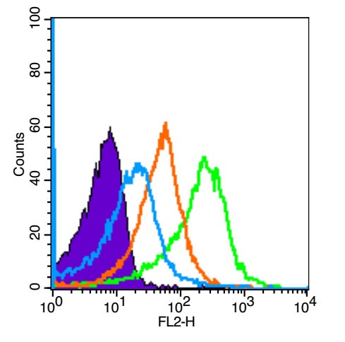 , ,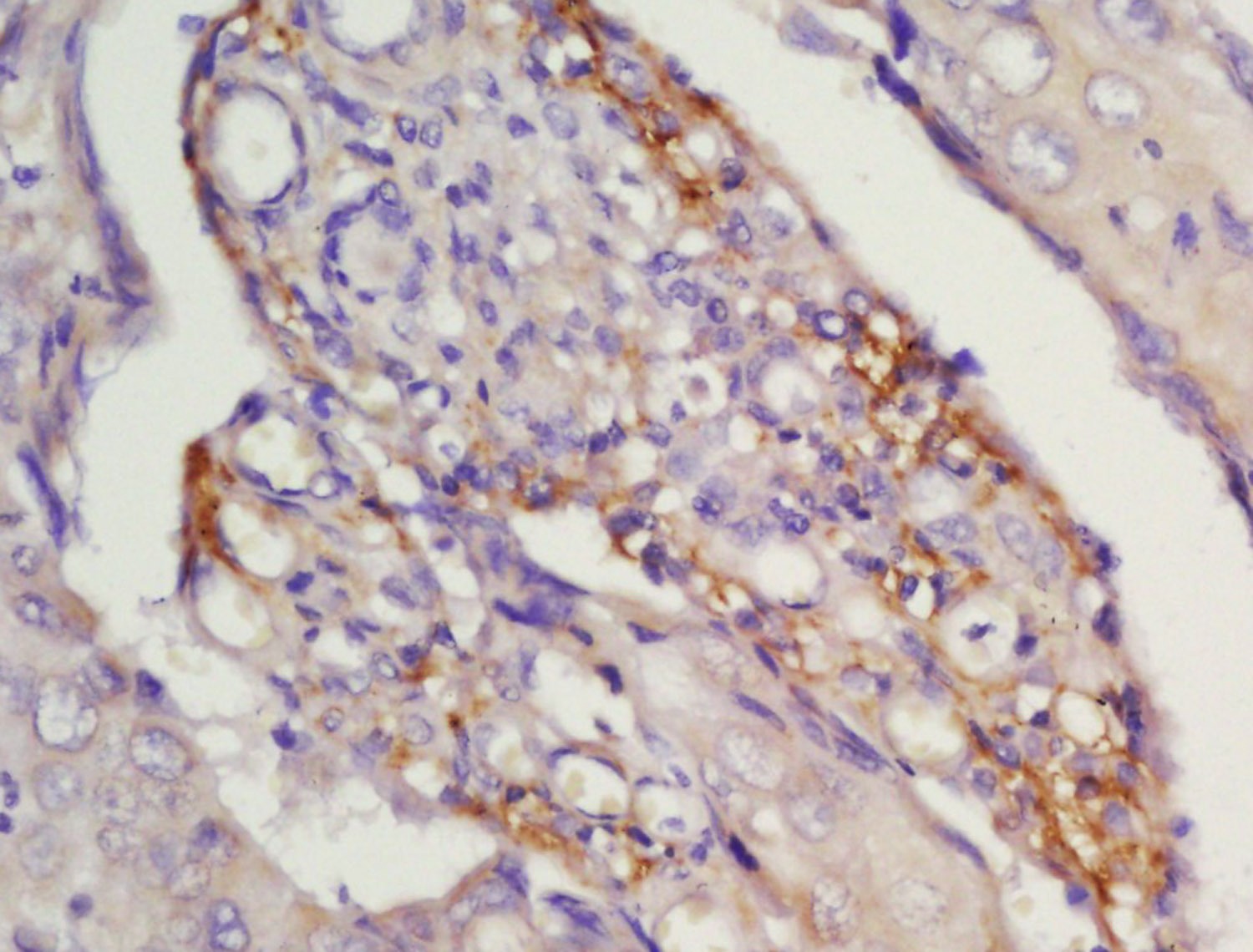 , ,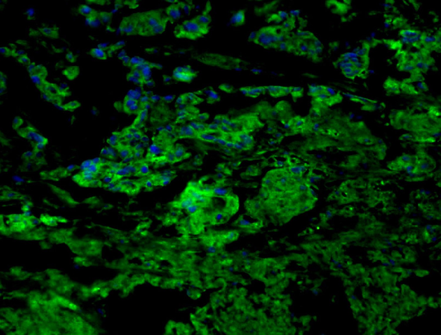 , ,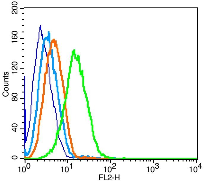 , ,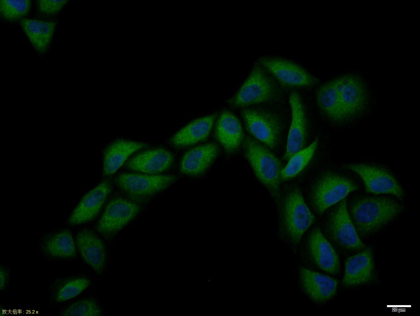 , ,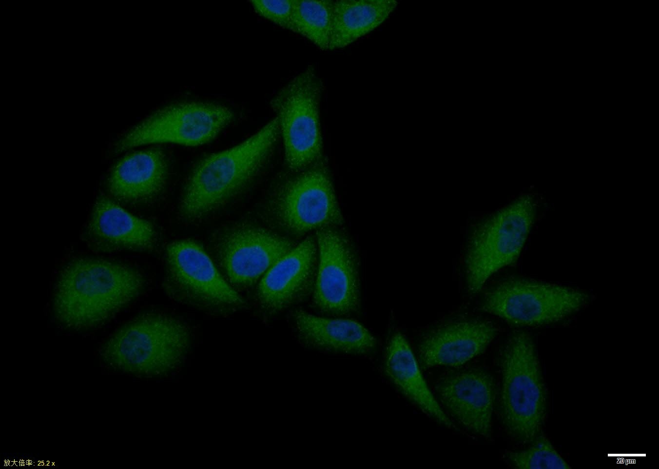 , ,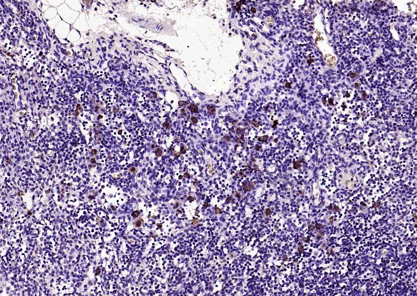 , ,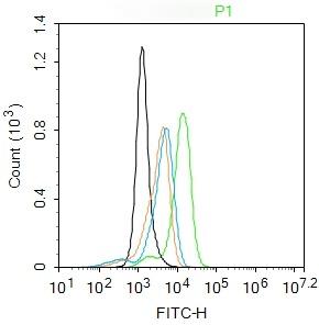 , ,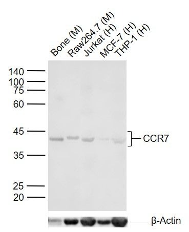 |
| Application | |
| Recommended Dose | FCM=1 μg/Test; ICC/IF=1:100-500; WB=1:500-2000 |
| Antibody Type | Polyclonal |
| Host Species | Rabbit |
| Subcellular Localization | Cell membrane; Multi-pass membrane protein. |
| Tissue Specificity | Expressed in various lymphoid tissues and activated B- and T-lymphocytes, strongly up-regulated in B-cells infected with Epstein-Barr virus and T-cells infected with herpesvirus 6 or 7. |
| Construction | Polyclonal Antibody |
| Purification | Protein A purified |
| Appearance | Liquid |
| Formulation | 0.01M TBS (pH7.4) with 1% BSA, 0.02% Proclin300 and 50% Glycerol. |
| Concentration | 1 mg/mL |
| Research Background | The protein encoded by this gene is a member of the G protein-coupled receptor family. This receptor was identified as a gene induced by the Epstein-Barr virus (EBV), and is thought to be a mediator of EBV effects on B lymphocytes. This receptor is expressed in various lymphoid tissues and activates B and T lymphocytes. It has been shown to control the migration of memory T cells to inflamed tissues, as well as stimulate dendritic cell maturation. The chemokine (C-C motif) ligand 19 (CCL19/ECL) has been reported to be a specific ligand of this receptor. [provided by RefSeq, Jul 2008] |
| Immunogen | KLH conjugated synthetic peptide: human CCR7 |
| Antigen Species | Human |
| Gene Name | CCR7 |
| Gene ID | |
| Protein Name | C-C chemokine receptor type 7 |
| Uniprot ID | |
| Biology Area | Beta Chemokine Rec. (CCR),SARS Coronavirus,GPCR |
| Function | Receptor for the MIP-3-beta chemokine. Probable mediator of EBV effects on B-lymphocytes or of normal lymphocyte functions. |
| Molecular Weight | Theoretical: 42 kDa. Actual: 43 kDa. |
| Stability & Storage | Store at -20°C or -80°C for 12 months. Avoid repeated freeze-thaw cycles. |
| Transport | Shipping with blue ice. |
| Size | Quantity | Unit Price | Amount | Operation |
|---|
 Hello! How can I help you today?
 Copyright © 2015-2026 TargetMol Chemicals Inc. All Rights Reserved.
