
Shopping Cart
Remove All Your shopping cart is currently empty

Synonyms: cyclin E1, CCNE
Anti-CCNE1 Polyclonal Antibody
| Pack Size | Price | USA Stock | Global Stock | Quantity |
|---|---|---|---|---|
| 50 µL | $221 | 7-10 days | 7-10 days | |
| 100 µL | $374 | 7-10 days | 7-10 days | |
| 200 µL | $528 | 7-10 days | 7-10 days |
| Description | Anti-CCNE1 Polyclonal Antibody is a Rabbit antibody targeting CCNE1. Anti-CCNE1 Polyclonal Antibody can be used in FCM,IF,IHC-Fr,IHC-P,WB. |
| Synonyms | cyclin E1, CCNE |
| Ig Type | IgG |
| Reactivity | Human,Mouse,Rat |
| Verified Activity | 1. Tissue/cell: rat testis tissue;4% Paraformaldehyde-fixed and paraffin-embedded; Antigen retrieval: citrate buffer (0.01M, pH6.0), Boiling bathing for 15 min; Blocking buffer (normal goat serum) at 37°C for 20 min; Incubation: Anti-Cyclin E Polyclonal Antibody, Unconjugated (TMAB-00319) 1:200, overnight at 4°C; The secondary antibody was Goat Anti-Rabbit IgG, Cy3 conjugated used at 1:200 dilution for 40 minutes at 37°C. 2. Tissue/cell: human laryngocarcinoma; 4% Paraformaldehyde-fixed and paraffin-embedded; Antigen retrieval: citrate buffer (0.01M, pH6.0), Boiling bathing for 15 min; Block endogenous peroxidase by 3% Hydrogen peroxide for 30 min; Blocking buffer (normal goat serum) at 37°C for 20 min; Incubation: Anti-Cyclin-E Polyclonal Antibody, Unconjugated (TMAB-00319) 1:200, overnight at 4°C, followed by conjugation to the secondary antibody and DAb staining. 3. Cell: NIH/3T3 Concentration:1:100 Host/Isotype: Rabbit/IgG Flow cytometric analysis of primary antibody (Cat#: TMAB-00319) on NIH/3T3 (green) compared with Rabbit IgG isotype control in the absence of primary antibody (blue) followed by Alexa Fluor 488-conjugated goat anti-rabbit Igg (H+L) secondary antibody. 4. Blank control (blue line): Mouse spleen cells (blue). Primary Antibody (green line): Rabbit Anti-Cyclin E1 antibody (TMAB-00319) Dilution: 1 μg/10^6 cells; Isotype Control Antibody (orange line): Rabbit IgG. Secondary Antibody (white blue line): Goat anti-rabbit IgG-FITC Dilution: 1 μg/test. Protocol The cells were fixed with 70% ethanol (overninght at 4°C) and then permeabilized with 0.1% PBS-Tween for 20 min at room temperature. Cells stained with Primary Antibody for 30 min at room temperature. The cells were then incubated in 1 X PBS/2% BSA/10% goat serum to block non-specific protein-protein interactions followed by the antibody for 15 min at room temperature. The secondary antibody used for 40 min at room temperature. 5. Blank control: MCF7. Primary Antibody (green line): Rabbit Anti-Cyclin E1 antibody (TMAB-00319) Dilution: 2 μg/10^6 cells; Isotype Control Antibody (orange line): Rabbit IgG. Secondary Antibody: Goat anti-rabbit IgG-AF647 Dilution: 1 μg/test. Protocol The cells were fixed with 4% PFA (10 min at room temperature) and then permeabilized with 90% ice-cold methanol for 20 min at-20°C. The cells were then incubated in 5% BSA to block non-specific protein-protein interactions for 30 min at room temperature. Cells stained with Primary Antibody for 30 min at room temperature. The secondary antibody used for 40 min at room temperature. 6. Sample: LOVO (Human) Cell Lysate at 30 μg Thymus (Mouse) Lysate at 40 μg U2os (Human) Cell Lysate at 30 μg K562 (Human) Cell Lysate at 30 μg Primary: Anti-Cyclin E1 (TMAB-00319) at 1/1000 dilution Secondary: IRDye800CW Goat Anti-Rabbit IgG at 1/20000 dilution Predicted band size: 45 kDa Observed band size: 50 kDa 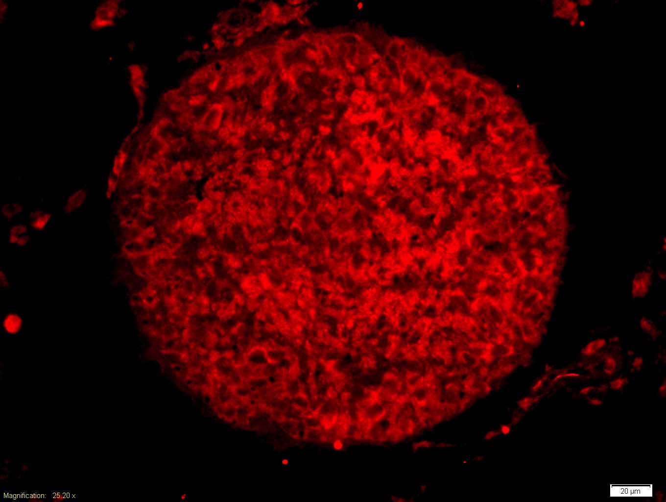 , ,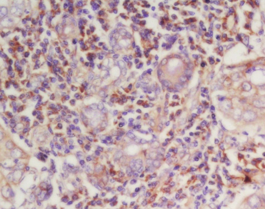 , ,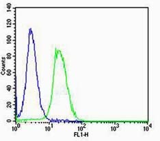 , ,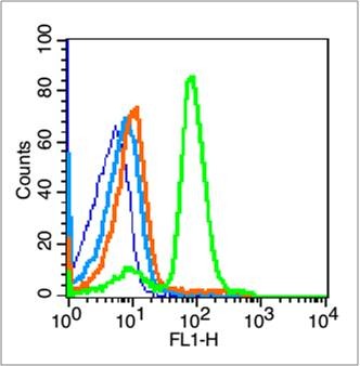 , ,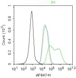 , ,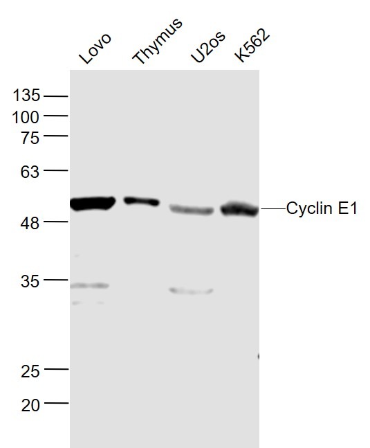 |
| Application | |
| Recommended Dose | WB: 1:500-2000; IHC-P: 1:100-500; IHC-Fr: 1:100-500; IF: 1:100-500; FCM: 1μg/Test |
| Antibody Type | Polyclonal |
| Host Species | Rabbit |
| Subcellular Localization | Nucleus. |
| Tissue Specificity | Highly expressed in testis and placenta. Low levels in bronchial epithelial cells. |
| Construction | Polyclonal Antibody |
| Purification | Protein A purified |
| Appearance | Liquid |
| Formulation | 0.01M TBS (pH7.4) with 1% BSA, 0.02% Proclin300 and 50% Glycerol. |
| Concentration | 1 mg/mL |
| Research Background | Cyclin E is a regulatory subunit of Cdk2 and controls G1 / S transition during the mammalian cell cycle. Multiple isoforms of Cyclin E are only expressed in tumors but not in normal tissue, suggesting a post transcriptional regulation of Cyclin E. In vitro analyses indicated that these truncated variant isoforms of Cyclin E are able to phosphorylate histone H1. Alterations in the Cyclin E protein have been implicated as indicators of worse prognosis in various cancers. |
| Immunogen | KLH conjugated synthetic peptide: rat Cyclin E |
| Antigen Species | Rat |
| Gene Name | CCNE1 |
| Gene ID | |
| Protein Name | G1/S-specific cyclin-E1 |
| Uniprot ID | |
| Biology Area | Cyclin E family,Cyclins,Cyclin E Family,Cyclin E Family |
| Function | Essential for the control of the cell cycle at the G1/S (start) transition. |
| Molecular Weight | Theoretical: 45 kDa. Actual: 50 kDa. |
| Stability & Storage | Store at -20°C or -80°C for 12 months. Avoid repeated freeze-thaw cycles. |
| Transport | Shipping with blue ice. |
| Size | Quantity | Unit Price | Amount | Operation |
|---|
 Hello! How can I help you today?
 Copyright © 2015-2026 TargetMol Chemicals Inc. All Rights Reserved.
