
Shopping Cart
Remove All Your shopping cart is currently empty

Synonyms: KIAA0413, CED4, CED 4, Apoptotic protease-activating factor 1, Apoptotic protease activating factor, Apoptotic peptidase activating factor 1, APAF1, APAF 1, APAF
Anti-Apaf-1 Antibody
(9E241)
| Pack Size | Price | USA Stock | Global Stock | Quantity |
|---|---|---|---|---|
| 50 µL | $298 | 7-10 days | 7-10 days | |
| 100 µL | $498 | 7-10 days | 7-10 days |
| Description | Anti-Apaf-1 Antibody (9E241) is a Rabbit antibody targeting Apaf-1. Anti-Apaf-1 Antibody (9E241) can be used in ICC/IF,IHC,WB. |
| Synonyms | KIAA0413, CED4, CED 4, Apoptotic protease-activating factor 1, Apoptotic protease activating factor, Apoptotic peptidase activating factor 1, APAF1, APAF 1, APAF |
| Ig Type | IgG |
| Clone | 9E241 |
| Reactivity | Human,Mouse |
| Verified Activity | 1. Western blot analysis of Apaf-1 on HUVEC cell lysates using anti-Apaf-1 antibody at 1/1,000 dilution. 2. Immunohistochemical analysis of paraffin-embedded human tonsil tissue using anti-Apaf-1 antibody. Counter stained with hematoxylin. 3. Immunohistochemical analysis of paraffin-embedded mouse colon tissue using anti-Apaf-1 antibody. Counter stained with hematoxylin. 4. Immunohistochemical analysis of paraffin-embedded mouse skin tissue using anti-Apaf-1 antibody. Counter stained with hematoxylin. 5. Immunohistochemical analysis of paraffin-embedded mouse heart tissue using anti-Apaf-1 antibody. Counter stained with hematoxylin. 6. ICC staining Apaf-1 in Hela cells (green). The nuclear counter stain is DAPI (blue). Cells were fixed in paraformaldehyde, permeabilised with 0.25% Triton X100/PBS. 7. ICC staining Apaf-1 in MCF-7 cells (green). The nuclear counter stain is DAPI (blue). Cells were fixed in paraformaldehyde, permeabilised with 0.25% Triton X100/PBS. 8. ICC staining Apaf-1 in Ags cells (green). The nuclear counter stain is DAPI (blue). Cells were fixed in paraformaldehyde, permeabilised with 0.25% Triton X100/PBS. 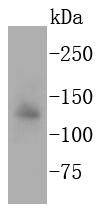 , ,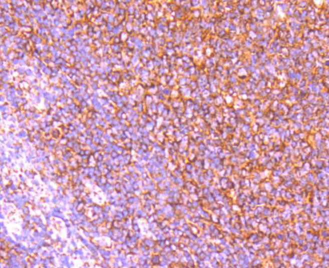 , ,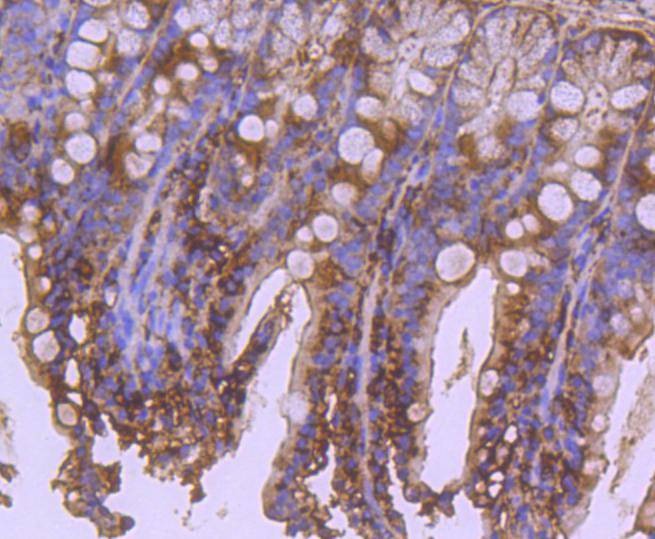 , ,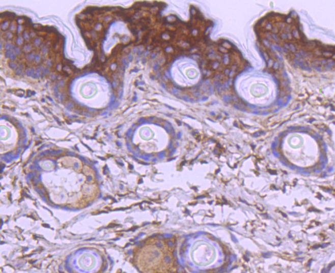 , ,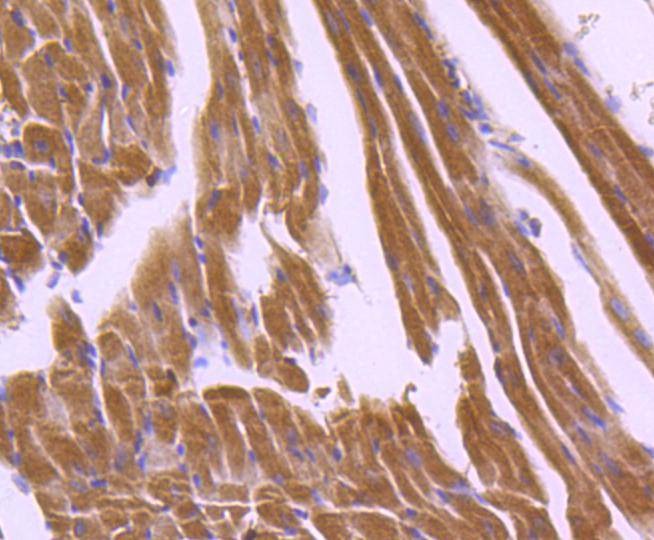 , ,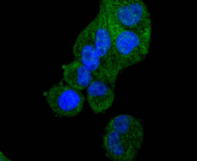 , ,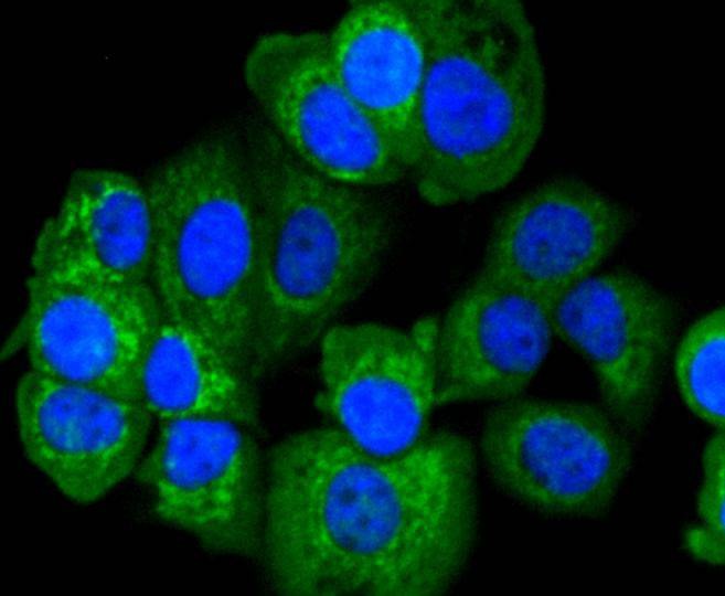 , ,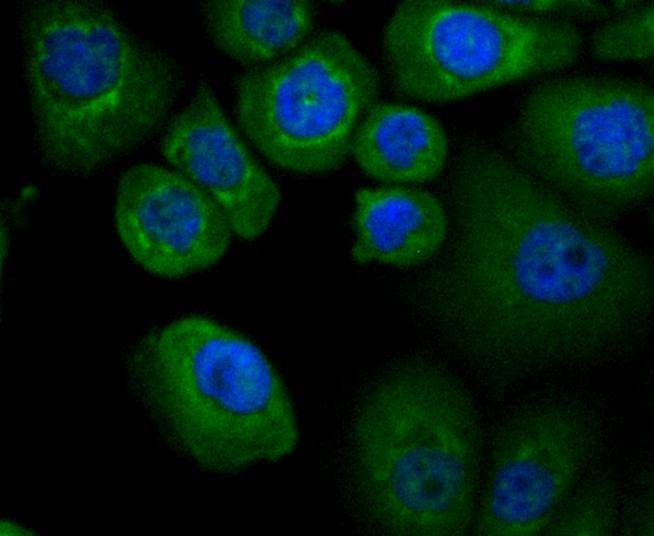 |
| Application | |
| Recommended Dose | WB: 1:1000; IHC: 1:50-200; ICC/IF: 1:50-200 |
| Antibody Type | Monoclonal |
| Host Species | Rabbit |
| Construction | Recombinant Antibody |
| Purification | ProA affinity purified |
| Appearance | Liquid |
| Formulation | 1*TBS (pH7.4), 1%BSA, 40%Glycerol. Preservative: 0.05% Sodium Azide. |
| Research Background | The mammalian homologs of the Ced-4 proteins, Apaf-1 (Ced-4), Nod1 (CARD4), and Nod2 contain a caspase recruitment domain (CARD) and a putative nucleotide binding domain, signified by a consensus Walker's A box (P-loop) and B box (Mg2+-binding site). Nod1 contains a putative regulatory domain and multiple leucine-rich repeats. Nod1 is a member of a growing family of intracellular proteins which share structural homology to the apoptosis regulator Apaf-1. Nod1 associates with the CARD-containing kinase RICK and activates NFkB. The self-association of Nod1 mediates proximity of RICK and the interaction of RICK with IKKg. In addition, Nod-1 binds to multiple caspases with long prodomains, but specifically activates caspase-9 and promotes caspase-9-induced apoptosis. Nod2 is composed of two N-terminal CARDs, a nucleotide-binding domain, and multiple C-terminal leucine-rich repeats. The expression of Nod2 is highly restricted to monocytes, and activates NFkB in response to bacterial lipopoly-saccharides. |
| Conjucates | Unconjugated |
| Immunogen | Recombinant Protein |
| Uniprot ID |
| Molecular Weight | Theoretical: 141 kDa. |
| Stability & Storage | Store at -20°C or -80°C for 12 months. Avoid repeated freeze-thaw cycles. |
| Transport | Shipping with blue ice. |
| Size | Quantity | Unit Price | Amount | Operation |
|---|
 Hello! How can I help you today?
 Copyright © 2015-2026 TargetMol Chemicals Inc. All Rights Reserved.
