Shopping Cart
Remove All Your shopping cart is currently empty
Anti-ACTA2 Antibody (3G548) is a Rabbit antibody targeting ACTA2. Anti-ACTA2 Antibody (3G548) can be used in FCM,ICC/IF,IHC,WB.
| Pack Size | Price | USA Warehouse | Global Warehouse | Quantity |
|---|---|---|---|---|
| 50 μL | $298 | 7-10 days | 7-10 days | |
| 100 μL | $498 | 7-10 days | 7-10 days |
| Description | Anti-ACTA2 Antibody (3G548) is a Rabbit antibody targeting ACTA2. Anti-ACTA2 Antibody (3G548) can be used in FCM,ICC/IF,IHC,WB. |
| Synonyms | Cell growth-inhibiting gene 46 protein, aortic smooth muscle, Alpha-actin-2, ACTVS, ACTSA, Actin, aortic smooth muscle, ACTA2 |
| Ig Type | IgG |
| Clone | 3G548 |
| Reactivity | Human,Mouse,Rat |
| Verified Activity | 1. Western blot analysis of alpha smooth muscle Actin on different lysates using anti-alpha smooth muscle Actin antibody at 1/1,000 dilution. Positive control: Lane 1: Hela, Lane 2: A431, Lane 3: NIH/3T3. 2. Immunohistochemical analysis of paraffin-embedded human tonsil tissue using anti-alpha smooth muscle Actin antibody. Counter stained with hematoxylin. 3. Immunohistochemical analysis of paraffin-embedded human liver tissue using anti-alpha smooth muscle Actin antibody. Counter stained with hematoxylin. 4. Immunohistochemical analysis of paraffin-embedded human gastric carcinoma tissue using anti-alpha smooth muscle Actin antibody. Counter stained with hematoxylin. 5. ICC staining alpha smooth muscle Actin in HepG2 cells (green). The nuclear counter stain is DAPI (blue). Cells were fixed in paraformaldehyde, permeabilised with 0.25% Triton X100/PBS. 6. ICC staining alpha smooth muscle Actin in RH-35 cells (green). The nuclear counter stain is DAPI (blue). Cells were fixed in paraformaldehyde, permeabilised with 0.25% Triton X100/PBS. 7. ICC staining alpha smooth muscle Actin in A431 cells (green). The nuclear counter stain is DAPI (blue). Cells were fixed in paraformaldehyde, permeabilised with 0.25% Triton X100/PBS. 8. ICC staining alpha smooth muscle Actin in A549 cells (green). The nuclear counter stain is DAPI (blue). Cells were fixed in paraformaldehyde, permeabilised with 0.25% Triton X100/PBS. 9. Flow cytometric analysis of Jurkat cells with alpha smooth muscle Actin antibody at 1/50 dilution (red) compared with an unlabelled control (cells without incubation with primary antibody; black). Alexa Fluor 488-conjugated goat anti rabbit IgG was used as the secondary antibody. 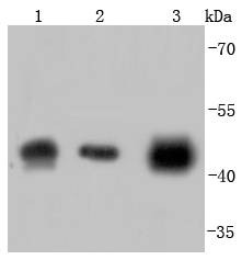 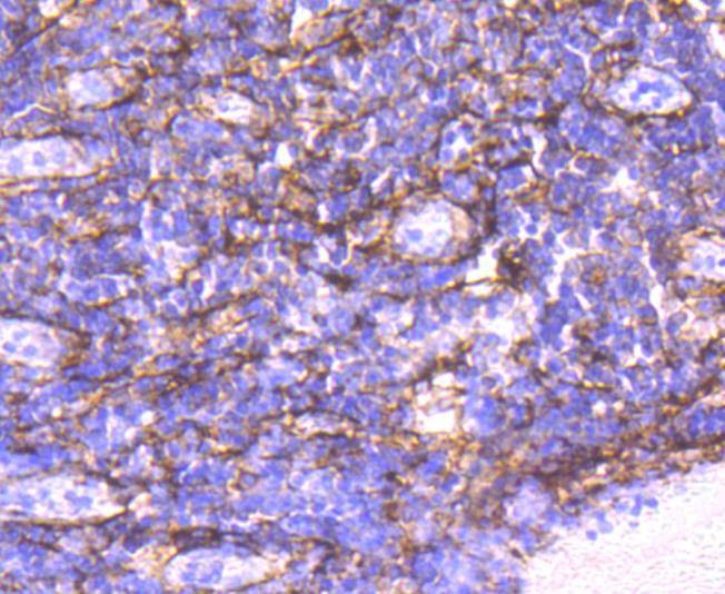 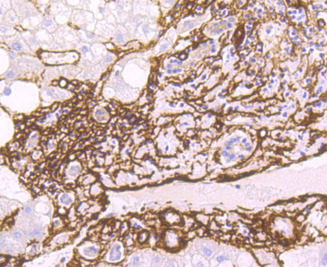 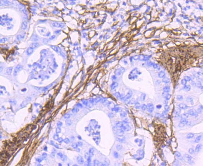 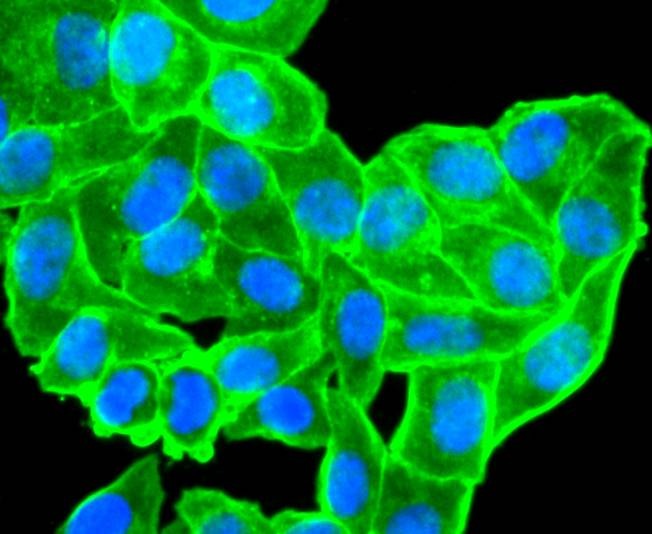 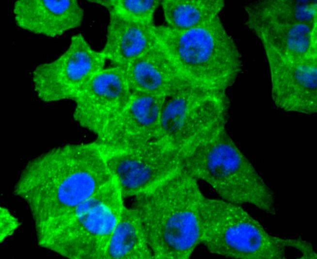 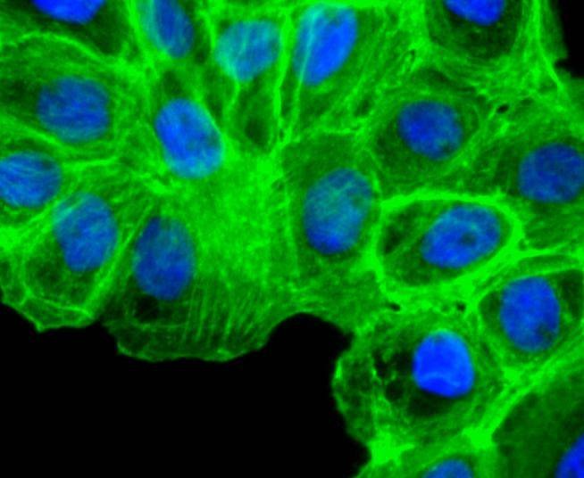 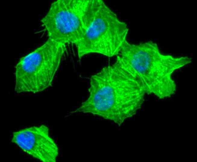 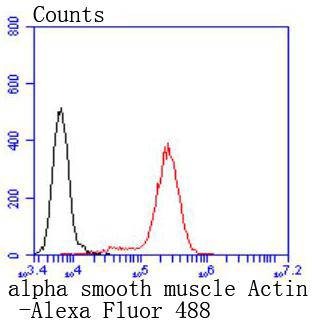 |
| Application | |
| Recommended Dose | WB: 1:1000-2000; IHC: 1:50-200; ICC/IF: 1:100-500; FCM: 1:50-100 |
| Antibody Type | Monoclonal |
| Host Species | Rabbit |
| Construction | Recombinant Antibody |
| Purification | ProA affinity purified |
| Appearance | Liquid |
| Formulation | 1*TBS (pH7.4), 1%BSA, 40%Glycerol. Preservative: 0.05% Sodium Azide. |
| Research Background | All eukaryotic cells express Actin, which often constitutes as much as 50% of total cellular protein. Actin filaments can form both stable and labile structures and are crucial components of microvilli and the contractile apparatus of muscle cells. While lower eukaryotes, such as yeast, have only one Actin gene, higher eukaryotes have several isoforms encoded by a family of genes. At least six types of Actin are present in mammalian tissues and fall into three classes. α-Actin expression is limited to various types of muscle, whereas β-Actin and γ-Actin are the principle constituents of filaments in other tissues. Members of the small GTPase family regulate the organization of the Actin cytoskeleton. Rho controls the assembly of Actin stress fibers and focal adhesion. Rac regulates Actin filament accumulation at the plasma membrane. Cdc42 stimulates formation of filopodia. |
| Conjucates | Unconjugated |
| Immunogen | Recombinant Protein |
| Uniprot ID |
| Molecular Weight | Theoretical: 42 kDa. |
| Stability & Storage | Store at -20°C or -80°C for 12 months. Avoid repeated freeze-thaw cycles. |
| Transport | Shipping with blue ice. |
| Size | Quantity | Unit Price | Amount | Operation |
|---|

Copyright © 2015-2026 TargetMol Chemicals Inc. All Rights Reserved.