
Shopping Cart
Remove All Your shopping cart is currently empty

Synonyms: WT33, WT1, Wilms tumor protein
Anti-Wilms tumor/WT1 Polyclonal Antibody
| Pack Size | Price | USA Stock | Global Stock | Quantity |
|---|---|---|---|---|
| 50 µL | $222 | 7-10 days | 7-10 days | |
| 100 µL | $372 | 7-10 days | 7-10 days | |
| 200 µL | $527 | 7-10 days | 7-10 days |
| Description | Anti-Wilms tumor/WT1 Polyclonal Antibody is a Rabbit antibody targeting Wilms tumor/WT1. Anti-Wilms tumor/WT1 Polyclonal Antibody can be used in FCM, IF, IHC-Fr, IHC-P, WB. |
| Synonyms | WT33, WT1, Wilms tumor protein |
| Ig Type | IgG |
| Reactivity | Human,Mouse (predicted:Rat,Chicken,Dog,Pig,Cow,Sheep) |
| Verified Activity | 1. Paraformaldehyde-fixed, paraffin embedded (Mouse testis); Antigen retrieval by boiling in sodium citrate buffer (pH6.0) for 15 min; Block endogenous peroxidase by 3% hydrogen peroxide for 20 min; Blocking buffer (normal goat serum) at 37°C for 30 min; Antibody incubation with (Wilms Tumor Protein) Polyclonal Antibody, Unconjugated (TMAB-01979) at 1:400 overnight at 4°C, followed by a conjugated Goat Anti-Rabbit IgG antibody for 90 minutes, and DAPI for nucleus staining. 2. Sample: TM4 Cell (Mouse) Lysate at 40 μg Primary: Anti-Wilms Tumor Protein (TMAB-01979) at 1/300 dilution Secondary: IRDye800CW Goat Anti-Rabbit IgG at 1/20000 dilution Predicted band size: 55 kDa Observed band size: 48 kDa 3. Blank control: Molt-4. Primary Antibody (green line): Rabbit Anti-Wilms Tumor Protein antibody (TMAB-01979) Dilution: 1 μg/10^6 cells; Isotype Control Antibody (orange line): Rabbit IgG. Secondary Antibody: Goat anti-rabbit IgG-AF647 Dilution: 1 μg/test. Protocol The cells were fixed with 4% PFA (10 min at room temperature) and then permeabilized with 90% ice-cold methanol for 20 min at-20°C. The cells were then incubated in 5% BSA to block non-specific protein-protein interactions for 30 min at at room temperature. Cells stained with Primary Antibody for 30 min at room temperature. The secondary antibody used for 40 min at room temperature. 4. Paraformaldehyde-fixed, paraffin embedded (Mouse kidney); Antigen retrieval by boiling in sodium citrate buffer (pH6.0) for 15 min; Block endogenous peroxidase by 3% hydrogen peroxide for 20 min; Blocking buffer (normal goat serum) at 37°C for 30 min; Antibody incubation with (Wilms Tumor Protein) Polyclonal Antibody, Unconjugated (TMAB-01979) at 1:400 overnight at 4°C, followed by operating according to SP Kit (Rabbit) instructionsand DAB staining. 5. Sample: Lane 1: Mouse Testis tissue lysates Lane 2: Mouse Ovary tissue lysates Lane 3: Mouse Liver tissue lysates Lane 4: Human HL-60 cell lysates Primary: Anti-WT1 (TMAB-01979) at 1/1000 dilution Secondary: IRDye800CW Goat Anti-Rabbit IgG at 1/20000 dilution Predicted band size: 55 kDa Observed band size: 50 kDa 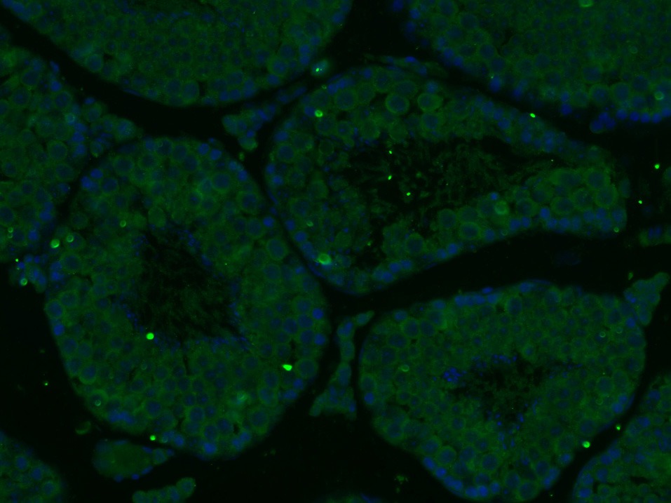 , ,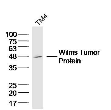 , ,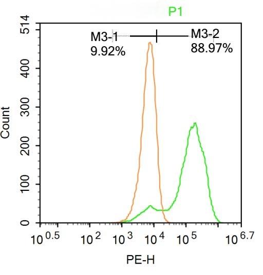 , ,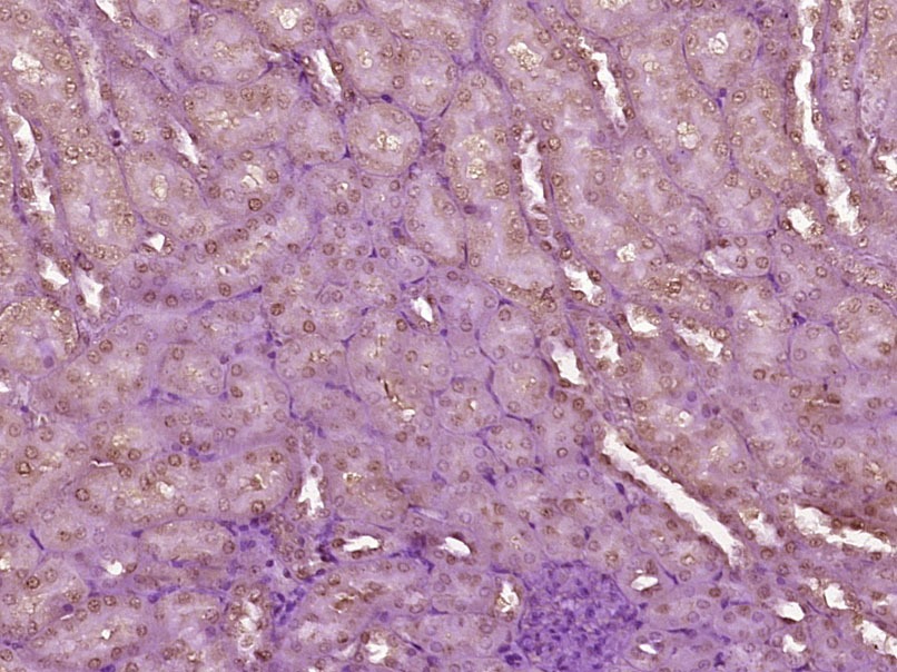 , ,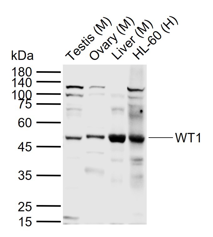 |
| Application | |
| Recommended Dose | WB: 1:500-2000; IHC-P: 1:100-500; IHC-Fr: 1:100-500; IF: 1:100-500; FCM: 1ug/test |
| Antibody Type | Polyclonal |
| Host Species | Rabbit |
| Subcellular Localization | Nucleus. Nucleus, nucleolus. Cytoplasm. Note=Shuttles between nucleus and cytoplasm.Isoform 1: Nucleus speckle.Isoform 4: Nucleus, nucleoplasm. |
| Tissue Specificity | Expressed in the kidney and a subset of hematopoietic cells. |
| Construction | Polyclonal Antibody |
| Purification | Protein A purified |
| Appearance | Liquid |
| Formulation | 0.01M TBS (pH7.4) with 1% BSA, 0.02% Proclin300 and 50% Glycerol. |
| Concentration | 1 mg/mL |
| Research Background | Transcription factor that plays an important role in cellular development and cell survival. Regulates the expression of numerous target genes, including EPO. Plays an essential role for development of the urogenital system. Recognizes and binds to the DNA sequence 5'-CGCCCCCGC-3'. It has a tumor suppressor as well as an oncogenic role in tumor formation. Function may be isoform-specific: isoforms lacking the KTS motif may act as transcription factors. Isoforms containing the KTS motif may bind mRNA and play a role in mRNA metabolism or splicing. Isoform 1 has lower affinity for DNA, and can bind RNA. |
| Immunogen | KLH conjugated synthetic peptide: human WT1 |
| Antigen Species | Human |
| Gene Name | WT1 |
| Gene ID | |
| Protein Name | Wilms tumor protein |
| Uniprot ID | |
| Biology Area | Tumor biomarkers,Kidney development,Other factors,Tumor Associated |
| Function | Transcription factor that plays an important role in cellular development and cell survival. Regulates the expression of numerous target genes, including EPO. Plays an essential role for development of the urogenital system. Recognizes and binds to the DNA sequence 5'-CGCCCCCGC-3'. It has a tumor suppressor as well as an oncogenic role in tumor formation. Function may be isoform-specific: isoforms lacking the KTS motif may act as transcription factors. Isoforms containing the KTS motif may bind mRNA and play a role in mRNA metabolism or splicing. Isoform 1 has lower affinity for DNA, and can bind RNA. |
| Molecular Weight | Theoretical: 55 kDa. Actual: 48 kDa. |
| Stability & Storage | Store at -20°C or -80°C for 12 months. Avoid repeated freeze-thaw cycles. |
| Transport | Shipping with blue ice. |
| Size | Quantity | Unit Price | Amount | Operation |
|---|
 Hello! How can I help you today?
 Copyright © 2015-2026 TargetMol Chemicals Inc. All Rights Reserved.
