
Shopping Cart
Remove All Your shopping cart is currently empty

Synonyms: Vitamin D-binding protein, VDB, Group-specific component, Gc-globulin, Gc, DBP
Anti-VDB Antibody
(2T657)
| Pack Size | Price | USA Stock | Global Stock | Quantity |
|---|---|---|---|---|
| 50 µL | $298 | 7-10 days | 7-10 days | |
| 100 µL | $497 | 7-10 days | 7-10 days |
| Description | Anti-VDB Antibody (2T657) is a Rabbit antibody targeting VDB. Anti-VDB Antibody (2T657) can be used in FCM,ICC/IF,IHC,WB. |
| Synonyms | Vitamin D-binding protein, VDB, Group-specific component, Gc-globulin, Gc, DBP |
| Ig Type | IgG |
| Clone | 2T657 |
| Reactivity | Human |
| Verified Activity | 1. Western blot analysis of DBP on human lung lysates using anti- DBP antibody at 1/1,000 dilution. 2. Immunohistochemical analysis of paraffin-embedded human kidney tissue using anti- DBP antibody. Counter stained with hematoxylin. 3. ICC staining DBP in Hela cells (red). The nuclear counter stain is DAPI (blue). Cells were fixed in paraformaldehyde, permeabilised with 0.25% Triton X100/PBS. 4. ICC staining DBP in HepG2 cells (red). The nuclear counter stain is DAPI (blue). Cells were fixed in paraformaldehyde, permeabilised with 0.25% Triton X100/PBS. 5. ICC staining DBP in SKOV-3 cells (red). The nuclear counter stain is DAPI (blue). Cells were fixed in paraformaldehyde, permeabilised with 0.25% Triton X100/PBS. 6. Flow cytometric analysis of HepG2 cells with DBP antibody at 1/50 dilution (red) compared with an unlabelled control (cells without incubation with primary antibody; black). Alexa Fluor 488-conjugated goat anti rabbit IgG was used as the secondary antibody. 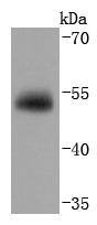 , ,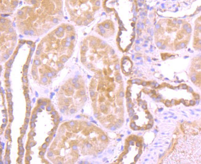 , ,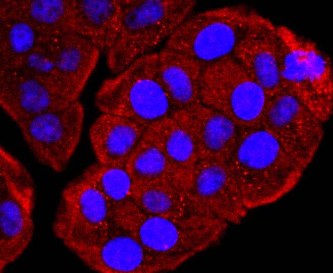 , ,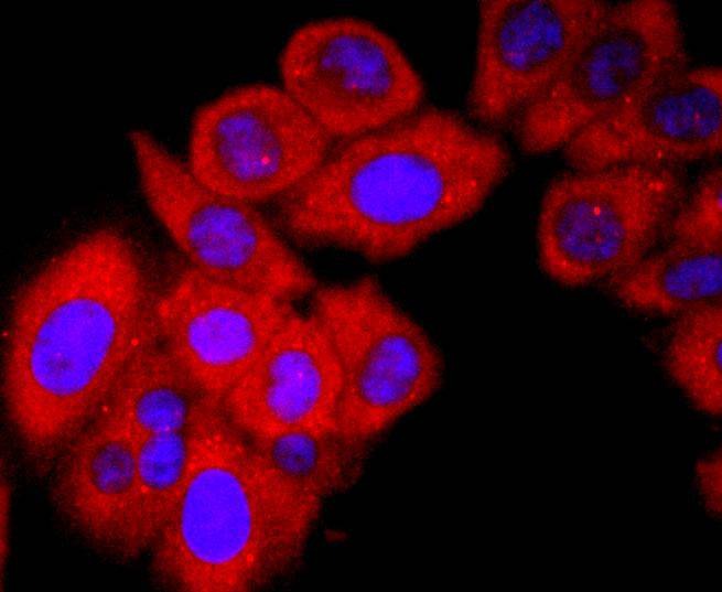 , ,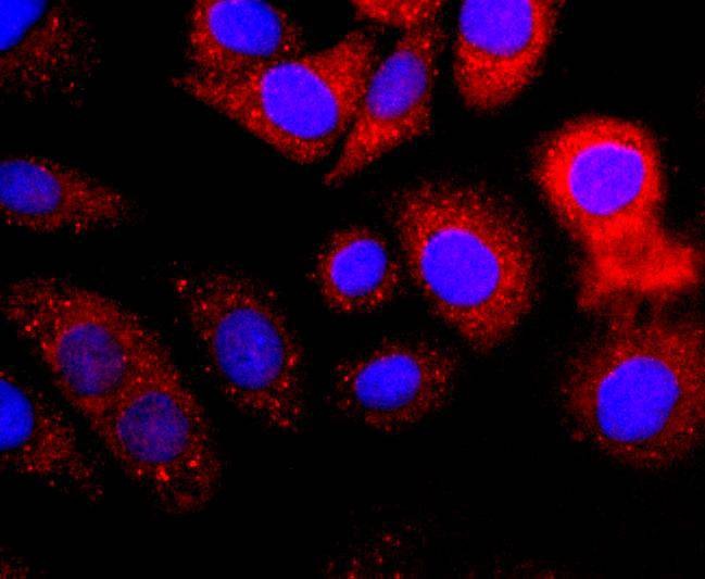 , ,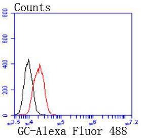 |
| Application | |
| Recommended Dose | WB: 1:1000-2000; IHC: 1:50-200; ICC/IF: 1:50-200; FCM: 1:50-100 |
| Antibody Type | Monoclonal |
| Host Species | Rabbit |
| Construction | Recombinant Antibody |
| Purification | ProA affinity purified |
| Appearance | Liquid |
| Formulation | 1*TBS (pH7.4), 1%BSA, 40%Glycerol. Preservative: 0.05% Sodium Azide. |
| Research Background | Vitamin D-binding protein (DBP) is a multi-functional serum protein that binds to the plasma membranes of numerous cell types and mediates a variety of cellular functions. The locus of the DBP protein (also known as group-specific component protein or GC) is located at human chromosome 4q13.3. DBP functions in organ-specific transportation of vitamin D and its metabolites to the various target organs of the vitamin D endocrine system. In addition, DBP has immunomodulatory properties and is able to bind to the surface of leukocytes. DBP binds to the plasma membrane through a chondroitin sulfate proteoglycan. DBP serves as a co-chemotactic factor for C5a to enhance the chemotactic activity of C5a. DBP can also bind to globular Actin with high affinity and is involved in the clearance of Actin from the blood. DBP plays an important role in osteoclast differentiation. The diverse cellular functions of DBP require its cell surface binding ability to mediate different biological processes. |
| Conjucates | Unconjugated |
| Immunogen | Recombinant Protein |
| Uniprot ID |
| Molecular Weight | Theoretical: 53 kDa. |
| Stability & Storage | Store at -20°C or -80°C for 12 months. Avoid repeated freeze-thaw cycles. |
| Transport | Shipping with blue ice. |
| Size | Quantity | Unit Price | Amount | Operation |
|---|
 Hello! How can I help you today?
 Copyright © 2015-2026 TargetMol Chemicals Inc. All Rights Reserved.
