
Shopping Cart
Remove All Your shopping cart is currently empty

Synonyms: tyrosine kinase with immunoglobulin-like and EGF-like domains 1, TIE, JTK14
Anti-TIE1 Polyclonal Antibody
| Pack Size | Price | USA Stock | Global Stock | Quantity |
|---|---|---|---|---|
| 50 µL | $222 | 7-10 days | 7-10 days | |
| 100 µL | $372 | 7-10 days | 7-10 days | |
| 200 µL | $529 | 7-10 days | 7-10 days |
| Description | Anti-TIE1 Polyclonal Antibody is a Rabbit antibody targeting TIE1. Anti-TIE1 Polyclonal Antibody can be used in IF,IHC-Fr,IHC-P,WB. |
| Synonyms | tyrosine kinase with immunoglobulin-like and EGF-like domains 1, TIE, JTK14 |
| Ig Type | IgG |
| Reactivity | Human (predicted:Mouse,Rat) |
| Verified Activity | 1. Sample: A549 (Human) Cell Lysate at 30 μg HepG2 (Human) Cell Lysate at 30 μg Primary: Anti-TIE1 (TMAB-01838) at 1/1000 dilution Secondary: IRDye800CW Goat Anti-Rabbit IgG at 1/20000 dilution Predicted band size: 123 kDa Observed band size: 123 kDa 2. Tissue/cell: human brain tissue; 4% Paraformaldehyde-fixed and paraffin-embedded; Antigen retrieval: citrate buffer (0.01M, pH6.0), Boiling bathing for 15 min; Block endogenous peroxidase by 3% Hydrogen peroxide for 30 min; Blocking buffer (normal goat serum) at 37°C for 20 min; Incubation: Anti-TIE1 Polyclonal Antibody, Unconjugated (TMAB-01838) 1:200, overnight at 4°C, followed by conjugation to the secondary antibody and DAb staining. 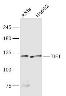 , ,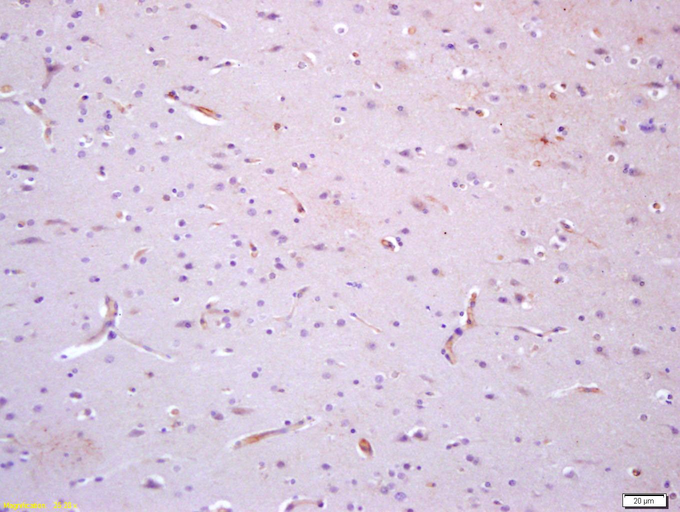 |
| Application | |
| Recommended Dose | WB: 1:500-2000; IHC-P: 1:100-500; IHC-Fr: 1:100-500; IF: 1:100-500 |
| Antibody Type | Polyclonal |
| Host Species | Rabbit |
| Subcellular Localization | Cell membrane; Single-pass type I membrane protein. |
| Tissue Specificity | Specifically expressed in developing vascular endothelial cells. |
| Construction | Polyclonal Antibody |
| Purification | Protein A purified |
| Appearance | Liquid |
| Formulation | 0.01M TBS (pH7.4) with 1% BSA, 0.02% Proclin300 and 50% Glycerol. |
| Concentration | 1 mg/mL |
| Research Background | TIE1/TIE (tyrosine kinase with Ig and EGF homology domains 1) and TIE2/Tek define a new class of the receptor tyrosine kinase (RTK) subfamily with unique structural characteristics: two immunoglobulin like domains flanking three epidermal growth factor (EFG) like domains followed by three fibronectin type III like repeats in the extracellular region and a split tyrosine kinase domain in the cytoplasmic region. Human TIE1 cDNA encodes a 1138 amino acid residue precursor protein with a putative signal peptide, an extracellular domain, and a cytoplasmic domain. Human TIE1/Fc, a disulfide linked homodimeric protein, has a calculated molecular mass of approximately 107 kDa. Due to glycosylation, the protein migrates to approximately 160 kDa in SDS PAGE under reducing conditions. TIE1 and TIE2, expressed primarily on endothelial and hematopoietic progenitor cells, play important roles in angiogenesis, vasculogenesis, and hematopoiesis. In developing vascular endothelial cells, TIE1 and TIE2 are specifically expressed. Two ligands that bind TIE have been identified, angiopoietin 1 and angiopoietin 2. Based on gene targeting studies, the in vivo functions of TIE1 are related to endothelial cell differentiation. The receptor tyrosine kinase TIE also plays a role in the survival and integrity of the endothelium. |
| Immunogen | KLH conjugated synthetic peptide: human Tie1 |
| Antigen Species | Human |
| Gene Name | TIE1 |
| Gene ID | |
| Protein Name | Tyrosine-protein kinase receptor Tie-1 |
| Uniprot ID | |
| Biology Area | Growth factor receptors,Endothelium,Mesoderm,Receptor Tyrosine Kinases,Endothelial Markers,Mesoderm |
| Function | Transmembrane tyrosine-protein kinase that may modulate TEK/TIE2 activity and contribute to the regulation of angiogenesis. |
| Molecular Weight | Theoretical: 123 kDa. Actual: 123 kDa. |
| Stability & Storage | Store at -20°C or -80°C for 12 months. Avoid repeated freeze-thaw cycles. |
| Transport | Shipping with blue ice. |
| Size | Quantity | Unit Price | Amount | Operation |
|---|
 Hello! How can I help you today?
 Copyright © 2015-2026 TargetMol Chemicals Inc. All Rights Reserved.
