
Shopping Cart
Remove All Your shopping cart is currently empty

Synonyms: SEMB, semaphorin 4A, SEMAB, RP35, CORD10
| Pack Size | Price | USA Stock | Global Stock | Quantity |
|---|---|---|---|---|
| 100 µL | $199 | 7-10 days | 7-10 days |
| Description | Anti-SEMA4A Antibody (9R816) is a Mouse antibody targeting SEMA4A. Anti-SEMA4A Antibody (9R816) can be used in ELISA. |
| Synonyms | SEMB, semaphorin 4A, SEMAB, RP35, CORD10 |
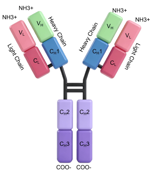
| Ig Type | Mouse IgG1 |
| Clone | 9R816 |
| Reactivity | Human |
| Specificity | Human Semaphorin 4A No cross-reactivity in ELISA with Human cell lysate (293 cell line) |
| Application | |
| Recommended Dose | ELISA: 1:1000-1:2000 |
| Antibody Type | Monoclonal |
| Host Species | Mouse |
| Construction | This antibody was produced from a hybridoma resulting from the fusion of a mouse myeloma with B cells obtained from a mouse immunized with purified, recombinant Human Semaphorin 4A / SEMA4A / Semaphorin B (rh Semaphorin 4A / SEMA4A / Semaphorin B; TMPY-01857; NP_071762.2; Met 1-His 683). The IgG fraction of the cell culture supernatant was purified by Protein A affinity chromatography. |
| Purification | Protein A |
| Appearance | Liquid |
| Formulation | 0.2 μm filtered solution in PBS |
| Research Background | Semaphorin-4A, also known as Semaphorin-B, SEMA4A, Sema B and SEMAB, is a single-pass type I membrane protein that belongs to the semaphorin family. It inhibits axonal extension by providing local signals to specify territories inaccessible for growing axons. Semaphorin-4A / SEMA4A contains one Ig-like C2-type (immunoglobulin-like) domain, one PSI domain and one Sema domain. Defects in SEMA4A are the cause of retinitis pigmentosa type 35 (RP35) which leads to degeneration of retinal photoreceptor cells. Patients typically have night vision blindness and loss of mid-peripheral visual field. As their condition progresses, they lose their far peripheral visual field and eventually central vision as well. Defects in SEMA4A are also the cause of cone-rod dystrophy type 1 (CORD1) which are inherited retinal dystrophies belonging to the group of pigmentary retinopathies. CORDs are characterized by retinal pigment deposits visible on fundus examination, predominantly in the macular region, and initial loss of cone photoreceptors followed by rod degeneration. Semaphorins are secreted, transmembrane, and GPI-linked proteins, defined by cysteine-rich semaphorin protein domains, that have important roles in a variety of tissues. Humans have 2 semaphorins, Drosophila has five, and two are known from DNA viruses. Semaphorins are found in nematodes and crustaceans but not in non-animals. They are grouped into eight classes on the basis of phylogenetic tree analyses and the presence of additional protein motifs. Semaphorins have been implicated in diverse developmental processes such as axon guidance during nervous system development and regulation of cell migration. |
| Conjucates | Unconjugated |
| Immunogen | Recombinant Protein: Human Semaphorin 4A / SEMA4A / Semaphorin B protein (TMPY-01857) |
| Antigen Species | Human |
| Stability & Storage | Store at 2°C-8°C for 1 month. Store at -20°C or -80°C for 12 months. Avoid repeated freeze-thaw cycles. Preservative-Free. |
| Transport | Shipping with blue ice. |
| Size | Quantity | Unit Price | Amount | Operation |
|---|
 Hello! How can I help you today?
 Copyright © 2015-2026 TargetMol Chemicals Inc. All Rights Reserved.
