
Shopping Cart
Remove All Your shopping cart is currently empty

Synonyms: Small ribosomal subunit protein uS2, RPSA, NEM/1CHD4, Multidrug resistance-associated protein MGr1-Ag, LAMR1, Laminin-binding protein precursor p40 (LBP/p40), Laminin receptor 1 (LamR), LAMBR, Colon carcinoma laminin-binding protein, 67 kDa laminin receptor (67LR), 40S ribosomal protein SA, 37/67 kDa laminin receptor (LRP/LR), 37 kDa laminin receptor precursor (37LRP)
Anti-RPSA Antibody
(6W769)
| Pack Size | Price | USA Stock | Global Stock | Quantity |
|---|---|---|---|---|
| 50 µL | $296 | 7-10 days | 7-10 days | |
| 100 µL | $497 | 7-10 days | 7-10 days |
| Description | Anti-RPSA Antibody (6W769) is a Rabbit antibody targeting RPSA. Anti-RPSA Antibody (6W769) can be used in FCM,ICC/IF,IHC,IP,WB. |
| Synonyms | Small ribosomal subunit protein uS2, RPSA, NEM/1CHD4, Multidrug resistance-associated protein MGr1-Ag, LAMR1, Laminin-binding protein precursor p40 (LBP/p40), Laminin receptor 1 (LamR), LAMBR, Colon carcinoma laminin-binding protein, 67 kDa laminin receptor (67LR), 40S ribosomal protein SA, 37/67 kDa laminin receptor (LRP/LR), 37 kDa laminin receptor precursor (37LRP) |
| Ig Type | IgG |
| Clone | 6W769 |
| Reactivity | Human,Mouse,Rat |
| Verified Activity | 1. Western blot analysis of RPSA on different lysates using anti-RPSA antibody at 1/1,000 dilution. Positive control: Lane 1: K562, Lane 2: HepG2, Lane 3: A431. 2. Immunohistochemical analysis of paraffin-embedded mouse brain tissue using anti-RPSA antibody. Counter stained with hematoxylin. 3. Immunohistochemical analysis of paraffin-embedded mouse kidney tissue using anti-RPSA antibody. Counter stained with hematoxylin. 4. Immunohistochemical analysis of paraffin-embedded human gastric carcinoma tissue using anti-RPSA antibody. Counter stained with hematoxylin. 5. ICC staining RPSA in Hela cells (green). The nuclear counter stain is DAPI (blue). Cells were fixed in paraformaldehyde, permeabilised with 0.25% Triton X100/PBS. 6. ICC staining RPSA in MCF-7 cells (green). The nuclear counter stain is DAPI (blue). Cells were fixed in paraformaldehyde, permeabilised with 0.25% Triton X100/PBS. 7. ICC staining RPSA in HepG2 cells (green). The nuclear counter stain is DAPI (blue). Cells were fixed in paraformaldehyde, permeabilised with 0.25% Triton X100/PBS. 8. ICC staining RPSA in RH-35 cells (green). The nuclear counter stain is DAPI (blue). Cells were fixed in paraformaldehyde, permeabilised with 0.25% Triton X100/PBS. 9. Flow cytometric analysis of MCF-7 cells with RPSA antibody at 1/50 dilution (red) compared with an unlabelled control (cells without incubation with primary antibody; black). Alexa Fluor 488-conjugated goat anti rabbit IgG was used as the secondary antibody. 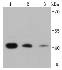 , ,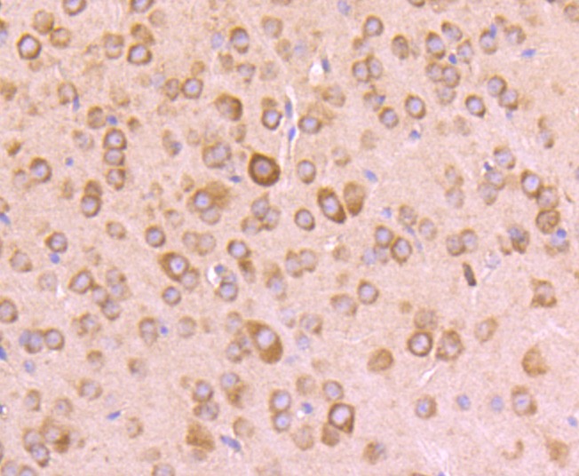 , ,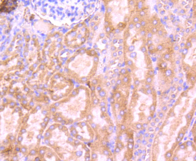 , ,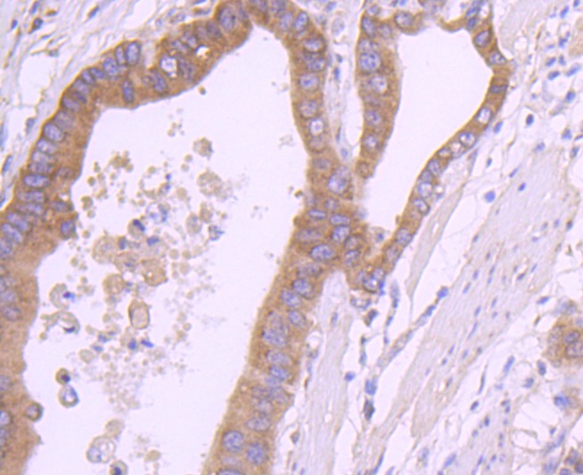 , ,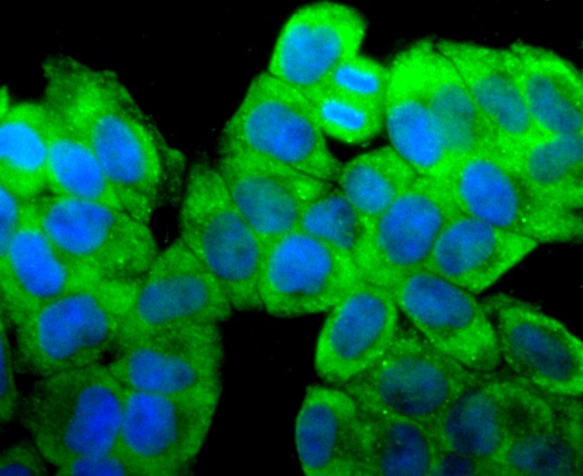 , ,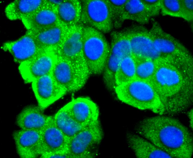 , ,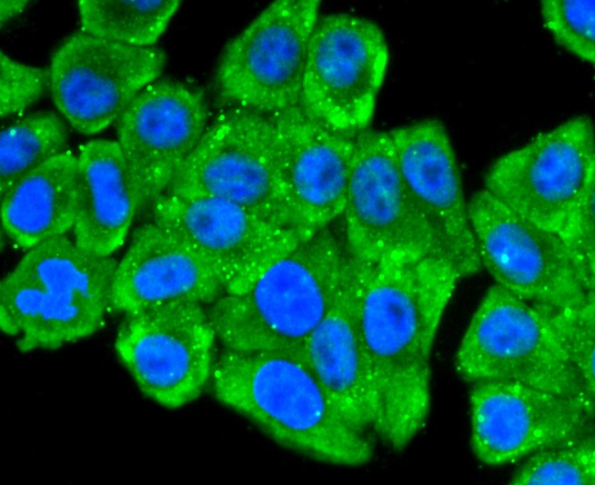 , ,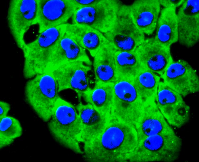 , ,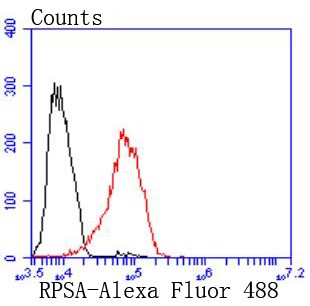 |
| Application | |
| Recommended Dose | WB: 1:1000-2000; IHC: 1:50-200; ICC/IF: 1:100-500; FCM: 1:50-100 |
| Antibody Type | Monoclonal |
| Host Species | Rabbit |
| Construction | Recombinant Antibody |
| Purification | ProA affinity purified |
| Appearance | Liquid |
| Formulation | 1*TBS (pH7.4), 1%BSA, 40%Glycerol. Preservative: 0.05% Sodium Azide. |
| Research Background | Laminin receptor (Laminin-R) has a heterodimeric structure similar to that of receptors for other extracellular matrix proteins such as Fibronectin and Vitronectin. Incorporation of Laminin-R into lysosomal membranes makes it possible for lysosomes to attach to surfaces coated with Laminin. This and other properties identify Laminin-R as a member of the integrin family of cell adhesion receptors. The Laminin-R precursor is a polypeptide whose expression is consistently upregulated in aggressive carcinoma. The precursor, which is also identified as p40 ribosome-associated protein, appears to be a multifunctional protein involved in the translational machinery. Laminin-R (also known as colon carcinoma laminin-binding protein) and is found at nine-fold higher levels in colon carcinoma than in adjacent normal colonic epithelium. Additionally, the level of the Laminin-R is higher in the lung cancer cell line than in the lung cell line. |
| Conjucates | Unconjugated |
| Immunogen | Recombinant Protein |
| Uniprot ID |
| Molecular Weight | Theoretical: 40 kDa. |
| Stability & Storage | Store at -20°C or -80°C for 12 months. Avoid repeated freeze-thaw cycles. |
| Transport | Shipping with blue ice. |
| Size | Quantity | Unit Price | Amount | Operation |
|---|
 Hello! How can I help you today?
 Copyright © 2015-2026 TargetMol Chemicals Inc. All Rights Reserved.
