
Shopping Cart
Remove All Your shopping cart is currently empty

Synonyms: Proviral integration site 2, Proviral integration site 1, Proto-oncogene serine/threonine-protein kinase Pim-1, Proto oncogene serine/threonine protein kinase Pim 1, PIM3, Pim2, PIM1_HUMAN, pim1 kinase 44 kDa isoform, PIM1, pim 1 oncogene (proviral integration site 1), Pim 1 oncogene, pim 1 kinase 44 kDa isoform, Pim 1 kinase, PIM 1, PIM, Oncogene PIM1, Oncogene PIM 1
Anti-PIM1 Antibody
(6F773)
| Pack Size | Price | USA Stock | Global Stock | Quantity |
|---|---|---|---|---|
| 50 µL | $297 | 7-10 days | 7-10 days | |
| 100 µL | $498 | 7-10 days | 7-10 days |
| Description | Anti-PIM1 Antibody (6F773) is a Rabbit antibody targeting PIM1. Anti-PIM1 Antibody (6F773) can be used in ICC/IF,IHC,WB. |
| Synonyms | Proviral integration site 2, Proviral integration site 1, Proto-oncogene serine/threonine-protein kinase Pim-1, Proto oncogene serine/threonine protein kinase Pim 1, PIM3, Pim2, PIM1_HUMAN, pim1 kinase 44 kDa isoform, PIM1, pim 1 oncogene (proviral integration site 1), Pim 1 oncogene, pim 1 kinase 44 kDa isoform, Pim 1 kinase, PIM 1, PIM, Oncogene PIM1, Oncogene PIM 1 |
| Ig Type | IgG |
| Clone | 6F773 |
| Reactivity | Human,Mouse |
| Verified Activity | 1. Western blot analysis of PIM1 on different lysates using anti-PIM1 antibody at 1/1,000 dilution. Positive control: Lane 1: BT-20, Lane 2: HepG2, Lane 3: SW480. 2. Immunohistochemical analysis of paraffin-embedded human gastric carcinoma tissue using anti-PIM1 antibody. Counter stained with hematoxylin. 3. Immunohistochemical analysis of paraffin-embedded human tonsil tissue using anti-PIM1 antibody. Counter stained with hematoxylin. 4. Immunohistochemical analysis of paraffin-embedded human colon cancer tissue using anti-PIM1 antibody. Counter stained with hematoxylin. 5. Immunohistochemical analysis of paraffin-embedded human spleen tissue using anti-PIM1 antibody. Counter stained with hematoxylin. 6. Immunohistochemical analysis of paraffin-embedded human breast carcinoma tissue using anti-PIM1 antibody. Counter stained with hematoxylin. 7. Immunohistochemical analysis of paraffin-embedded human gastric carcinoma tissue using anti-PIM1 antibody. Counter stained with hematoxylin. 8. ICC staining PIM1 in BT-20 cells (green). The nuclear counter stain is DAPI (blue). Cells were fixed in paraformaldehyde, permeabilised with 0.25% Triton X100/PBS. 9. ICC staining PIM1 in HepG2 cells (green). The nuclear counter stain is DAPI (blue). Cells were fixed in paraformaldehyde, permeabilised with 0.25% Triton X100/PBS. 10. ICC staining PIM1 in SW480 cells (green). The nuclear counter stain is DAPI (blue). Cells were fixed in paraformaldehyde, permeabilised with 0.25% Triton X100/PBS. 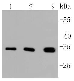 , ,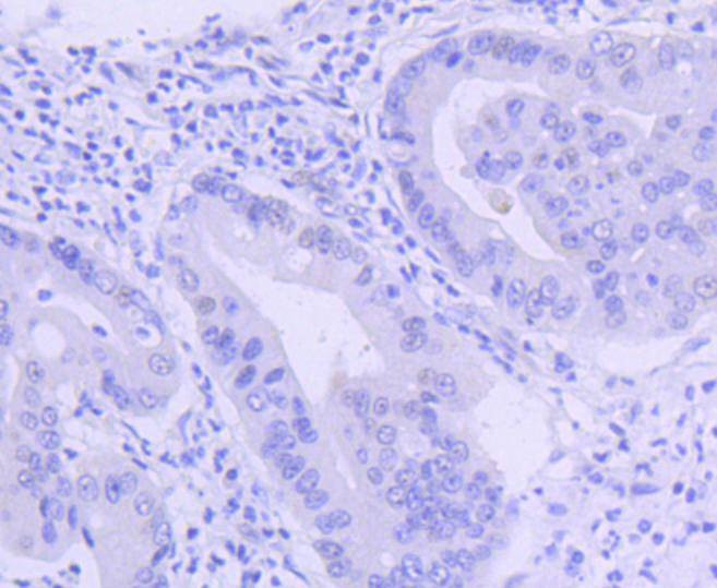 , ,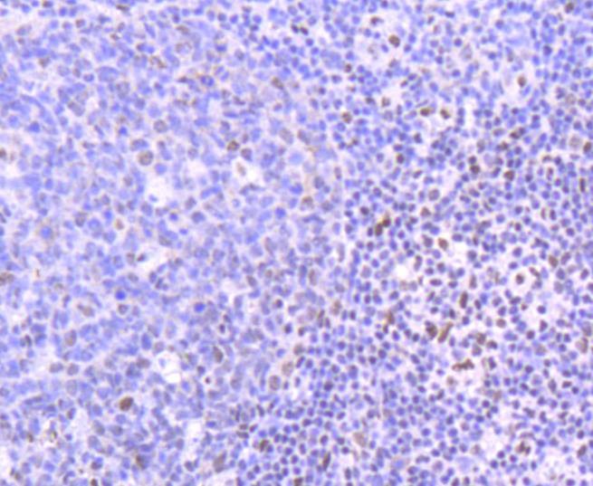 , ,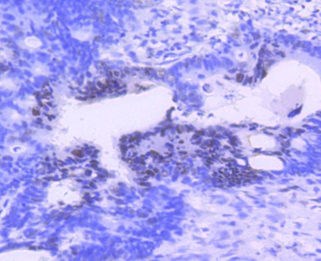 , ,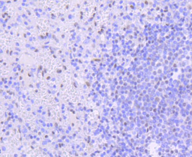 , ,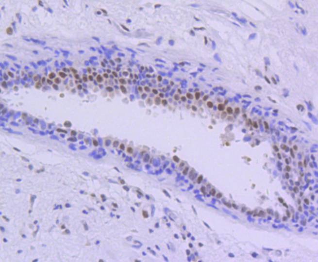 , ,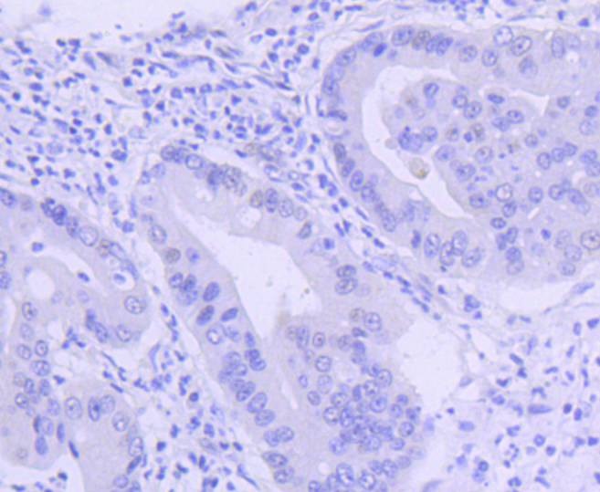 , ,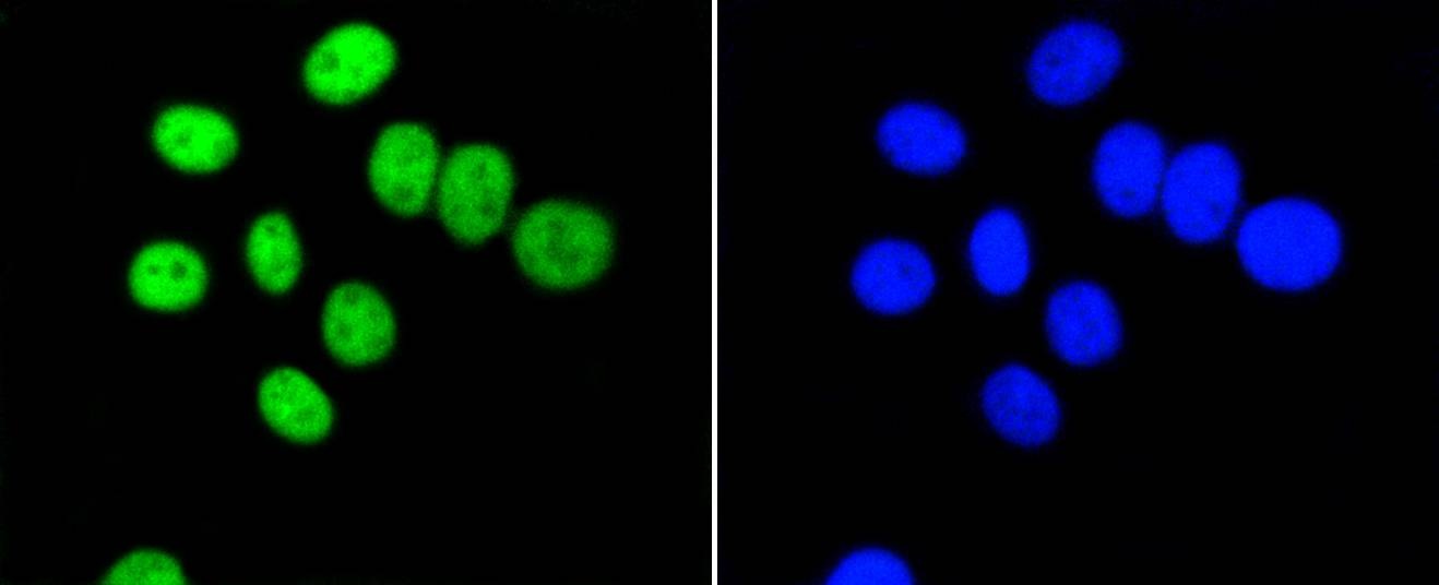 , ,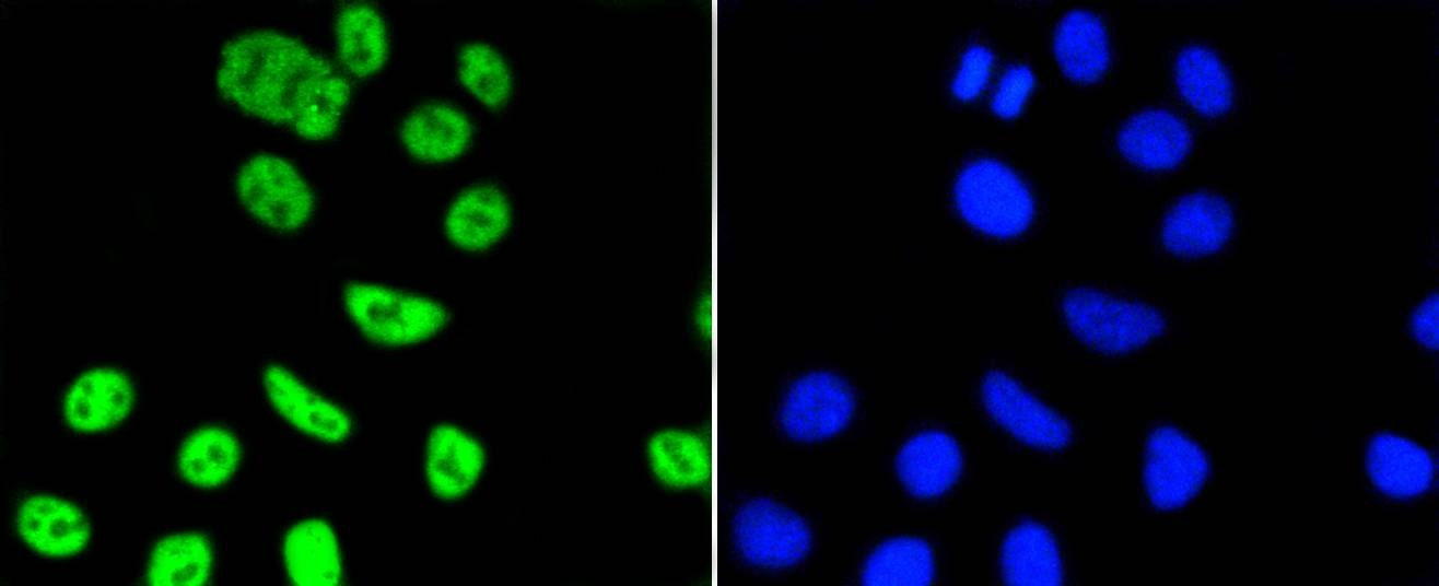 , ,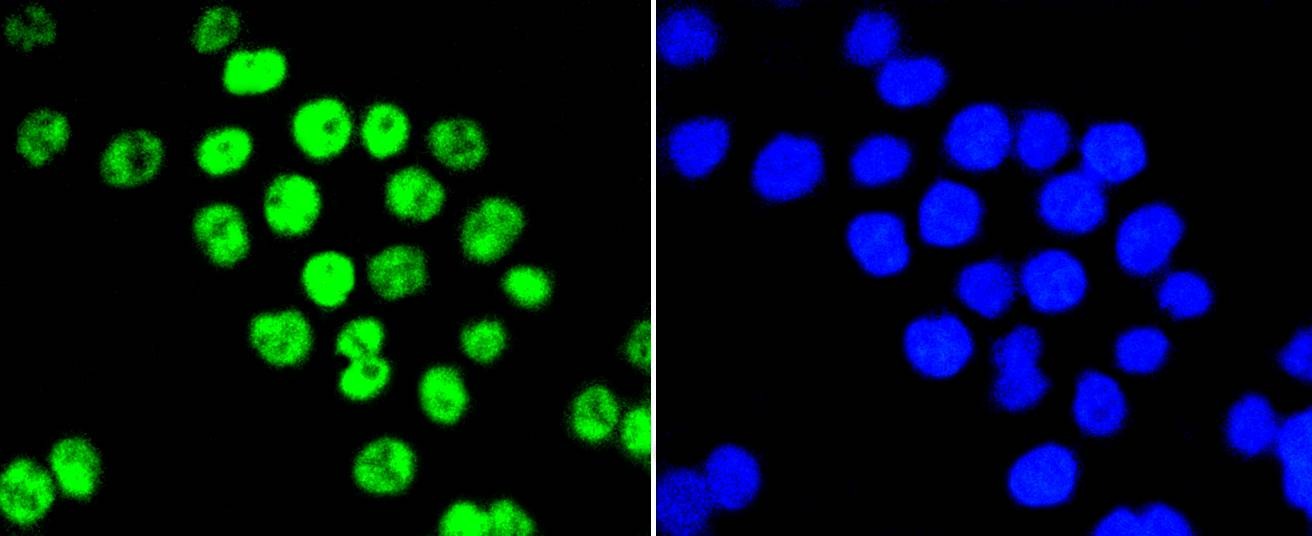 |
| Application | |
| Recommended Dose | WB: 1:1000-2000; IHC: 1:50-200; ICC/IF: 1:50-200 |
| Antibody Type | Monoclonal |
| Host Species | Rabbit |
| Construction | Recombinant Antibody |
| Purification | ProA affinity purified |
| Appearance | Liquid |
| Formulation | 1*TBS (pH7.4), 1%BSA, 40%Glycerol. Preservative: 0.05% Sodium Azide. |
| Research Background | Pim-1 is a serine/threonine kinase that cooperates with c-Myc in lymphoid cell transformation. The expression of pim-1 increases during the progression from early to late G1, remaining high at the G1/S boundary and G2 phases of the cell cycle. Pim-1 is regulated at both the transcriptional and translational level, and it has been shown to be induced by IL-2 stimulation. Pim-1 also plays a role in T cell differentiation, and it has been shown to stimulate c-Myc-mediated apoptosis upstream of caspase-3-like proteases. |
| Conjucates | Unconjugated |
| Immunogen | Recombinant Protein |
| Uniprot ID |
| Molecular Weight | Theoretical: 32 kDa. |
| Stability & Storage | Store at -20°C or -80°C for 12 months. Avoid repeated freeze-thaw cycles. |
| Transport | Shipping with blue ice. |
| Size | Quantity | Unit Price | Amount | Operation |
|---|
 Hello! How can I help you today?
 Copyright © 2015-2026 TargetMol Chemicals Inc. All Rights Reserved.
