
Shopping Cart
Remove All Your shopping cart is currently empty

Synonyms: PAR2, GPR11
Anti-PAR2 Polyclonal Antibody
| Pack Size | Price | USA Stock | Global Stock | Quantity |
|---|---|---|---|---|
| 50 µL | $220 | 7-10 days | 7-10 days | |
| 100 µL | $372 | 7-10 days | 7-10 days | |
| 200 µL | $528 | 7-10 days | 7-10 days |
| Description | Anti-PAR2 Polyclonal Antibody is a Rabbit antibody targeting PAR2. Anti-PAR2 Polyclonal Antibody can be used in IF,IHC-Fr,IHC-P,WB. |
| Synonyms | PAR2, GPR11 |
| Ig Type | IgG |
| Reactivity | Human,Rat (predicted:Mouse,Rabbit) |
| Verified Activity | 1. Tissue/cell: human colon carcinoma; 4% Paraformaldehyde-fixed and paraffin-embedded; Antigen retrieval: citrate buffer (0.01M, pH6.0), Boiling bathing for 15 min; Block endogenous peroxidase by 3% Hydrogen peroxide for 30 min; Blocking buffer (normal goat serum) at 37°C for 20 min; Incubation: Anti-APR-2 Polyclonal Antibody, Unconjugated (TMAB-01331) 1:500, overnight at 4°C, followed by conjugation to the secondary antibody and DAb staining. 2. Sample: Lane 1: HepG2 (Human) Cell Lysate at 30 μg Lane 2: MCF-7 (Human) Cell Lysate at 30 μg Primary: Anti-PAR-2 (TMAB-01331) at 1/500 dilution Secondary: IRDye800CW Goat Anti-Rabbit IgG at 1/20000 dilution Predicted band size: 40 kDa Observed band size: 60 kDa 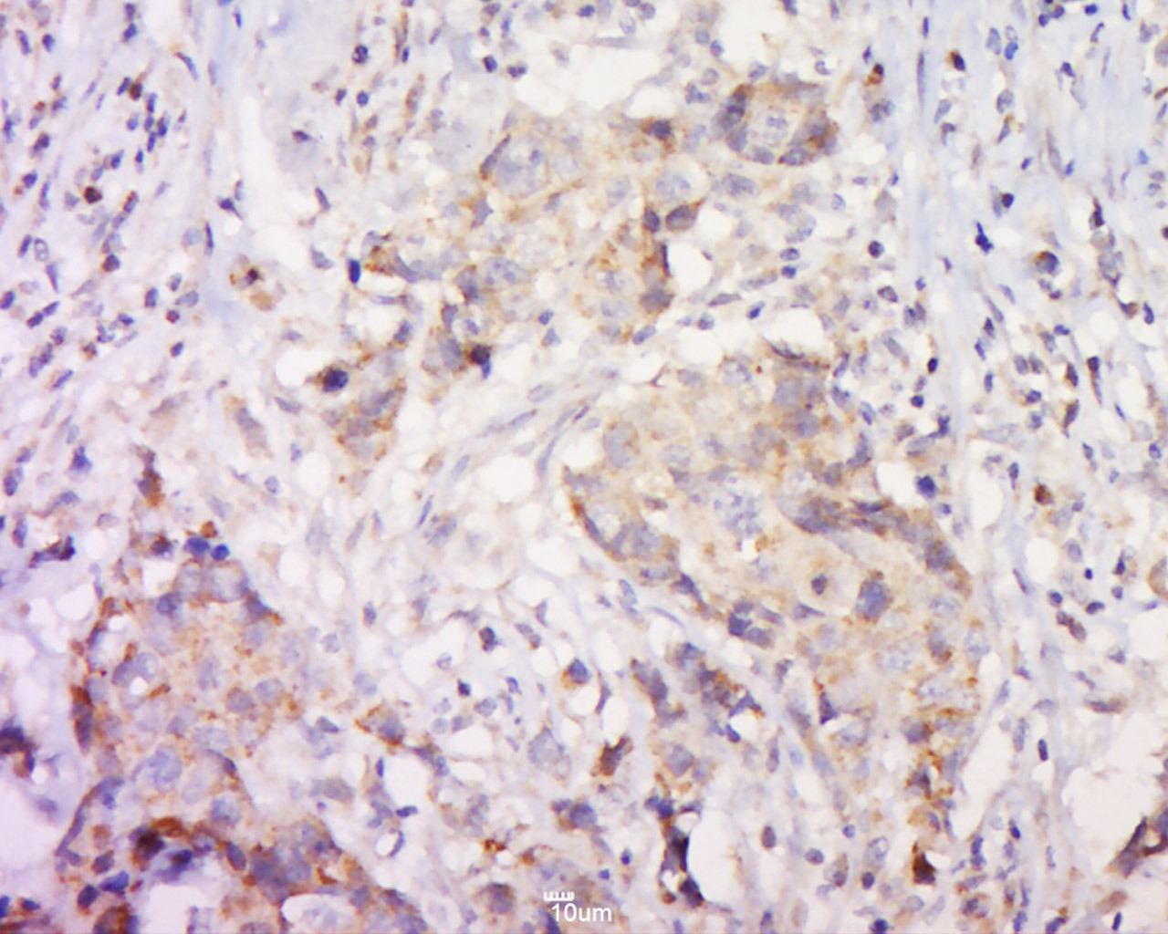 , ,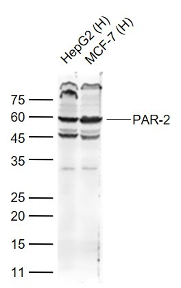 |
| Application | |
| Recommended Dose | WB: 1:500-2000; IHC-P: 1:100-500; IHC-Fr: 1:100-500; IF: 1:100-500 |
| Antibody Type | Polyclonal |
| Host Species | Rabbit |
| Subcellular Localization | Cell membrane; Multi-pass membrane protein. |
| Tissue Specificity | Widely expressed in tissues with especially high levels in pancreas, liver, kidney, small intestine, and colon. Moderate expression is detected in many organs, but none in brain or skeletal muscle. |
| Construction | Polyclonal Antibody |
| Purification | Protein A purified |
| Appearance | Liquid |
| Formulation | 0.01M TBS (pH7.4) with 1% BSA, 0.02% Proclin300 and 50% Glycerol. |
| Concentration | 1 mg/mL |
| Research Background | The Proteinase-activated receptor 2 (PAR2) is a member of the proteinase-activated receptor subfamily. It is activated through proteolytic exposure of an occult tethered ligand by trypsin and trypsin-like proteases. This is in contrast to other members of the subfamily which are activated by the protease thrombin. PAR2 has been implicated in acute inflammatory response, asthma, and pain transmission. PAR2 expression has been documented in the periphery. ESTs have been isolated from adrenal, brain, breast, heart/melanocyte/uterus, kidney, lung, and vessel libraries. Coagulation factor II (thrombin) receptor-like 1 (F2RL1)is a member of the large family of 7-transmembrane-region receptors that couple to guanosine-nucleotide-binding proteins. F2RL1 is also a member of the protease-activated receptor family. It is activated by trypsin, but not by thrombin. It is activated by proteolytic cleavage of its extracellular amino terminus. The new amino terminus functions as a tethered ligand and activates the receptor. The F2RL1 gene contains two exons and is widely expressed in human tissues. The predicted protein sequence is 83% identical to the mouse receptor sequence. [provided by RefSeq]. |
| Immunogen | KLH conjugated synthetic peptide: human PAR2 |
| Antigen Species | Human |
| Gene Name | F2RL1 |
| Gene ID | |
| Protein Name | Proteinase-activated receptor 2 |
| Uniprot ID | |
| Function | Receptor for trypsin and trypsin-like enzymes coupled to G proteins. Its function is mediated through the activation of several signaling pathways including phospholipase C (PLC), intracellular calcium, mitogen-activated protein kinase (MAPK), I-kappaB kinase/NF-kappaB and Rho. Can also be transactivated by cleaved F2R/PAR1. Involved in modulation of inflammatory responses and regulation of innate and adaptive immunity, and acts as a sensor for proteolytic enzymes generated during infection. Generally is promoting inflammation. Can signal synergistically with TLR4 and probably TLR2 in inflammatory responses and modulates TLR3 signaling. Has a protective role in establishing the endothelial barrier; the activity involves coagulation factor X. Proposed to have a bronchoprotective role in airway epithelium, but also shown to compromise the airway epithelial barrier by interrupting E-cadherin adhesion. Involved in the regulation of vascular tone; activation results in hypotension presumably mediated by vasodilation. Associates with a subset of G proteins alpha subunits such as G alpha-q, G alpha-11, G alpha-14, G alpha-12 and G alpha-13, but probably not with G(o) alpha, G(i) subunit alpha-1 and G(i) subunit alpha-2. However, according to PubMed:21627585 can signal through G(i) subunit alpha. Believed to be a class B receptor which internalizes as a complex with arrestin and traffic with it to endosomal vesicles, presumably as desensitized receptor, for extended periods of time. Mediates inhibition of TNF-alpha stimulated JNK phosphorylation via coupling to G alpha-q/11; the function involves dissociation of RIPK1 and TRADD from TNFR1. Mediates phosphorylation of nuclear factor NF-kappa-B RELA subunit at 'Ser-536'; the function involves IKBKB and is predominantly independent of G proteins. Involved in cellular migration. Involved in cytoskeletal rearrangement and chemotaxis through beta-arrestin-promoted scaffolds; the function is independent of G alpha-q/11 and involves promotion of cofilin dephosphoryltaion and actin filament severing. Induces redistribution of COPS5 from the plasma membrane to the cytosol and activation of the JNK cascade is mediated by COPS5. Involved in the recruitment of leukocytes to the sites of inflammation and is the major PAR receptor capable of modulating eosinophil function such as proinflammatory cytokine secretion, superoxide production and degranulation. During inflammation promotes dendritic cell maturation, trafficking to the lymph nodes and subsequent T-cell activation. Involved in antimicrobial response of innate immnune cells; activation enhances phagocytosis of Gram-positive and killing of Gram-negative bacteria. Acts synergistically with interferon-gamma in enhancing antiviral responses. Implicated in a number of acute and chronic inflammatory diseases such as of the joints, lungs, brain, gastrointestinal tract, periodontium, skin, and vascular systems, and in autoimmune disorders. |
| Molecular Weight | Theoretical: 40 kDa. Actual: 60 kDa. |
| Stability & Storage | Store at -20°C or -80°C for 12 months. Avoid repeated freeze-thaw cycles. |
| Transport | Shipping with blue ice. |
| Size | Quantity | Unit Price | Amount | Operation |
|---|
 Hello! How can I help you today?
 Copyright © 2015-2026 TargetMol Chemicals Inc. All Rights Reserved.
