
Shopping Cart
Remove All Your shopping cart is currently empty

Synonyms: wide spectrum Cytokeratin, P-CK, pan-cytokeratin, pan-CK, Cytokeratins, CK-PAN, [cytokeratins 13, 14, 16, 17, 19, 24]
Anti-Pan Cytokeratin Polyclonal Antibody 2
| Pack Size | Price | USA Stock | Global Stock | Quantity |
|---|---|---|---|---|
| 50 µL | $220 | 7-10 days | 7-10 days | |
| 100 µL | $374 | 7-10 days | 7-10 days | |
| 200 µL | $529 | 7-10 days | 7-10 days |
| Description | Anti-Pan Cytokeratin Polyclonal Antibody 2 is a Rabbit antibody targeting Pan Cytokeratin. Anti-Pan Cytokeratin Polyclonal Antibody 2 can be used in FCM, ICC/IF, IF, IHC-Fr, IHC-P, WB. |
| Synonyms | wide spectrum Cytokeratin, P-CK, pan-cytokeratin, pan-CK, Cytokeratins, CK-PAN, [cytokeratins 13, 14, 16, 17, 19, 24] |
| Ig Type | IgG |
| Reactivity | Human,Mouse,Rat (predicted:Chicken,Dog,Pig,Cow,Horse,Rabbit) |
| Verified Activity | 1. Paraformaldehyde-fixed, paraffin embedded (Rat bladder); Antigen retrieval by boiling in sodium citrate buffer (pH6.0) for 15 min; Block endogenous peroxidase by 3% hydrogen peroxide for 20 min; Blocking buffer (normal goat serum) at 37°C for 30 min; Antibody incubation with (Pan Cytokeratin) Polyclonal Antibody, Unconjugated (TMAB-01330) at 1:400 overnight at 4°C, followed by operating according to SP Kit (Rabbit) instructionsand DAB staining. 2. Sample: Bladder (Mouse) Lysate at 40 μg Primary: Anti-Pan Cytokeratin (TMAB-01330) at 1/300 dilution Secondary: IRDye800CW Goat Anti-Rabbit IgG at 1/20000 dilution Predicted band size: 42-64 kDa Observed band size: 60 kDa 3. Blank control (blue line): Hela (blue). Primary Antibody (green line): Rabbit Anti-Pan Cytokeratin antibody (TMAB-01330) Dilution: 1 μg/10^6 cells; Isotype Control Antibody (orange line): Rabbit IgG. Secondary Antibody (white blue line): Goat anti-rabbit IgG-PE Dilution: 1 μg/test. Protocol The cells were fixed with 70% methanol (Overnight at 4°C) and then permeabilized with 90% ice-cold methanol for 20 min at-20°C. Cells stained with Primary Antibody for 30 min at room temperature. The cells were then incubated in 1 X PBS/2% BSA/10% goat serum to block non-specific protein-protein interactions followed by the antibody for 15 min at room temperature. The secondary antibody used for 40 min at room temperature. 4. Paraformaldehyde-fixed, paraffin embedded (Human stomach carcinoma); Antigen retrieval by boiling in sodium citrate buffer (pH6.0) for 15 min; Block endogenous peroxidase by 3% hydrogen peroxide for 20 min; Blocking buffer (normal goat serum) at 37°C for 30 min; Antibody incubation with (Pan Cytokeratin) Polyclonal Antibody, Unconjugated (TMAB-01330) at 1:400 overnight at 4°C, followed by operating according to SP Kit (Rabbit) instructionsand DAB staining. 5. Paraformaldehyde-fixed, paraffin embedded (human liver); Antigen retrieval by boiling in sodium citrate buffer (pH6.0) for 15 min; Block endogenous peroxidase by 3% hydrogen peroxide for 20 min; Blocking buffer (normal goat serum) at 37°C for 30 min; Antibody incubation with (Pan Cytokeratin) Polyclonal Antibody, Unconjugated (TMAB-01330) at 1:200 overnight at 4°C, followed by operating according to SP Kit (Rabbit) instructionsand DAB staining. 6. Paraformaldehyde-fixed, paraffin embedded (Human kidney); Antigen retrieval by boiling in sodium citrate buffer (pH6.0) for 15 min; Block endogenous peroxidase by 3% hydrogen peroxide for 20 min; Blocking buffer (normal goat serum) at 37°C for 30 min; Antibody incubation with (Pan Cytokeratin) Polyclonal Antibody, Unconjugated (TMAB-01330) at 1:200 overnight at 4°C, followed by operating according to SP Kit (Rabbit) instructionsand DAB staining. 7. Sample: Lane 1: Hela (Human) Cell Lysate at 30 μg Lane 2: A549 (Human) Cell Lysate at 30 μg Lane 3: A673 (Human) Cell Lysate at 30 μg Primary: Anti-Pan Cytokeratin (TMAB-01330) at 1/1000 dilution Secondary: IRDye800CW Goat Anti-Rabbit IgG at 1/20000 dilution Predicted band size: 42-64 kDa Observed band size: 46,60 kDa 8. Paraformaldehyde-fixed, paraffin embedded (human cervical cancer); Antigen retrieval by boiling in sodium citrate buffer (pH6.0) for 15 min; Block endogenous peroxidase by 3% hydrogen peroxide for 20 min; Blocking buffer (normal goat serum) at 37°C for 30 min; Antibody incubation with (Pan Cytokeratin) Polyclonal Antibody, Unconjugated (TMAB-01330) at 1:200 overnight at 4°C, followed by operating according to SP Kit (Rabbit) instructionsand DAB staining. 9. Paraformaldehyde-fixed, paraffin embedded (rat uterus); Antigen retrieval by boiling in sodium citrate buffer (pH6.0) for 15 min; Block endogenous peroxidase by 3% hydrogen peroxide for 20 min; Blocking buffer (normal goat serum) at 37°C for 30 min; Antibody incubation with (Pan Cytokeratin) Polyclonal Antibody, Unconjugated (TMAB-01330) at 1:200 overnight at 4°C, followed by operating according to SP Kit (Rabbit) instructionsand DAB staining. 10. Hela cell; 4% Paraformaldehyde-fixed; Triton X-100 at room temperature for 20 min; Blocking buffer (normal goat serum) at 37°C for 20 min; Antibody incubation with (Pan Cytokeratin) polyclonal Antibody, Unconjugated (TMAB-01330) 1:100, 90 minutes at 37°C; followed by a conjugated Goat Anti-Rabbit IgG antibody at 37°C for 90 minutes, DAPI (blue) was used to stain the cell nucleus. 11. Paraformaldehyde-fixed, paraffin embedded (human breast carcinoma); Antigen retrieval by boiling in sodium citrate buffer (pH6.0) for 15 min; Block endogenous peroxidase by 3% hydrogen peroxide for 20 min; Blocking buffer (normal goat serum) at 37°C for 30 min; Antibody incubation with (Pan Cytokeratin) Polyclonal Antibody, Unconjugated (TMAB-01330) at 1:200 overnight at 4°C, followed by operating according to SP Kit (Rabbit) instructionsand DAB staining. 12. Paraformaldehyde-fixed, paraffin embedded (human colon carcinoma); Antigen retrieval by boiling in sodium citrate buffer (pH6.0) for 15 min; Blocking buffer (normal goat serum) at 37°C for 30 min; Antibody incubation with (Pan Cytokeratin) Polyclonal Antibody, Unconjugated (TMAB-01330) at 1:200 overnight at 4°C, followed by a conjugated Goat Anti-Rabbit IgG antibody for 90 minutes, and DAPI for nucleus staining. 13. Paraformaldehyde-fixed, paraffin embedded (human cervical carcinoma); Antigen retrieval by boiling in sodium citrate buffer (pH6.0) for 15 min; Block endogenous peroxidase by 3% hydrogen peroxide for 20 min; Blocking buffer (normal goat serum) at 37°C for 30 min; Antibody incubation with (Pan Cytokeratin) Polyclonal Antibody, Unconjugated (TMAB-01330) at 1:200 overnight at 4°C, followed by operating according to SP Kit (Rabbit) instructionsand DAB staining. 14. Blank control: Hela. Primary Antibody (green line): Rabbit Anti-Pan Cytokeratin antibody (TMAB-01330) Dilution: 2 μg/Test; Secondary Antibody: Goat anti-rabbit IgG-FITC Dilution: 0.5 μg/Test. Protocol The cells were fixed with 4% PFA (10 min at room temperature) and then permeabilized with 0.1% PBST for 20 min at room temperature. The cells were then incubated in 5% BSA to block non-specific protein-protein interactions for 30 min at room temperature. Cells stained with Primary Antibody for 30 min at room temperature. The secondary antibody used for 40 min at room temperature. 15. Blank control (black line): A549. Primary Antibody (green line): Rabbit Anti-Pan Cytokeratin antibody (TMAB-01330) Dilution: 1 μg/Test; Secondary Antibody (white blue line): Goat anti-rabbit IgG-AF488 Dilution: 0.5 μg/Test. Isotype control (orange line): Normal Rabbit IgG Protocol The cells were fixed with 4% PFA (10 min at room temperature) and then permeabilized with 90% ice-cold methanol for 20 min at-20°C, The cells were then incubated in 5% BSA to block non-specific protein-protein interactions for 30 min at room temperature. Cells stained with Primary Antibody for 30 min at room temperature. The secondary antibody used for 40 min at room temperature. 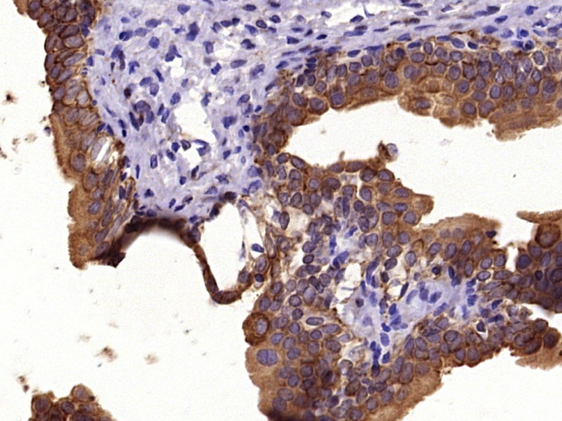 , ,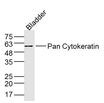 , ,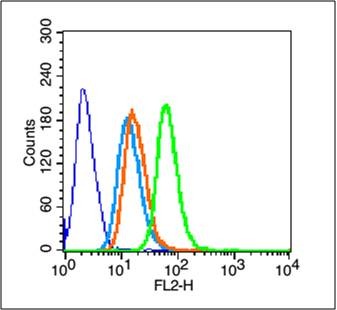 , ,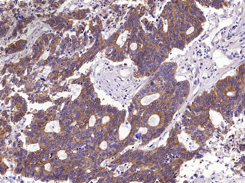 , ,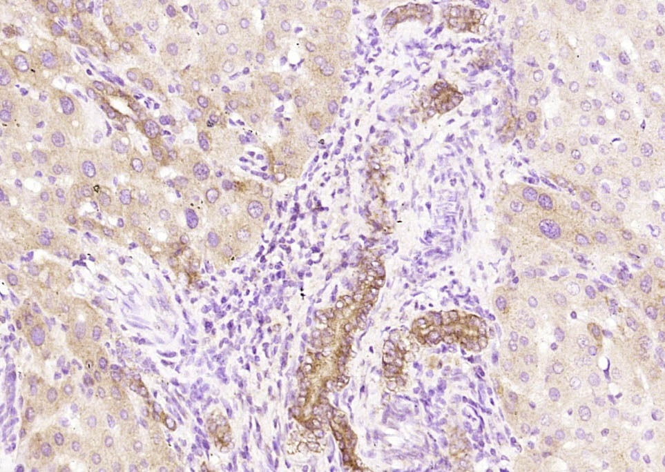 , ,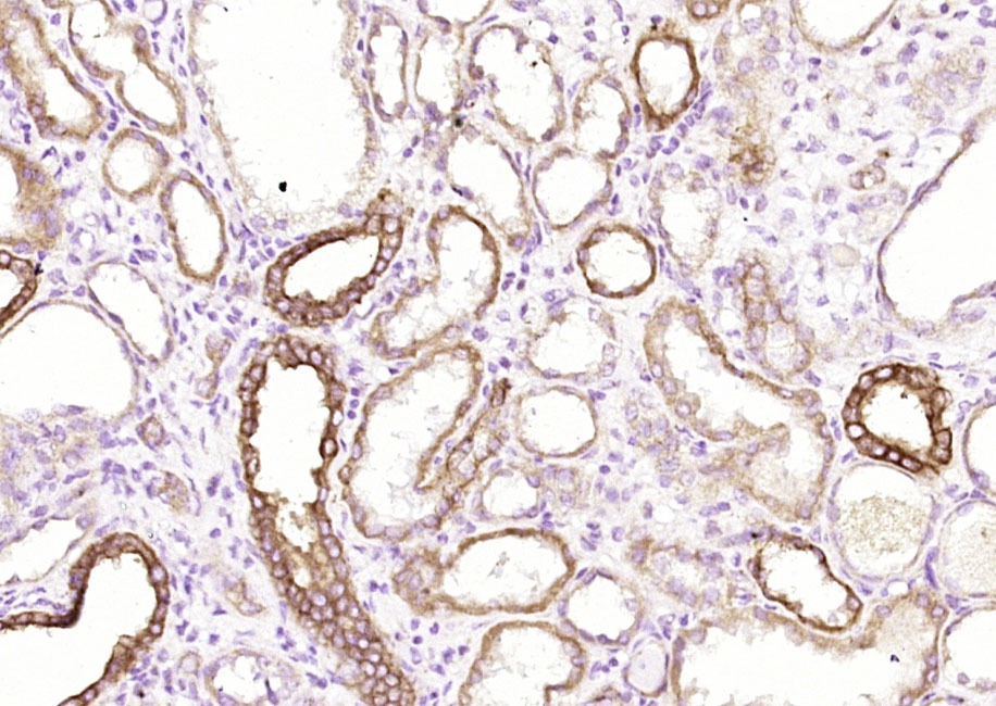 , ,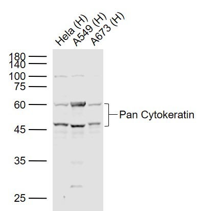 , ,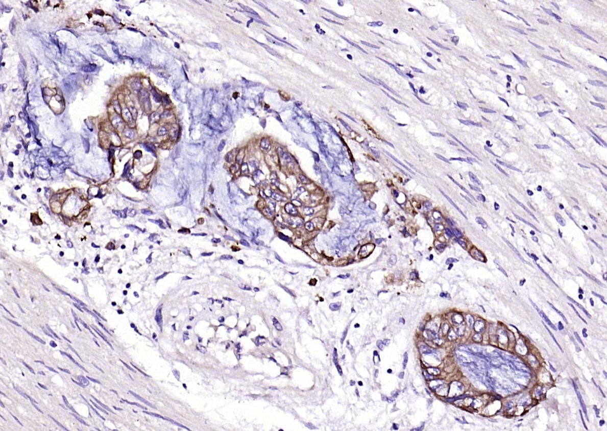 , ,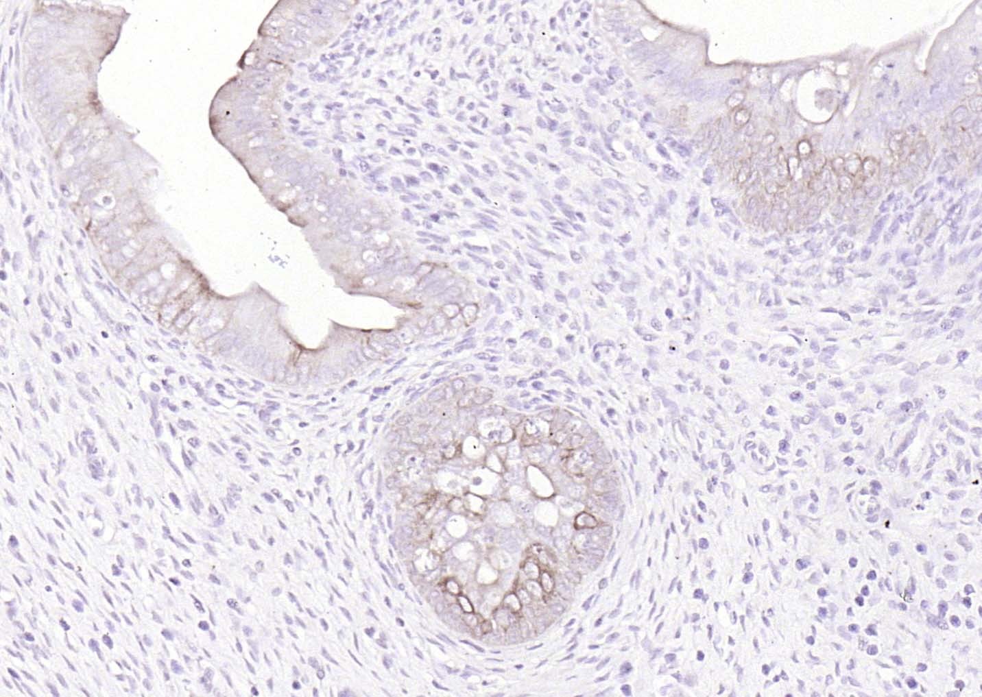 , ,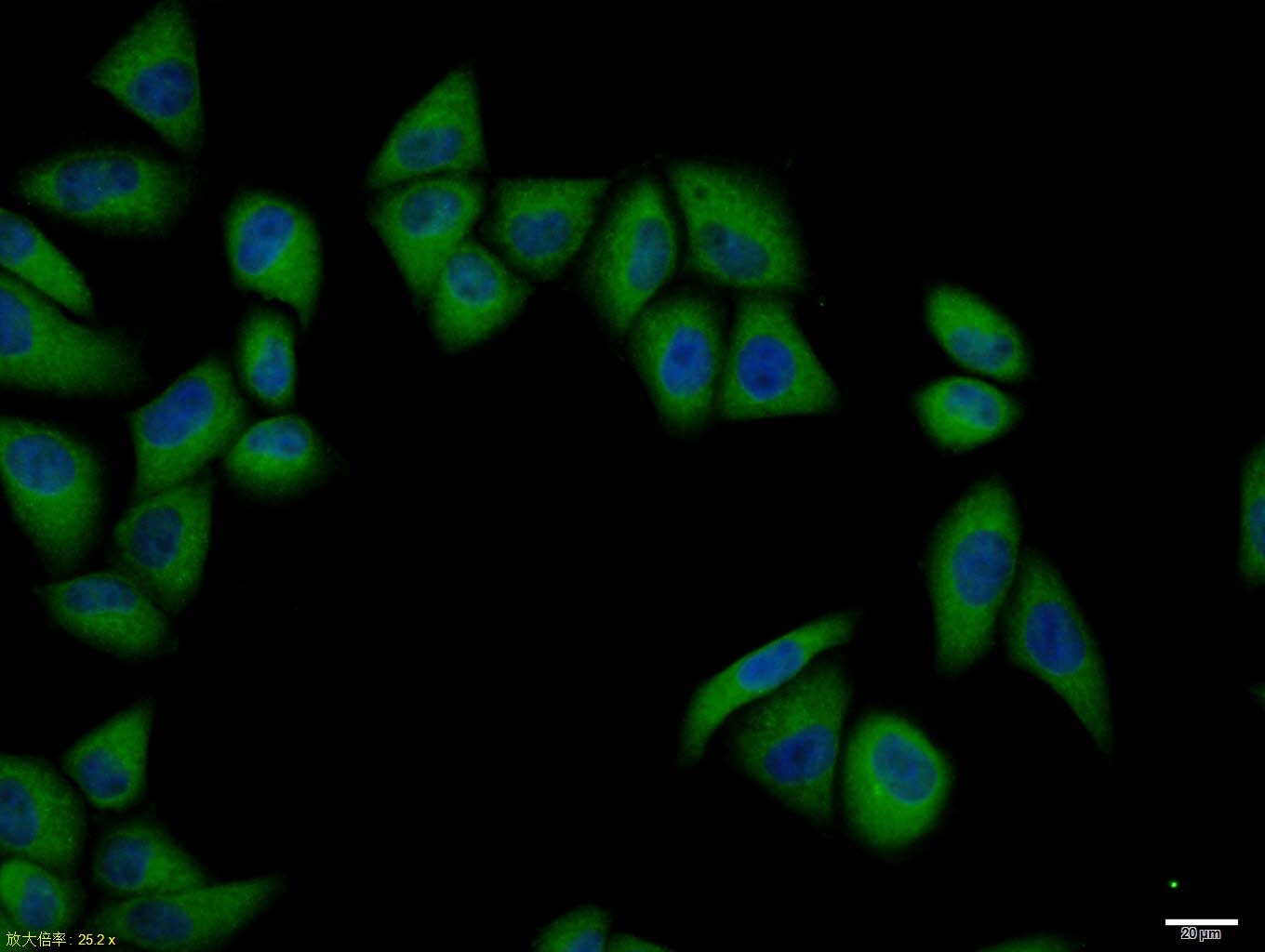 , ,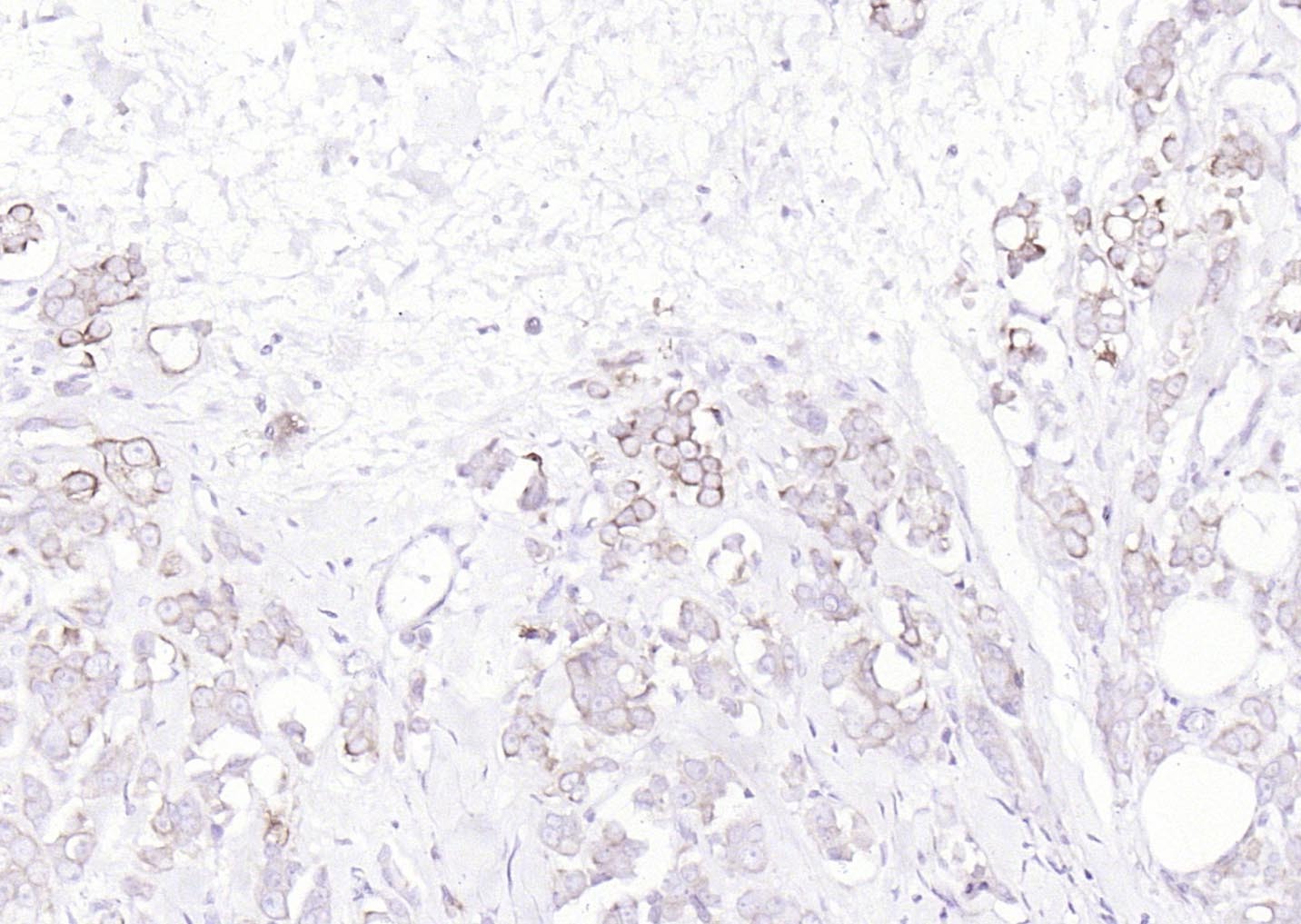 , ,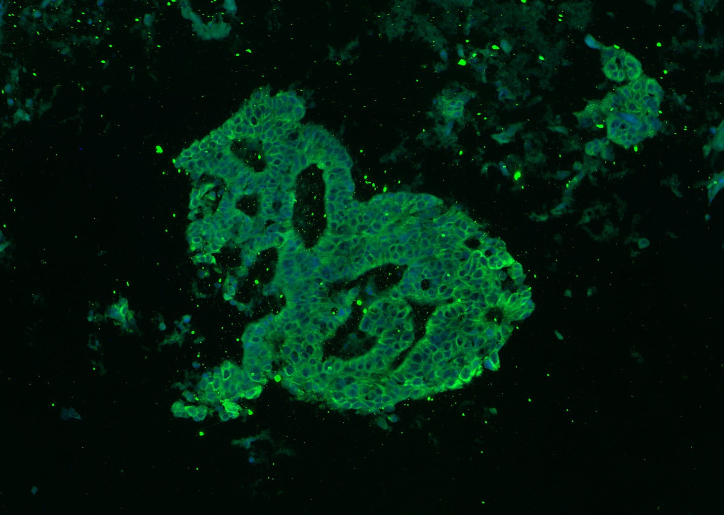 , ,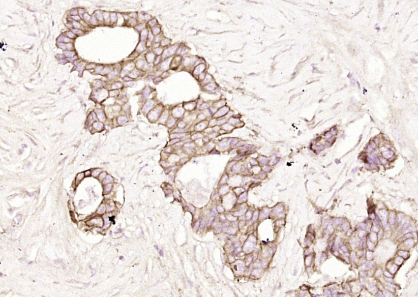 , ,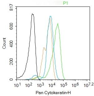 , ,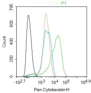 |
| Application | |
| Recommended Dose | FCM=1 μg/Test; ICC/IF=1:100-500; IF=1:100-500; IHC-Fr=1:100-500; IHC-P=1:100-500; WB=1:500-2000 |
| Antibody Type | Polyclonal |
| Host Species | Rabbit |
| Subcellular Localization | Cytoplasmic. |
| Tissue Specificity | epithelial cells |
| Construction | Polyclonal Antibody |
| Purification | Protein A purified |
| Appearance | Liquid |
| Formulation | 0.01M TBS (pH7.4) with 1% BSA, 0.02% Proclin300 and 50% Glycerol. |
| Concentration | 1 mg/mL |
| Research Background | Cytokeratins are proteins of keratin-containing intermediate filaments found in the intracytoplasmic cytoskeleton of epithelial tissue. The cytokeratins are encoded by a family encompassing 30 genes. Among them, 20 are epithelial genes and the remaining 10 are specific for trichocytes. In the cytoplasm, the keratin filaments conform a complex network which extends from the surface of the nucleus to the cell membrane. Numerous accessory proteins are involved in the genesis and maintenance of such structure. This association between the plasma membrane and the nuclear surface provides important implications for the organization of the cytoplasm and cellular communication mechanisms. Apart from the relatively static functions provided in terms of supporting the nucleus and providing tensile strength to the cell, the cytokeratin networks undergo rapid phosphate exchanges mediated depolymerization, with important implications in the more dynamic cellular processes such as mitosis and post-mitotic period, cell movement and differentiation. Cytokeratins interact with desmosomes and hemidesmosomes, thus collaborating to cell-cell adhesion and basal cell-underlying connective tissue connection. |
| Immunogen | KLH conjugated synthetic peptide: human Cytokeratin-1 |
| Antigen Species | Human |
| Gene Name | KRT24 |
| Gene ID | |
| Protein Name | Keratin, type I cytoskeletal 24 |
| Uniprot ID | |
| Biology Area | Cytokeratins |
| Molecular Weight | Theoretical: 42-64 kDa. Actual: 46,60 kDa. |
| Stability & Storage | Store at -20°C or -80°C for 12 months. Avoid repeated freeze-thaw cycles. |
| Transport | Shipping with blue ice. |
| Size | Quantity | Unit Price | Amount | Operation |
|---|
 Hello! How can I help you today?
 Copyright © 2015-2026 TargetMol Chemicals Inc. All Rights Reserved.
