
Shopping Cart
Remove All Your shopping cart is currently empty

Synonyms: secreted phosphoprotein 1, OPN, ETA-1, BSPI, BNSP
Anti-Osteopontin Polyclonal Antibody 3
| Pack Size | Price | USA Stock | Global Stock | Quantity |
|---|---|---|---|---|
| 50 µL | $220 | 7-10 days | 7-10 days | |
| 100 µL | $373 | 7-10 days | 7-10 days | |
| 200 µL | $527 | 7-10 days | 7-10 days |
| Description | Anti-Osteopontin Polyclonal Antibody 3 is a Rabbit antibody targeting Osteopontin. Anti-Osteopontin Polyclonal Antibody 3 can be used in FCM,IF,IHC-Fr,IHC-P,WB. |
| Synonyms | secreted phosphoprotein 1, OPN, ETA-1, BSPI, BNSP |
| Ig Type | IgG |
| Reactivity | Human,Rat |
| Verified Activity | 1. Sample: Lane 1: HepG2 (Human) Cell Lysate at 30 μg Lane 2: A549 (Human) Cell Lysate at 30 μg Lane 3: U251 (Human) Cell Lysate at 30 μg Primary: Anti-Osteopontin (TMAB-01299) at 1/1000 dilution Secondary: IRDye800CW Goat Anti-Rabbit IgG at 1/20000 dilution Predicted band size: 60-65 kDa Observed band size: 60 kDa 2. Blank control: HepG2. Primary Antibody (green line): Rabbit Anti-Osteopontin antibody (TMAB-01299) Dilution: 1 μg/10^6 cells; Isotype Control Antibody (orange line): Rabbit IgG. Secondary Antibody: Goat anti-rabbit IgG-AF647 Dilution: 1 μg/test. Protocol The cells were fixed with 4% PFA (10 min at room temperature) and then permeabilized with 0.1% PBST for 20 min at room temperature. The cells were then incubated in 5% BSA to block non-specific protein-protein interactions for 30 min at room temperature. Cells stained with Primary Antibody for 30 min at room temperature. The secondary antibody used for 40 min at room temperature. 3. Lane 1: Human HepG2 cell lysates Lane 2: Human U87MG cell lysates Lane 3: Human MG63 cell lysates Lane 4: Human U2OS Cell Lysates Primary: Anti-Osteopontin (TMAB-01299) at 1/1000 dilution Secondary: IRDye800CW Goat Anti-Rabbit IgG at 1/20000 dilution Predicted band size: 60 kDa Observed band size: 60 kDa 4. Paraformaldehyde-fixed, paraffin embedded (Rat lung); Antigen retrieval by boiling in sodium citrate buffer (pH6.0) for 15 min; Block endogenous peroxidase by 3% hydrogen peroxide for 20 min; Blocking buffer (normal goat serum) at 37°C for 30 min; Antibody incubation with (osteopontin) Polyclonal Antibody, Unconjugated (TMAB-01299) at 1:400 overnight at 4°C, followed by a conjugated secondary for 20 min and DAB staining. 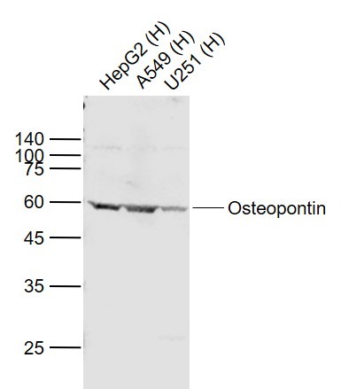 , ,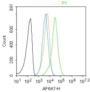 , ,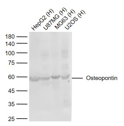 , ,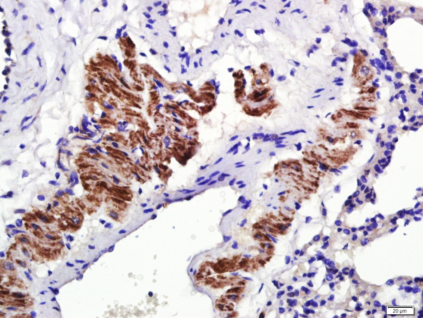 |
| Application | |
| Recommended Dose | WB: 1:500-2000; IHC-P: 1:500-2000; IHC-Fr: 1:100-500; IF: 1:100-500; FCM: 1ug/Test |
| Antibody Type | Polyclonal |
| Host Species | Rabbit |
| Subcellular Localization | Secreted. |
| Tissue Specificity | Bone. Found in plasma. |
| Construction | Polyclonal Antibody |
| Purification | Protein A purified |
| Appearance | Liquid |
| Formulation | 0.01M TBS (pH7.4) with 1% BSA, 0.02% Proclin300 and 50% Glycerol. |
| Concentration | 1 mg/mL |
| Research Background | Osteopontin is the principal phosphorylated glycoprotein of bone and is expressed in a limited number of other tissues including dentine. Osteopontin is produced by osteoblasts under stimulation by calcitriol and binds tightly to hydroxyapatite. It is also involved in the anchoring of osteoclasts to the mineral of bone matrix via the vitronectin receptor, which has specificity for osteopontin. Osteopontin is overexpressed in a variety of cancers, including lung, breast, colorectal, stomach, ovarian, melanoma and mesothelioma. |
| Immunogen | KLH conjugated synthetic peptide: rat osteopontin |
| Antigen Species | Rat |
| Gene Name | SPP1 |
| Gene ID | |
| Protein Name | Osteopontin |
| Uniprot ID | |
| Function | Binds tightly to hydroxyapatite. Appears to form an integral part of the mineralized matrix. Probably important to cell-matrix interaction. Acts as a cytokine involved in enhancing production of interferon-gamma and interleukin-12 and reducing production of interleukin-10 and is essential in the pathway that leads to type I immunity (By similarity). |
| Molecular Weight | Theoretical: 34 kDa. |
| Stability & Storage | Store at -20°C or -80°C for 12 months. Avoid repeated freeze-thaw cycles. |
| Transport | Shipping with blue ice. |
| Size | Quantity | Unit Price | Amount | Operation |
|---|
 Hello! How can I help you today?
 Copyright © 2015-2026 TargetMol Chemicals Inc. All Rights Reserved.
