
Shopping Cart
Remove All Your shopping cart is currently empty

Synonyms: newcastle disease virus(NDV), Newcastle disease virus HN, Newcastle disease virus hemagglutinin neuraminidase, HN_NDVB, HN, hemagglutinin-neuraminidase protein [Newcastle disease virus], hemagglutinin-neuraminidase
Anti-NDV HN Polyclonal Antibody
| Pack Size | Price | USA Stock | Global Stock | Quantity |
|---|---|---|---|---|
| 50 µL | $221 | 7-10 days | 7-10 days | |
| 100 µL | $374 | 7-10 days | 7-10 days | |
| 200 µL | $527 | 7-10 days | 7-10 days |
| Description | Anti-NDV HN Polyclonal Antibody is a Rabbit antibody targeting NDV HN. Anti-NDV HN Polyclonal Antibody can be used in ELISA, WB. |
| Synonyms | newcastle disease virus(NDV), Newcastle disease virus HN, Newcastle disease virus hemagglutinin neuraminidase, HN_NDVB, HN, hemagglutinin-neuraminidase protein [Newcastle disease virus], hemagglutinin-neuraminidase |
| Ig Type | IgG |
| Reactivity | NDV |
| Verified Activity | Sample: Lane 1: Recombinant NDV HN protein, His Primary: Anti-NDV HN (TMAB-01210) at 1/1000 dilution Secondary: IRDye800CW Goat Anti-Rabbit IgG at 1/20000 dilution Predicted band size: 63 kDa Observed band size: 61 kDa 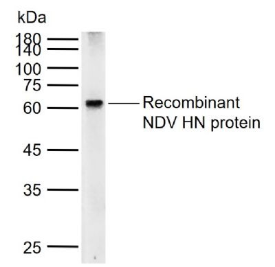 |
| Application | |
| Recommended Dose | WB: 1:500-2000; ELISA: 1:5000-10000 |
| Antibody Type | Polyclonal |
| Host Species | Rabbit |
| Subcellular Localization | Virion membrane {ECO:0000250}; Single-pass type I membrane protein {ECO:0000250}. Host cell membrane {ECO:0000250}; Single-pass membrane protein {ECO:0000250}. |
| Construction | Polyclonal Antibody |
| Purification | Protein A purified |
| Appearance | Liquid |
| Formulation | 0.01M TBS (pH7.4) with 1% BSA, 0.02% Proclin300 and 50% Glycerol. |
| Concentration | 1 mg/mL |
| Research Background | The entry of Newcastle disease virus (NDV), a prototype paramyxovirus, is directed by two virion glycoproteins, the hemagglutinin-neuraminidase (HN) protein and the fusion (F) protein . HN protein, the virus attachment protein, binds to sialic acid-containing receptors, and F protein mediates membrane fusion. In contrast to many viral fusion proteins, paramyxovirus F proteins do not require the acid pH of endosomes to activate fusion activity. As a consequence, infected cells expressing both attachment proteins and F proteins can fuse with adjacent cells to form multinuclear cells, or syncytia, a process that is assumed to mimic virus-cell fusion . |
| Immunogen | KLH conjugated synthetic peptide: NDV HN protein |
| Gene Name | HN |
| Protein Name | Hemagglutinin-neuraminidase |
| Biology Area | Other Viruses |
| Function | Class I viral fusion protein. Under the current model, the protein has at least 3 conformational states: pre-fusion native state, pre-hairpin intermediate state, and post-fusion hairpin state. During viral and plasma cell membrane fusion, the heptad repeat (HR) regions assume a trimer-of-hairpins structure, positioning the fusion peptide in close proximity to the C-terminal region of the ectodomain. The formation of this structure appears to drive apposition and subsequent fusion of viral and plasma cell membranes. Directs fusion of viral and cellular membranes leading to delivery of the nucleocapsid into the cytoplasm. This fusion is pH independent and occurs directly at the outer cell membrane. The trimer of F1-F2 (F protein) probably interacts with HN at the virion surface. Upon HN binding to its cellular receptor, the hydrophobic fusion peptide is unmasked and interacts with the cellular membrane, inducing the fusion between cell and virion membranes. Later in infection, F proteins expressed at the plasma membrane of infected cells could mediate fusion with adjacent cells to form syncytia, a cytopathic effect that could lead to tissue necrosis. |
| Molecular Weight | Theoretical: 63 kDa. Actual: 61 kDa. |
| Stability & Storage | Store at -20°C or -80°C for 12 months. Avoid repeated freeze-thaw cycles. |
| Transport | Shipping with blue ice. |
| Size | Quantity | Unit Price | Amount | Operation |
|---|
 Hello! How can I help you today?
 Copyright © 2015-2026 TargetMol Chemicals Inc. All Rights Reserved.
