
Shopping Cart
Remove All Your shopping cart is currently empty

Synonyms: neural cell adhesion molecule 1, NCAM, MSK39, CD56
Anti-NCAM1 Polyclonal Antibody 2
| Pack Size | Price | USA Stock | Global Stock | Quantity |
|---|---|---|---|---|
| 50 µL | $221 | 7-10 days | 7-10 days | |
| 100 µL | $374 | 7-10 days | 7-10 days | |
| 200 µL | $529 | 7-10 days | 7-10 days |
| Description | Anti-NCAM1 Polyclonal Antibody 2 is a Rabbit antibody targeting NCAM1. Anti-NCAM1 Polyclonal Antibody 2 can be used in FCM, ICC/IF, IF, IHC-Fr, IHC-P, WB. |
| Synonyms | neural cell adhesion molecule 1, NCAM, MSK39, CD56 |
| Ig Type | IgG |
| Reactivity | Human,Mouse,Rat (predicted:Chicken,Dog,Pig,Cow,Horse,Rabbit,GuineaPig) |
| Verified Activity | 1. Tissue/cell: rat brain tissue; 4% Paraformaldehyde-fixed and paraffin-embedded; Antigen retrieval: citrate buffer (0.01M, pH6.0), Boiling bathing for 15 min; Block endogenous peroxidase by 3% Hydrogen peroxide for 30 min; Blocking buffer (normal goat serum) at 37°C for 20 min; Incubation: Anti-CD56/NCAM1 Polyclonal Antibody, Unconjugated (TMAB-01205) 1:200, overnight at 4°C, followed by conjugation to the secondary antibody and DAb staining. 2. Blank control: Jurkat cells (blue). Primary Antibody: Rabbit Anti-CD56 antibody (TMAB-01205), Dilution: 1 μg in 100 μL 1X PBS containing 0.5% BSA; Isotype Control Antibody: Rabbit Igg (orange),used under the same conditions); Secondary Antibody: Goat anti-rabbit IgG-Pe (white blue), Dilution: 1:200 in 1 X PBS containing 0.5% BSA. Protocol The cells were fixed with 2% paraformaldehyde (10 min). Primary antibody (TMAB-01205, 1 μg/1x10^6 cells) were incubated for 30 min on the ice, followed by 1 X PBS containing 0.5% BSA + 10% goat serum (15 min) to block non-specific protein-protein interactions. Then the Goat Anti-rabbit IgG/PE antibody was added into the blocking buffer mentioned above to react with the primary antibody at 1/200 dilution for 30 min on ice. 3. Paraformaldehyde-fixed, paraffin embedded (Mouse brain); Antigen retrieval by boiling in sodium citrate buffer (pH6.0) for 15 min; Block endogenous peroxidase by 3% hydrogen peroxide for 20 min; Blocking buffer (normal goat serum) at 37°C for 30 min; Antibody incubation with (CD56) Polyclonal Antibody, Unconjugated (TMAB-01205) at 1:500 overnight at 4°C, followed by a conjugated secondary for 20 min and DAB staining. 4. Tissue/cell: SH-SY5Y cell; 4% Paraformaldehyde-fixed; Triton X-100 at room temperature for 20 min; Blocking buffer (normal goat serum) at 37°C for 20 min; Antibody incubation with (CD56) polyclonal Antibody, Unconjugated (TMAB-01205) 1:100, 90 minutes at 37°C; followed by a FITC conjugated Goat Anti-Rabbit IgG antibody at 37°C for 90 minutes, DAPI (blue) was used to stain the cell nucleus. 5. Sample: Cerebral cortex (Mouse) Lysate at 40 μg Primary: Anti-CD56 (TMAB-01205) at 1/1000 dilution Secondary: IRDye800CW Goat Anti-Rabbit IgG at 1/20000 dilution Predicted band size: 92 kDa Observed band size: 95 kDa 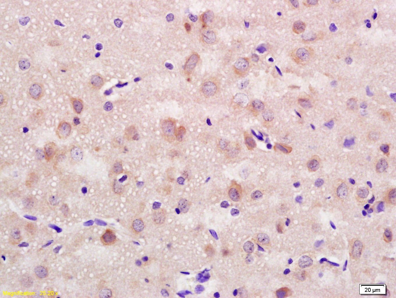 , , , ,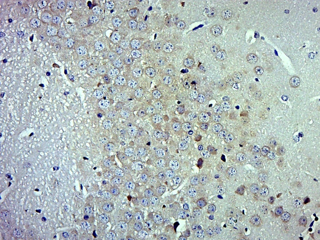 , ,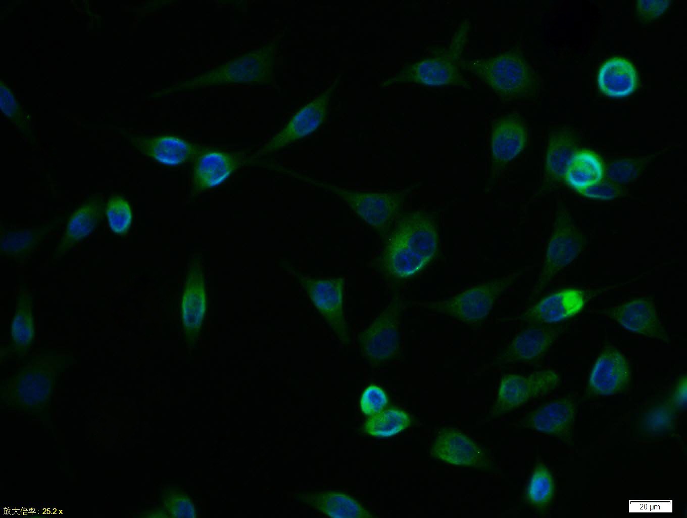 , ,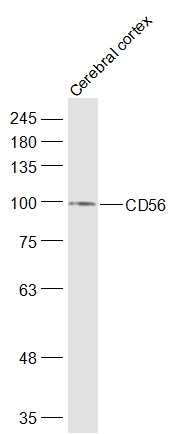 |
| Application | |
| Recommended Dose | FCM=1 μg/Test; ICC/IF=1:100-500; IF=1:100-500; IHC-Fr=1:100-500; IHC-P=1:100-500; WB=1:500-2000 |
| Antibody Type | Polyclonal |
| Host Species | Rabbit |
| Subcellular Localization | Isoform 1, 2,: Cell membrane; Single-pass type I membrane protein. Isoform 3, 4: Cell membrane; Lipid-anchor, GPI-anchor. Isoform 5, 6: Secreted. |
| Construction | Polyclonal Antibody |
| Purification | Protein A purified |
| Appearance | Liquid |
| Formulation | 0.01M TBS (pH7.4) with 1% BSA, 0.02% Proclin300 and 50% Glycerol. |
| Concentration | 1 mg/mL |
| Research Background | This gene encodes a cell adhesion protein which is a member of the immunoglobulin superfamily. The encoded protein is involved in cell-to-cell interactions as well as cell-matrix interactions during development and differentiation. The encoded protein has been shown to be involved in development of the nervous system, and for cells involved in the expansion of T cells and dendritic cells which play an important role in immune surveillance. Alternative splicing results in multiple transcript variants. [provided by RefSeq, Jun 2011] |
| Immunogen | KLH conjugated synthetic peptide: human CD56 |
| Antigen Species | Human |
| Gene Name | NCAM1 |
| Gene ID | |
| Protein Name | Neural cell adhesion molecule 1 |
| Uniprot ID | |
| Biology Area | ECM Proteins,Growth Cone,Neural Signal Transduction,Human Lineage Negative |
| Function | This protein is a cell adhesion molecule involved in neuron-neuron adhesion, neurite fasciculation, outgrowth of neurites, etc. Subcellular Location : Isoform 1, 2,: Cell membrane; Single-pass type I membrane protein. Isoform 3, 4: Cell membrane; Lipid-anchor, GPI-anchor. Isoform 5, 6: Secreted. |
| Molecular Weight | Theoretical: 92 kDa. Actual: 95 kDa. |
| Stability & Storage | Store at -20°C or -80°C for 12 months. Avoid repeated freeze-thaw cycles. |
| Transport | Shipping with blue ice. |
| Size | Quantity | Unit Price | Amount | Operation |
|---|
 Hello! How can I help you today?
 Copyright © 2015-2026 TargetMol Chemicals Inc. All Rights Reserved.
