
Shopping Cart
Remove All Your shopping cart is currently empty

Synonyms: WD 40 repeat protein MSI2, RNA-binding protein Musashi homolog 2, RNA binding protein Musashi homolog 2, Musashi-2, Musashi, Drosophila, homolog of, 2, Musashi RNA binding protein 2, Musashi homolog 2, Musashi 2, MSI2H_HUMAN, MSI2H, MSI2/HOXA9 fusion gene, included, Msi 2, MGC3245, FLJ36569
Anti-MSI2 Antibody
(1Q704)
| Pack Size | Price | USA Stock | Global Stock | Quantity |
|---|---|---|---|---|
| 50 µL | $298 | 7-10 days | 7-10 days | |
| 100 µL | $496 | 7-10 days | 7-10 days |
| Description | Anti-MSI2 Antibody (1Q704) is a Rabbit antibody targeting MSI2. Anti-MSI2 Antibody (1Q704) can be used in FCM,ICC/IF,IHC,WB. |
| Synonyms | WD 40 repeat protein MSI2, RNA-binding protein Musashi homolog 2, RNA binding protein Musashi homolog 2, Musashi-2, Musashi, Drosophila, homolog of, 2, Musashi RNA binding protein 2, Musashi homolog 2, Musashi 2, MSI2H_HUMAN, MSI2H, MSI2/HOXA9 fusion gene, included, Msi 2, MGC3245, FLJ36569 |
| Ig Type | IgG |
| Clone | 1Q704 |
| Reactivity | Human,Mouse,Rat |
| Verified Activity | 1. Western blot analysis of MSI2 on different lysates using anti-MSI2 antibody at 1/1,000 dilution. Positive control: Lane 1: Lovo, Lane 2: PC-12. 2. Immunohistochemical analysis of paraffin-embedded human liver tissue using anti-MSI2 antibody. Counter stained with hematoxylin. 3. Immunohistochemical analysis of paraffin-embedded human pancreas tissue using anti-MSI2 antibody. Counter stained with hematoxylin. 4. Immunohistochemical analysis of paraffin-embedded mouse liver tissue using anti-MSI2 antibody. Counter stained with hematoxylin. 5. Immunohistochemical analysis of paraffin-embedded mouse prostate tissue using anti-MSI2 antibody. Counter stained with hematoxylin. 6. Immunohistochemical analysis of paraffin-embedded mouse heart tissue using anti-MSI2 antibody. Counter stained with hematoxylin. 7. ICC staining MSI2 in Hela cells (green). The nuclear counter stain is DAPI (blue). Cells were fixed in paraformaldehyde, permeabilised with 0.25% Triton X100/PBS. 8. ICC staining MSI2 in Lovo cells (green). The nuclear counter stain is DAPI (blue). Cells were fixed in paraformaldehyde, permeabilised with 0.25% Triton X100/PBS. 9. ICC staining MSI2 in PC-12 cells (green). The nuclear counter stain is DAPI (blue). Cells were fixed in paraformaldehyde, permeabilised with 0.25% Triton X100/PBS. 10. Flow cytometric analysis of Hela cells with MSI2 antibody at 1/50 dilution (red) compared with an unlabelled control (cells without incubation with primary antibody; black). Alexa Fluor 488-conjugated goat anti rabbit IgG was used as the secondary antibody. 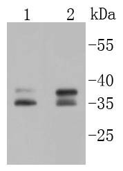 , ,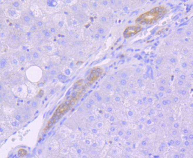 , ,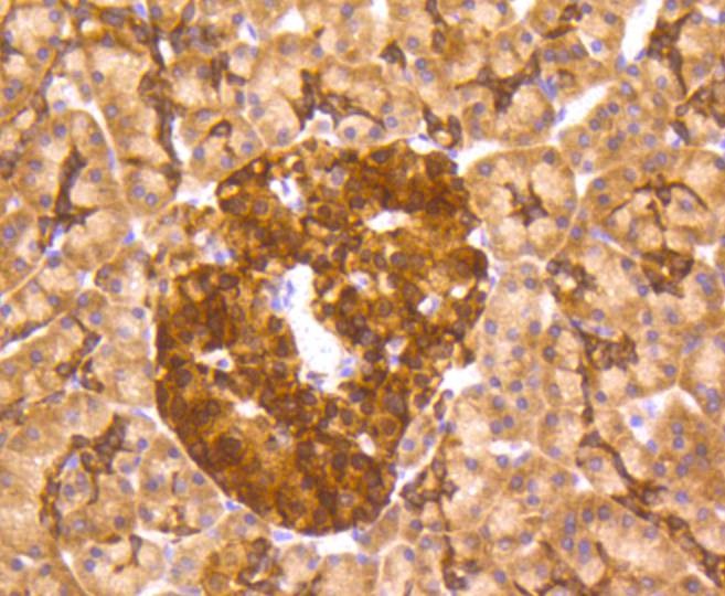 , ,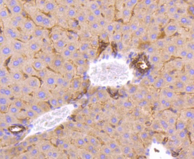 , ,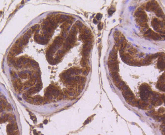 , ,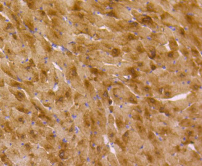 , ,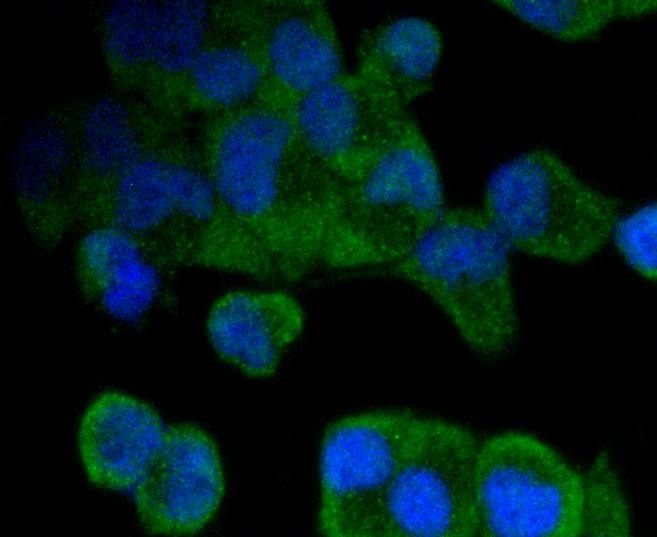 , ,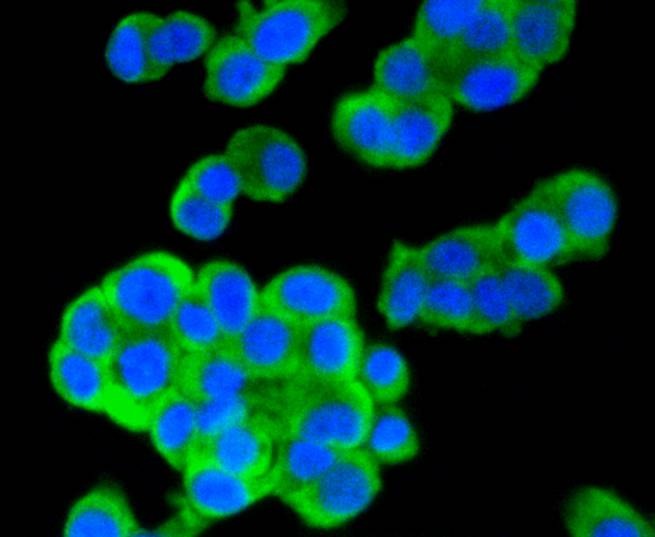 , ,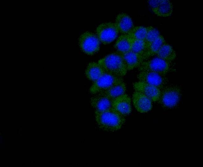 , ,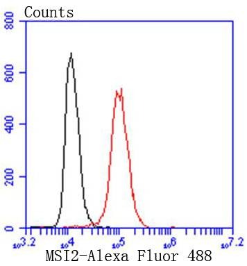 |
| Application | |
| Recommended Dose | WB: 1:1000; IHC: 1:50-200; ICC/IF: 1:50-200; FCM: 1:50-100 |
| Antibody Type | Monoclonal |
| Host Species | Rabbit |
| Construction | Recombinant Antibody |
| Purification | ProA affinity purified |
| Appearance | Liquid |
| Formulation | 1*TBS (pH7.4), 1%BSA, 40%Glycerol. Preservative: 0.05% Sodium Azide. |
| Research Background | Msi2 (musashi homolog 2), also known as MSI2H, is a 328 amino acid protein that localizes to the cytoplasm and contains two RRM (RNA recognition motif) domains. Expressed ubiquitously at low levels, Msi2 functions as an RNA binding protein that, by regulating the expression of target mRNAs, is thought to play a role in the proliferation and maintenance of stem cells within the central nervous system. Msi2 is subject to post-translational phosphorylation and is upregulated in response to brain injury, suggesting a role in healing and brain tissue regeneration. Chromosomal aberrations involving the Msi2 gene are associated with the progression of chronic myeloid leukemia. Multiple isoforms of Msi2 exist due to alternative splicing events. |
| Conjucates | Unconjugated |
| Immunogen | Recombinant Protein |
| Uniprot ID |
| Molecular Weight | Theoretical: 35/37 kDa. |
| Stability & Storage | Store at -20°C or -80°C for 12 months. Avoid repeated freeze-thaw cycles. |
| Transport | Shipping with blue ice. |
| Size | Quantity | Unit Price | Amount | Operation |
|---|
 Hello! How can I help you today?
 Copyright © 2015-2026 TargetMol Chemicals Inc. All Rights Reserved.
