
Shopping Cart
Remove All Your shopping cart is currently empty

Synonyms: TBE-1, MONA, MMP-II, MMP-2, matrix metallopeptidase 2, CLG4A, CLG4
Anti-MMP-2 Polyclonal Antibody
| Pack Size | Price | USA Stock | Global Stock | Quantity |
|---|---|---|---|---|
| 50 µL | $220 | 7-10 days | 7-10 days | |
| 100 µL | $372 | 7-10 days | 7-10 days | |
| 200 µL | $527 | 7-10 days | 7-10 days |
| Description | Anti-MMP-2 Polyclonal Antibody is a Rabbit antibody targeting MMP-2. Anti-MMP-2 Polyclonal Antibody can be used in FCM, ICC/IF, IF, IHC-Fr, IHC-P, WB. |
| Synonyms | TBE-1, MONA, MMP-II, MMP-2, matrix metallopeptidase 2, CLG4A, CLG4 |
| Ig Type | IgG |
| Reactivity | Human (predicted:Mouse,Rat,Pig,Cow,Horse,Rabbit,Sheep) |
| Verified Activity | 1. Paraformaldehyde-fixed, paraffin embedded (Rat brain); Antigen retrieval by boiling in sodium citrate buffer (pH6.0) for 15 min; Block endogenous peroxidase by 3% hydrogen peroxide for 20 min; Blocking buffer (normal goat serum) at 37°C for 30 min; Antibody incubation with (MMP2) Polyclonal Antibody, Unconjugated (TMAB-01154) at 1:400 overnight at 4°C, followed by operating according to SP Kit (Rabbit) instructions and DAB staining. 2. Blank control (blue line): Hela (blue). Primary Antibody (green line): Rabbit Anti-MMP2 antibody (TMAB-01154) Dilution: 1 μg/10^6 cells; Isotype Control Antibody (orange line): Rabbit IgG. Secondary Antibody (white blue line): Goat anti-rabbit IgG-FITC Dilution: 1 μg/test. Protocol The cells were fixed with 80% methanol (5 min at-20°C) and then permeabilized with 0.1% PBS-Tween for 20 min at room temperature. Cells stained with Primary Antibody for 30 min at room temperature. The cells were then incubated in 1 X PBS/2% BSA/10% goat serum to block non-specific protein-protein interactions followed by the antibody for 15 min at room temperature. The secondary antibody used for 40 min at room temperature. 3. Paraformaldehyde-fixed, paraffin embedded (Mouse brain); Antigen retrieval by boiling in sodium citrate buffer (pH6.0) for 15 min; Block endogenous peroxidase by 3% hydrogen peroxide for 20 min; Blocking buffer (normal goat serum) at 37°C for 30 min; Antibody incubation with (MMP2) Polyclonal Antibody, Unconjugated (TMAB-01154) at 1:400 overnight at 4°C, followed by operating according to SP Kit (Rabbit) instructions and DAB staining. 4. Sample: SY5Y (Human) Cell Lysate at 30 μg U2os (Human) Cell Lysate at 30 μg U251 (Human) Cell Lysate at 30 μg Siha (Human) Cell Lysate at 30 μg Primary: Anti-MMP-2 (TMAB-01154) at 1/1000 dilution Secondary: IRDye800CW Goat Anti-Rabbit IgG at 1/20000 dilution Predicted band size: 62 kDa Observed band size: 60 kDa 5. Sample: A431 (Human) Cell Lysate at 30 μg Mcf-7 (Human) Cell Lysate at 30 μg Primary: Anti-MMP-2 (TMAB-01154) at 1/300 dilution Secondary: IRDye800CW Goat Anti-Rabbit IgG at 1/20000 dilution Predicted band size: 62 kDa Observed band size: 62 kDa 6. Tissue/cell: U-87MG cell; 4% Paraformaldehyde-fixed; Triton X-100 at room temperature for 20 min; Blocking buffer (normal goat serum) at 37°C for 20 min; Antibody incubation with (MMP2) polyclonal Antibody, Unconjugated (TMAB-01154) 1:100, 90 minutes at 37°C; followed by a conjugated Goat Anti-Rabbit IgG antibody at 37°C for 90 minutes, DAPI (blue) was used to stain the cell nucleus. 7. Paraformaldehyde-fixed, paraffin embedded Human Glioma; Antigen retrieval by boiling in sodium citrate buffer (pH6.0) for 15 min; Antibody incubation with MMP2 Polyclonal Antibody, Unconjugated (TMAB-01154) at 1:400 overnight at 4°C, followed by conjugation to the SP Kit (Rabbit) and DAB staining. 8. Paraformaldehyde-fixed, paraffin embedded Mouse Placenta; Antigen retrieval by boiling in sodium citrate buffer (pH6.0) for 15 min; Antibody incubation with MMP2 Polyclonal Antibody, Unconjugated (TMAB-01154) at 1:400 overnight at 4°C, followed by conjugation to the SP Kit (Rabbit) and DAB staining. 9. Blank control (blue line): Hela (fixed with 80% methanol (5 min at-20°C) and then permeabilized with 0.1% PBS-Tween for 20 min at room temperature). Primary Antibody (green line): Rabbit Anti-MMP2 antibody (TMAB-01154), Dilution: 1 μg/10^6 cells; Isotype Control Antibody (orange line): Rabbit IgG. Secondary Antibody (white blue line): Goat anti-rabbit IgG-FITC, Dilution: 1 μg/test. 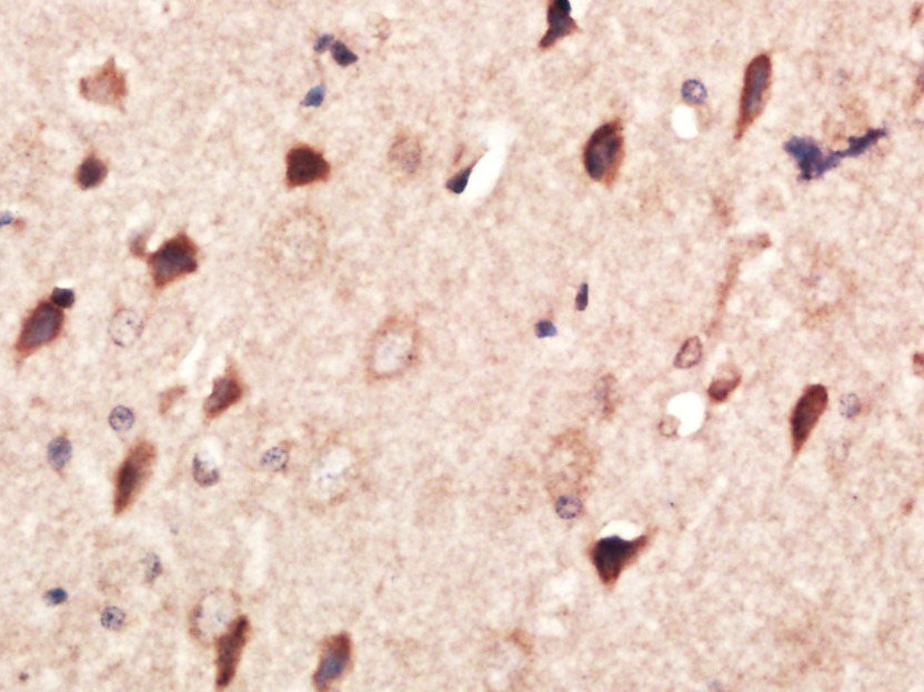 , ,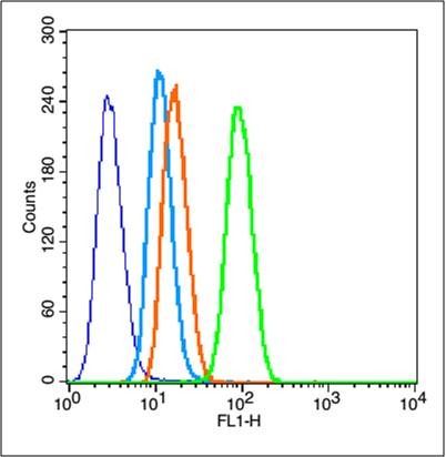 , ,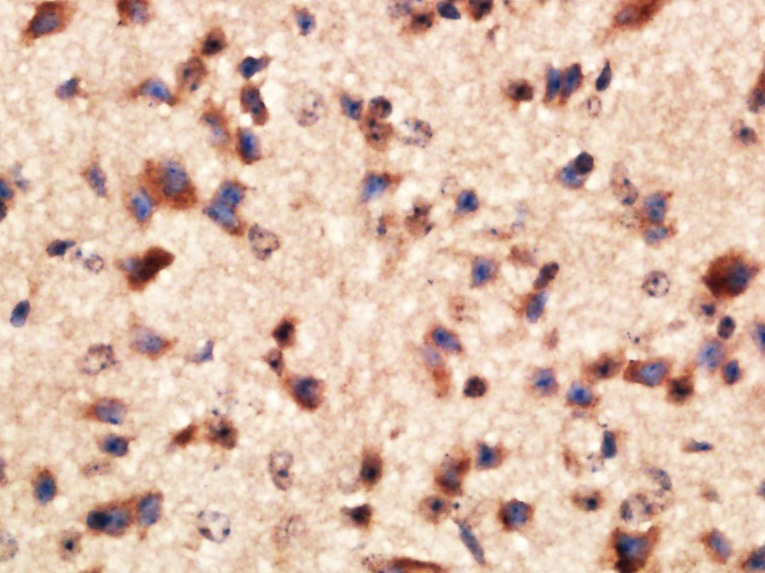 , ,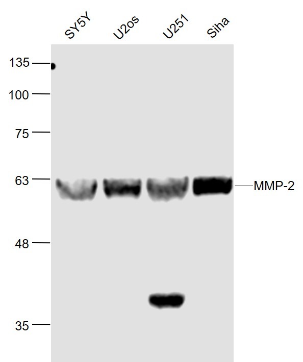 , ,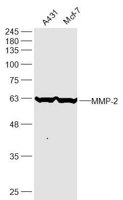 , ,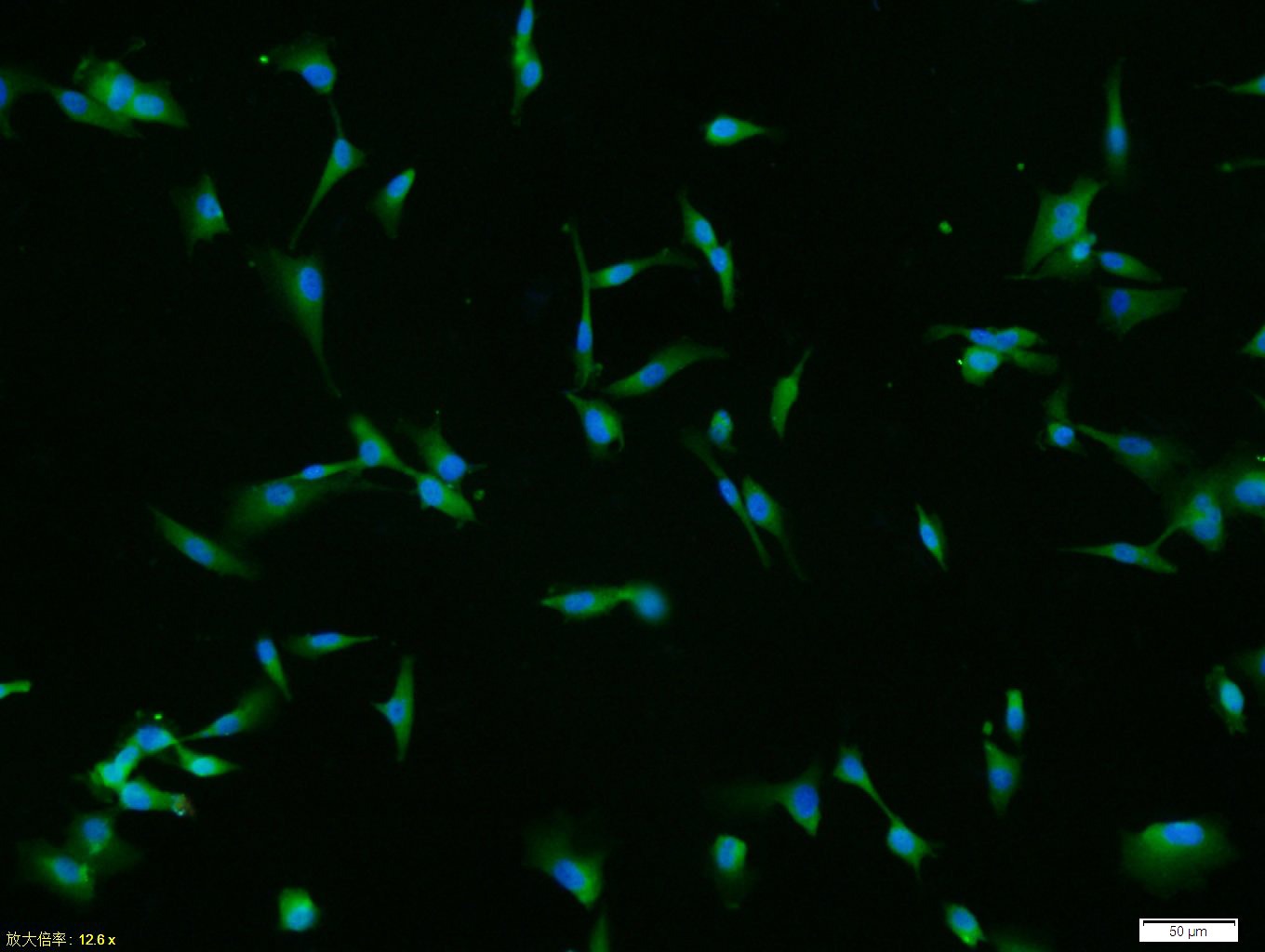 , ,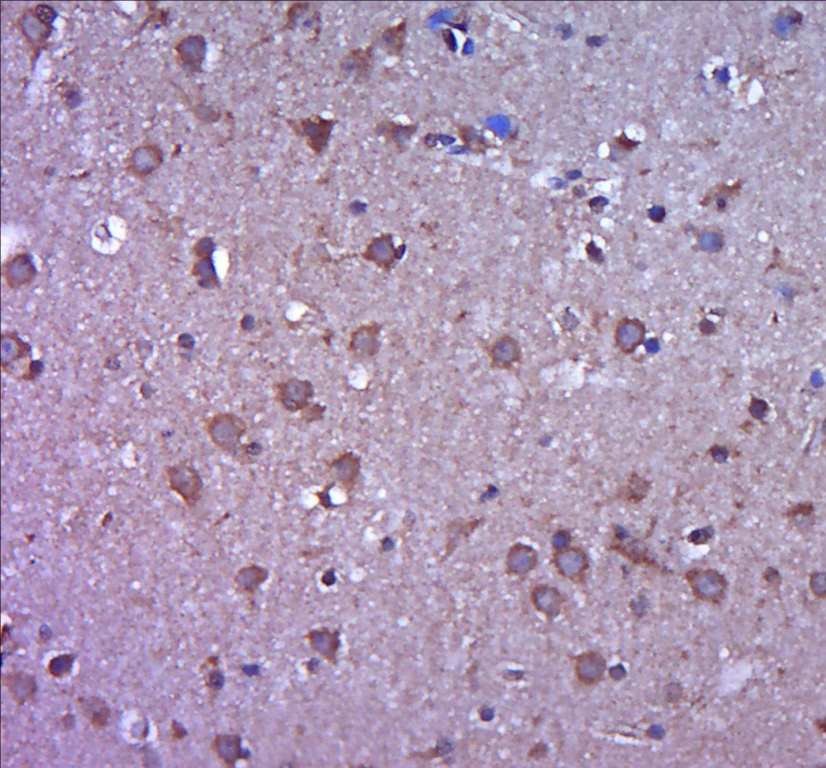 , ,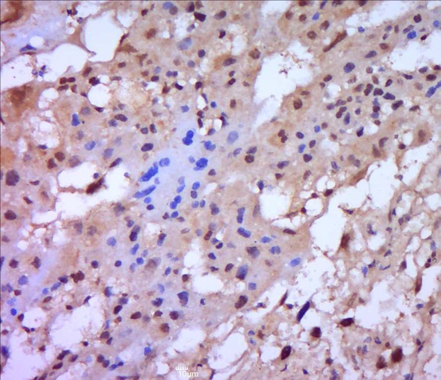 , ,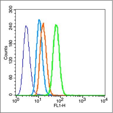 |
| Application | |
| Recommended Dose | FCM=1 μg/Test; ICC/IF=1:100-500; IF=1:200-800; IHC-Fr=1:200-800; IHC-P=1:200-800; WB=1:1000-5000 |
| Antibody Type | Polyclonal |
| Host Species | Rabbit |
| Subcellular Localization | Isoform 1: Secreted, extracellular space, extracellular matrix. Membrane. Nucleus. Note=Colocalizes with integrin alphaV/beta3 at the membrane surface in angiogenic blood vessels and melanomas. Found in mitochondria, along microfibrils, and in nuclei of cardiomyocytes. Isoform 2: Cytoplasm. Mitochondrion. |
| Tissue Specificity | Produced by normal skin fibroblasts. PEX is expressed in a number of tumors including gliomas, breast and prostate. |
| Construction | Polyclonal Antibody |
| Purification | Protein A purified |
| Appearance | Liquid |
| Formulation | 0.01M TBS (pH7.4) with 1% BSA, 0.02% Proclin300 and 50% Glycerol. |
| Concentration | 1 mg/mL |
| Research Background | Proteins of the matrix metalloproteinase (MMP) family are involved in the breakdown of extracellular matrix in normal physiological processes, such as embryonic development, reproduction, and tissue remodeling, as well as in disease processes, such as arthritis and metastasis. Most MMP's are secreted as inactive proproteins which are activated when cleaved by extracellular proteinases. This gene encodes an enzyme which degrades type IV collagen, the major structural component of basement membranes. The enzyme plays a role in endometrial menstrual breakdown, regulation of vascularization and the inflammatory response. Mutations in this gene have been associated with Winchester syndrome and Nodulosis-Arthropathy-Osteolysis (NAO) syndrome. Two transcript variants encoding different isoforms have been found for this gene. [provided by RefSeq]. |
| Immunogen | KLH conjugated synthetic peptide: human MMP2 |
| Antigen Species | Human |
| Gene Name | MMP2 |
| Gene ID | |
| Protein Name | 72 kDa type IV collagenase |
| Uniprot ID | |
| Biology Area | Response to hypoxia,MMPs,MMPs,MMP,MMPs,Hypoxia,Cancer |
| Function | Ubiquitinous metalloproteinase that is involved in diverse functions such as remodeling of the vasculature, angiogenesis, tissue repair, tumor invasion, inflammation, and atherosclerotic plaque rupture. As well as degrading extracellular matrix proteins, can also act on several nonmatrix proteins such as big endothelial 1 and beta-type CGRP promoting vasoconstriction. Also cleaves KISS at a Gly-|-Leu bond. Appears to have a role in myocardial cell death pathways. Contributes to myocardial oxidative stress by regulating the activity of GSK3beta. Cleaves GSK3beta in vitro. PEX, the C-terminal non-catalytic fragment of MMP2, posseses anti-angiogenic and anti-tumor properties and inhibits cell migration and cell adhesion to FGF2 and vitronectin. Ligand for integrinv/beta3 on the surface of blood vessels. Isoform 2: Mediates the proteolysis of CHUK/IKKA and initiates a primary innate immune response by inducing mitochondrial-nuclear stress signaling with activation of the pro-inflammatory NF-kappaB, NFAT and IRF transcriptional pathways. |
| Molecular Weight | Theoretical: 72 kDa. Actual: 60 kDa. |
| Stability & Storage | Store at -20°C or -80°C for 12 months. Avoid repeated freeze-thaw cycles. |
| Transport | Shipping with blue ice. |
| Size | Quantity | Unit Price | Amount | Operation |
|---|
 Hello! How can I help you today?
 Copyright © 2015-2026 TargetMol Chemicals Inc. All Rights Reserved.
