
Shopping Cart
Remove All Your shopping cart is currently empty

Synonyms: Protein Melan-A, MLANA, Melanoma antigen recognized by T-cells 1, MART-1, MART1, Antigen SK29-AA, Antigen LB39-AA
Anti-MLANA Polyclonal Antibody
| Pack Size | Price | USA Stock | Global Stock | Quantity |
|---|---|---|---|---|
| 50 µL | $221 | 7-10 days | 7-10 days | |
| 100 µL | $374 | 7-10 days | 7-10 days | |
| 200 µL | $529 | 7-10 days | 7-10 days |
| Description | Anti-MLANA Polyclonal Antibody is a Rabbit antibody targeting MLANA. Anti-MLANA Polyclonal Antibody can be used in WB. |
| Synonyms | Protein Melan-A, MLANA, Melanoma antigen recognized by T-cells 1, MART-1, MART1, Antigen SK29-AA, Antigen LB39-AA |
| Ig Type | IgG |
| Reactivity | Human |
| Verified Activity | 25 μg total protein per Lane of various lysates probed with Melan A polyclonal antibody, unconjugated (TMAB-01148) at 1:1000 dilution and 4°C overnight incubation. Followed by conjugated secondary antibody incubation at RT for 60 min. 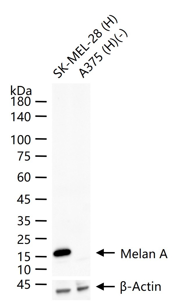 |
| Application | |
| Recommended Dose | WB: 1:500-2000 |
| Antibody Type | Polyclonal |
| Host Species | Rabbit |
| Subcellular Localization | Endoplasmic reticulum membrane; Single-pass type III membrane protein. Golgi apparatus. Golgi apparatus, trans-Golgi network membrane. Melanosome. Note=Also found in small vesicles and tubules dispersed over the entire cytoplasm. A small fraction of the protein is inserted into the membrane in an inverted orientation. Inversion of membrane topology results in the relocalization of the protein from a predominant Golgi/post-Golgi area to the endoplasmic reticulum. Melanoma cells expressing the protein with an inverted membrane topology are more effectively recognized by specific cytolytic T-lymphocytes than those expressing the protein in its native membrane orientation. |
| Tissue Specificity | Expression is restricted to melanoma and melanocyte cell lines and retina. |
| Construction | Polyclonal Antibody |
| Purification | Protein A purified |
| Appearance | Liquid |
| Formulation | 0.01M TBS (pH7.4) with 1% BSA, 0.02% Proclin300 and 50% Glycerol. |
| Concentration | 1 mg/mL |
| Research Background | Melan A, a product of the MART-1 gene, is a melanocyte differentiation marker recognized by autologous cytotoxic T lymphocytes. Other melanoma-associated markers recognized by autologous cytotoxic T cells are reported to include MAGE-1, MAGE-3, tyrosinase, gp100, gp75, BAGE-1 and GAGE-1. The analysis of these different molecules and their expression in individual melanomas may be of help in the study of their particular molecular roles in melanocyte differentiation and tumorigenesis. |
| Immunogen | KLH conjugated synthetic peptide: human Melan A |
| Antigen Species | Human |
| Gene Name | MLANA |
| Gene ID | |
| Protein Name | Melanoma antigen recognized by T-cells 1 |
| Uniprot ID | |
| Biology Area | Tumor-associated antigens,Tumor Associated |
| Function | Involved in melanosome biogenesis by ensuring the stability of GPR143. Plays a vital role in the expression, stability, trafficking, and processing of melanocyte protein PMEL, which is critical to the formation of stage II melanosomes. |
| Molecular Weight | Theoretical: 13 kDa. Actual: 17 kDa. |
| Stability & Storage | Store at -20°C or -80°C for 12 months. Avoid repeated freeze-thaw cycles. |
| Transport | Shipping with blue ice. |
| Size | Quantity | Unit Price | Amount | Operation |
|---|
 Hello! How can I help you today?
 Copyright © 2015-2026 TargetMol Chemicals Inc. All Rights Reserved.
