
Shopping Cart
Remove All Your shopping cart is currently empty

Synonyms: Proliferation marker protein Ki-67, MKI67, Antigen identified by monoclonal antibody Ki-67 (Antigen KI-67;Antigen Ki67)
Anti-MKI67 Antibody
(3H14)
| Pack Size | Price | USA Stock | Global Stock | Quantity |
|---|---|---|---|---|
| 25 µL | $155 | 7-10 days | 7-10 days | |
| 50 µL | $271 | 7-10 days | 7-10 days | |
| 100 µL | $488 | 7-10 days | 7-10 days |
| Description | Anti-MKI67 Antibody (3H14) is a Rabbit antibody targeting MKI67. Anti-MKI67 Antibody (3H14) can be used in FCM, ICC/IF, IF, IHC-Fr, IHC-P. |
| Synonyms | Proliferation marker protein Ki-67, MKI67, Antigen identified by monoclonal antibody Ki-67 (Antigen KI-67;Antigen Ki67) |
| Ig Type | IgG |
| Clone | 3H14 |
| Reactivity | Human,Mouse,Rat |
| Verified Activity | 1. Paraformaldehyde-fixed, paraffin embedded (rat spleen); Antigen retrieval by boiling in sodium EDTA buffer (Ph9.0) for 15 min; Block endogenous peroxidase by 3% hydrogen peroxide for 20 min; Blocking buffer (normal goat serum) at 37°C for 30 min; Incubation with (Ki67) Monoclonal Antibody, Unconjugated (TMAB-01142) at 1:200 overnight at 4°C, followed by operating according to SP Kit (Rabbit) instructionsand DAB staining. 2. Paraformaldehyde-fixed, paraffin embedded (mouse thymus); Antigen retrieval by boiling in sodium EDTA buffer (Ph9.0) for 15 min; Block endogenous peroxidase by 3% hydrogen peroxide for 20 min; Blocking buffer (normal goat serum) at 37°C for 30 min; Incubation with (Ki67) Monoclonal Antibody, Unconjugated (TMAB-01142) at 1:200 overnight at 4°C, followed by operating according to SP Kit (Rabbit) instructionsand DAB staining. 3. Blank control (black line): Hela. Primary Antibody (green line): Rabbit Anti-Ki67 antibody (TMAB-01142) Dilution: 0.5 μg/Test; Secondary Antibody (white blue line): Goat anti-rabbit IgG-FITC Dilution: 0.5 μg/Test. Isotype control (orange line): Normal Rabbit IgG Protocol The cells were fixed with 4% PFA (10 min at room temperature) and then permeabilized with 90% ice-cold methanol for 20 min at-20°C, The cells were then incubated in 5% BSA to block non-specific protein-protein interactions for 30 min at room temperature. Cells stained with Primary Antibody for 30 min at room temperature. The secondary antibody used for 40 min at room temperature. 4. 4% Paraformaldehyde-fixed Hela (H) cell; Triton X-100 at RT for 20 min; Antibody incubation with (Ki-67) monoclonal Antibody, unconjugated (TMAB-01142) 1:200, 90 min at 37°C; followed by conjugated Goat Anti-Rabbit IgG antibody (green) at 37°C for 90 min, DAPI (blue) was used to stain the cell nucleus. PBS instead of the primary antibody was used as the blank control. 5. (Negative control) Paraformaldehyde-fixed, paraffin embedded Mouse Heart; Antigen retrieval by boiling in sodium citrate buffer (pH6.0) for 15 min; Antibody incubation with Ki67 Monoclonal Antibody, Unconjugated (TMAB-01142) at 1:500 overnight at 4°C, followed by conjugation to the SP Kit (Rabbit) and DAB staining. 6. (Negative control) Paraformaldehyde-fixed, paraffin embedded Rat Heart; Antigen retrieval by boiling in sodium citrate buffer (pH6.0) for 15 min; Antibody incubation with Ki67 Monoclonal Antibody, Unconjugated (TMAB-01142) at 1:500 overnight at 4°C, followed by conjugation to the SP Kit (Rabbit) and DAB staining. 7. (Negative control) Paraformaldehyde-fixed, paraffin embedded Human Heart; Antigen retrieval by boiling in sodium citrate buffer (pH6.0) for 15 min; Antibody incubation with Ki67 Monoclonal Antibody, Unconjugated (TMAB-01142) at 1:500 overnight at 4°C, followed by conjugation to the SP Kit (Rabbit) and DAB staining. 8. Paraformaldehyde-fixed, paraffin embedded Rat Spleen; Antigen retrieval by boiling in sodium citrate buffer (pH6.0) for 15 min; Antibody incubation with Ki67 Monoclonal Antibody, Unconjugated (TMAB-01142) at 1:500 overnight at 4°C, followed by conjugation to the SP Kit (Rabbit) and DAB staining. 9. Paraformaldehyde-fixed, paraffin embedded Human Colon Cancer; Antigen retrieval by boiling in sodium citrate buffer (pH6.0) for 15 min; Antibody incubation with Ki67 Monoclonal Antibody, Unconjugated (TMAB-01142) at 1:500 overnight at 4°C, followed by conjugation to the SP Kit (Rabbit) and DAB staining. 10. Paraformaldehyde-fixed, paraffin embedded Human Ovarian Cancer; Antigen retrieval by boiling in sodium citrate buffer (pH6.0) for 15 min; Antibody incubation with Ki67 Monoclonal Antibody, Unconjugated (TMAB-01142) at 1:500 overnight at 4°C, followed by conjugation to the SP Kit (Rabbit) and DAB staining. 11. Paraformaldehyde-fixed, paraffin embedded Human Tonsil; Antigen retrieval by boiling in sodium citrate buffer (pH6.0) for 15 min; Antibody incubation with Ki67 Monoclonal Antibody, Unconjugated (TMAB-01142) at 1:500 overnight at 4°C, followed by conjugation to the SP Kit (Rabbit) and DAB staining. 12. Paraformaldehyde-fixed, paraffin embedded Human Colon; Antigen retrieval by boiling in sodium citrate buffer (pH6.0) for 15 min; Antibody incubation with Ki67 Monoclonal Antibody, Unconjugated (TMAB-01142) at 1:500 overnight at 4°C, followed by conjugation to the SP Kit (Rabbit) and DAB staining. 13. Paraformaldehyde-fixed, paraffin embedded Human Gastric Cancer; Antigen retrieval by boiling in sodium citrate buffer (pH6.0) for 15 min; Antibody incubation with Ki67 Monoclonal Antibody, Unconjugated (TMAB-01142) at 1:500 overnight at 4°C, followed by conjugation to the SP Kit (Rabbit) and DAB staining. 14. Paraformaldehyde-fixed, paraffin embedded Rat Colon; Antigen retrieval by boiling in sodium citrate buffer (pH6.0) for 15 min; Antibody incubation with Ki67 Monoclonal Antibody, Unconjugated (TMAB-01142) at 1:200 overnight at 4°C. Followed by conjugated Goat Anti-Rabbit IgG antibody (green), DAPI (blue) was used to stain the cell nucleus. 15. Paraformaldehyde-fixed, paraffin embedded Mouse Colon; Antigen retrieval by boiling in sodium citrate buffer (pH6.0) for 15 min; Antibody incubation with Ki67 Monoclonal Antibody, Unconjugated (TMAB-01142) at 1:200 overnight at 4°C. Followed by conjugated Goat Anti-Rabbit IgG antibody (green), DAPI (blue) was used to stain the cell nucleus. 16. Paraformaldehyde-fixed, paraffin embedded Human Esophageal Cancer; Antigen retrieval by boiling in sodium citrate buffer (pH6.0) for 15 min; Antibody incubation with Ki67 Monoclonal Antibody, Unconjugated (TMAB-01142) at 1:200 overnight at 4°C. Followed by conjugated Goat Anti-Rabbit IgG antibody (green), DAPI (blue) was used to stain the cell nucleus. 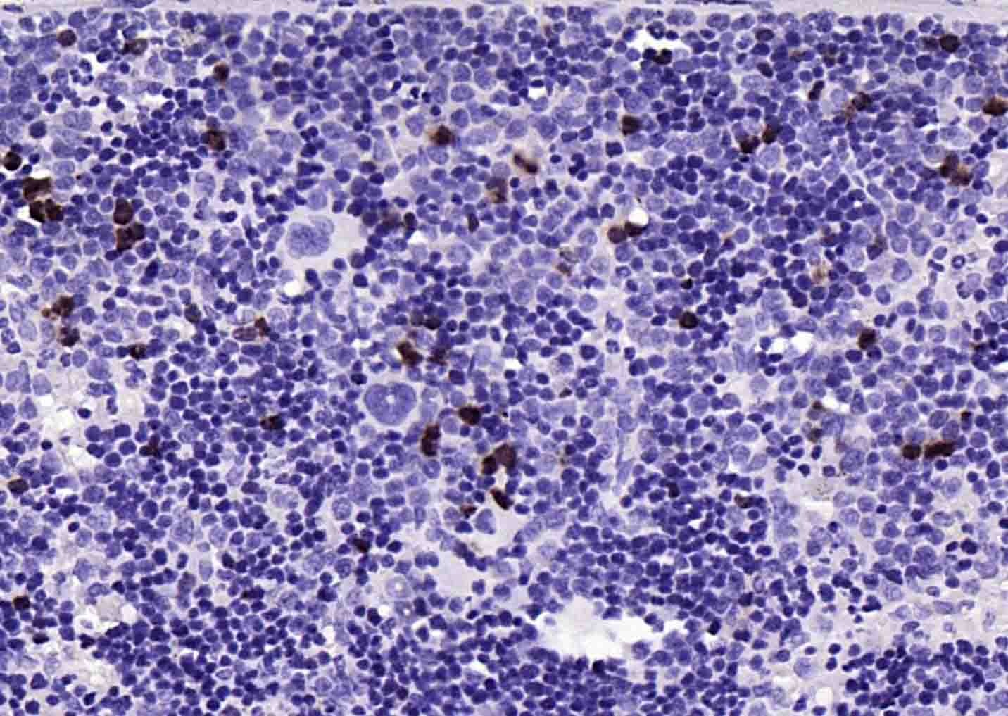 , ,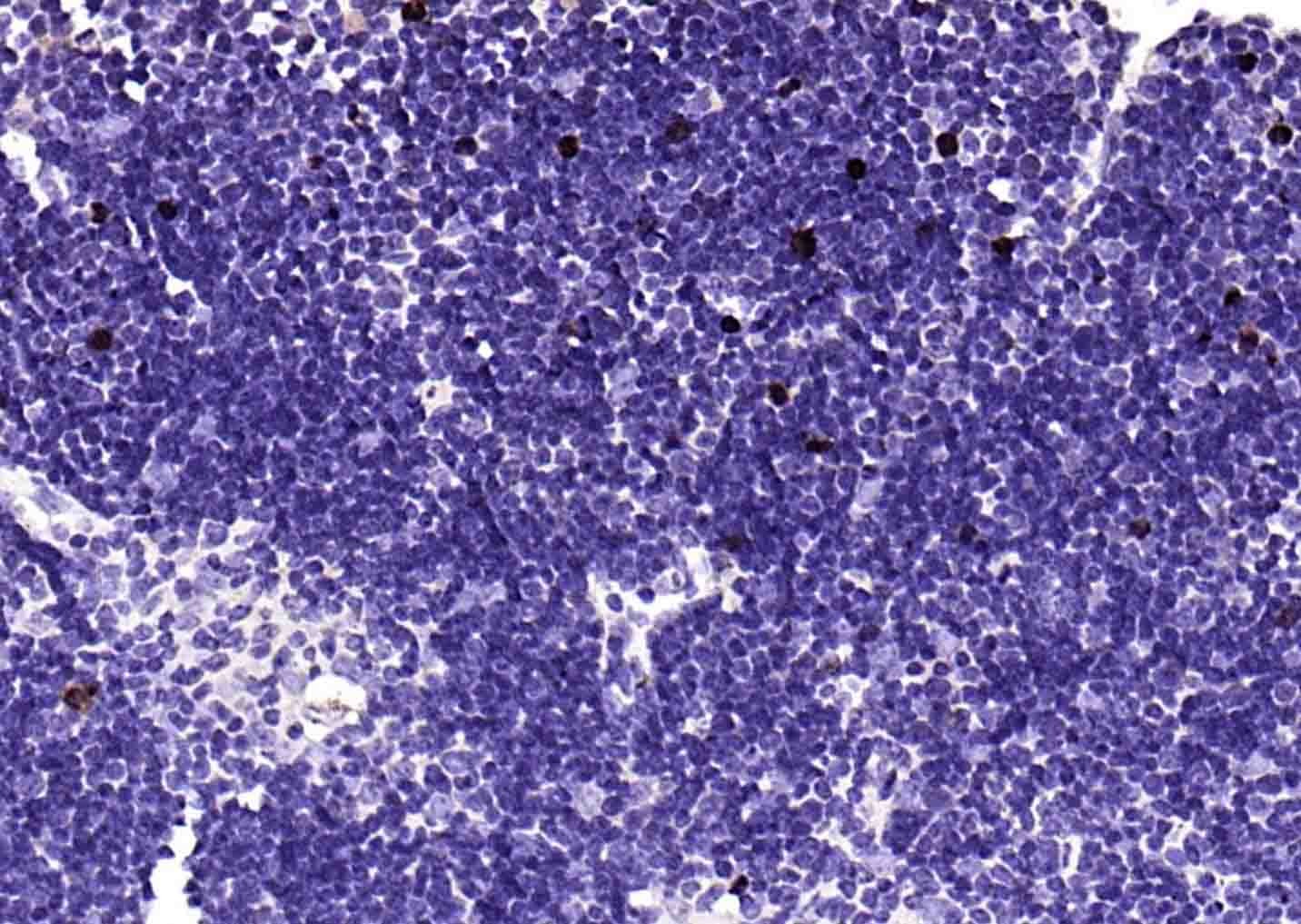 , ,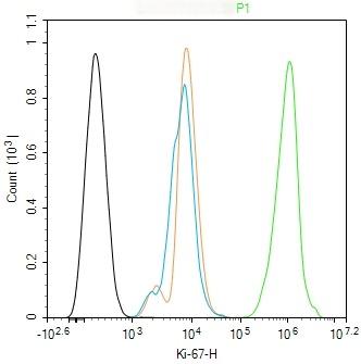 , ,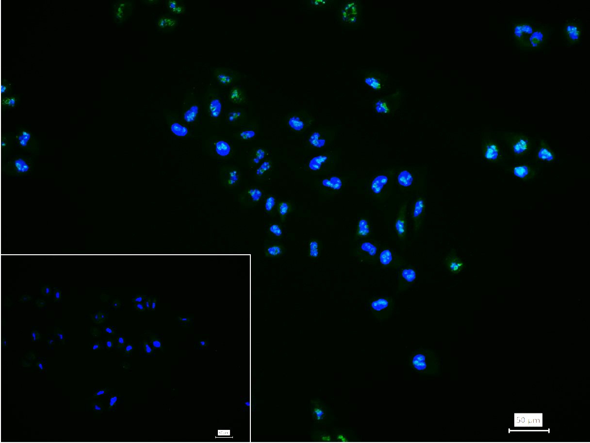 , ,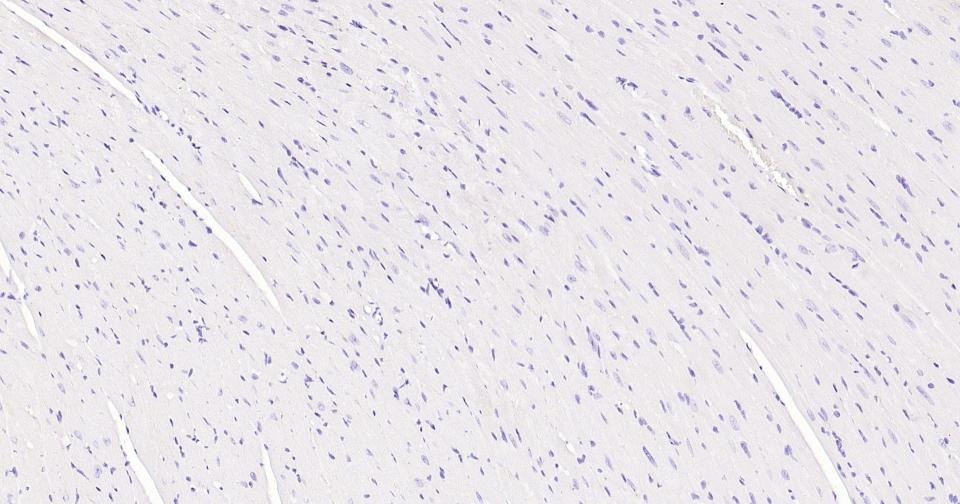 , ,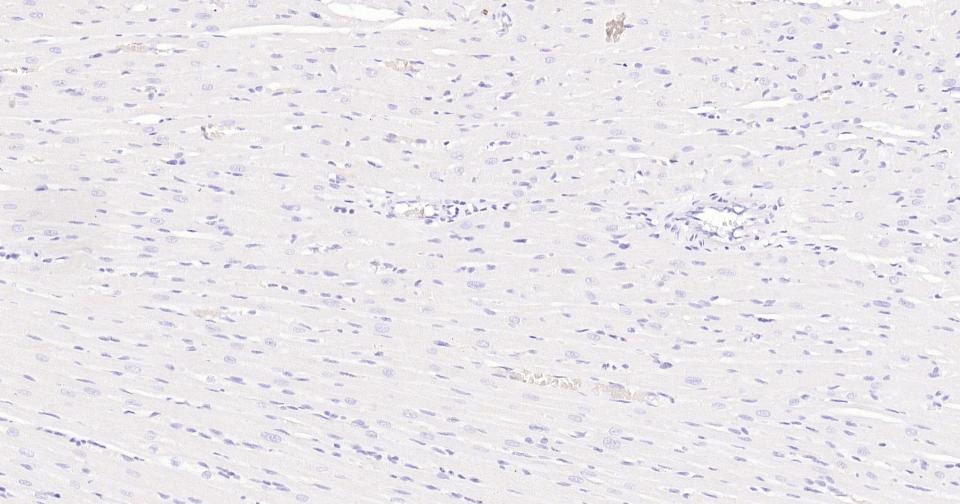 , ,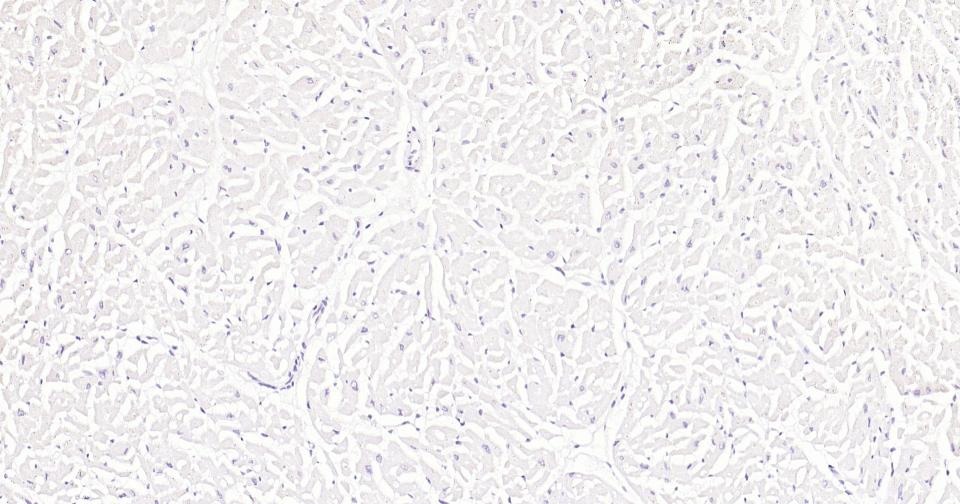 , ,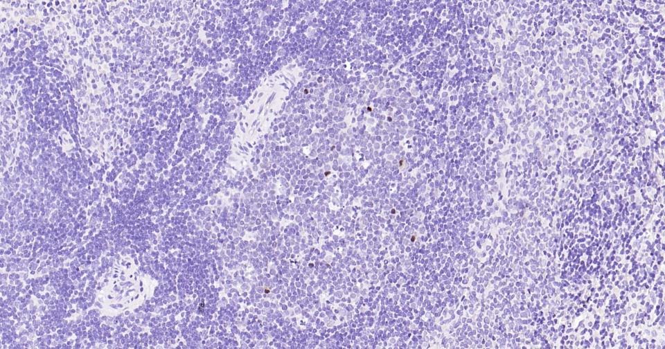 , ,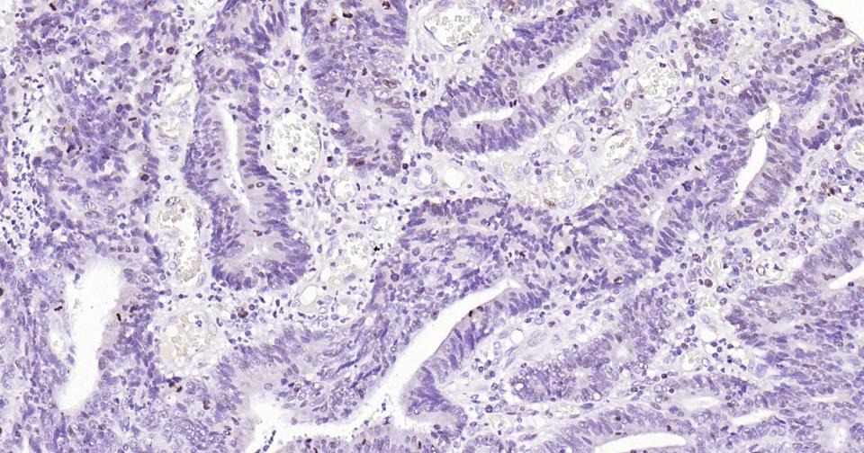 , ,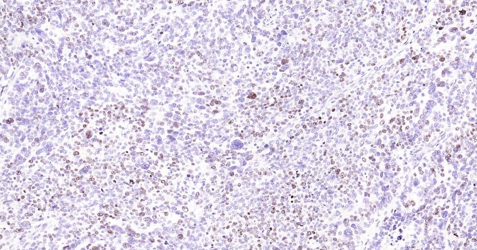 , ,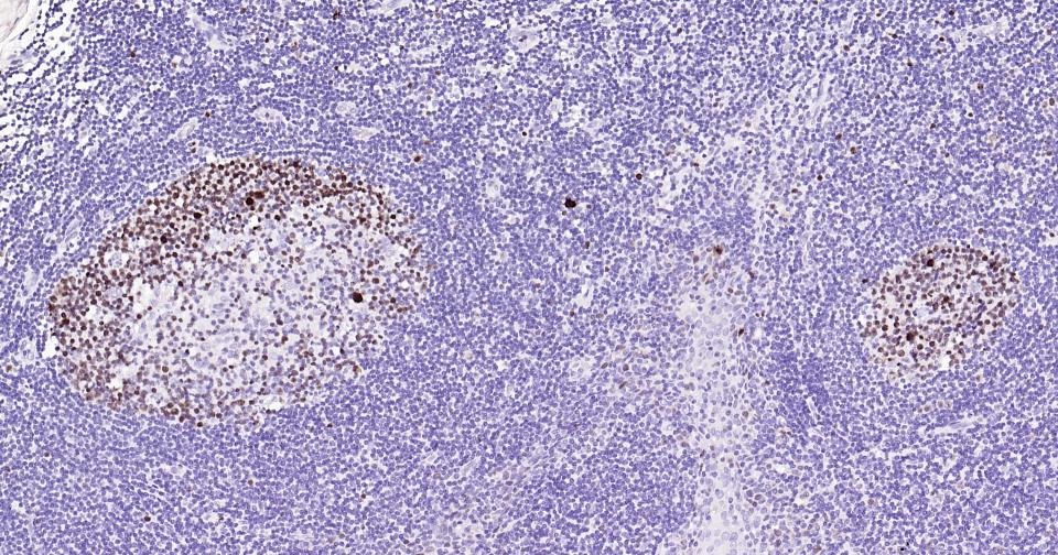 , ,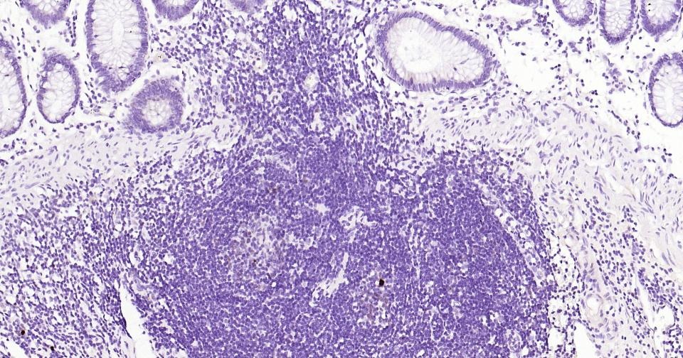 , ,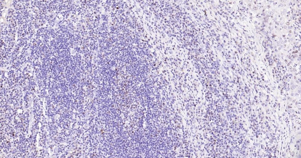 , ,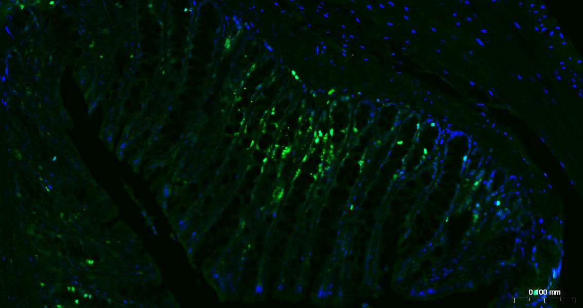 , ,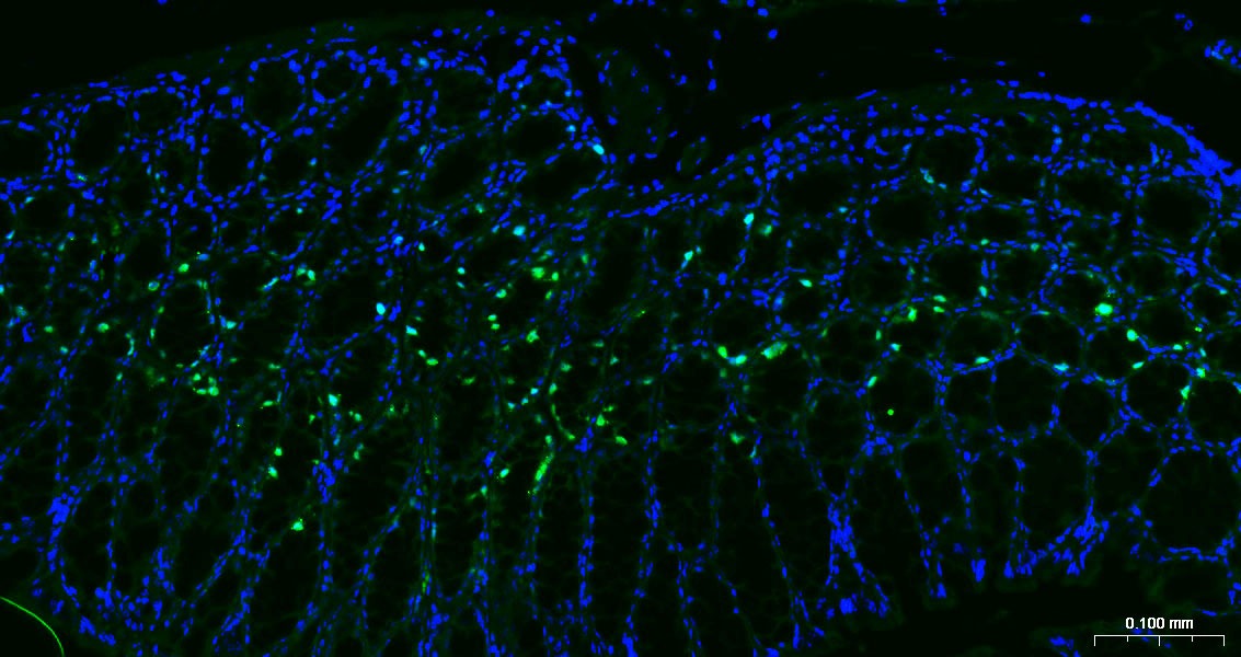 , ,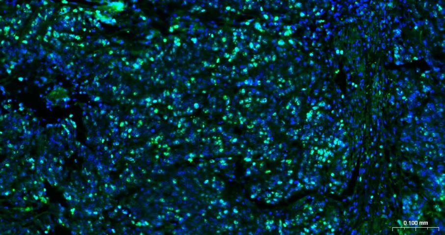 |
| Application | |
| Recommended Dose | FCM=1 μg/Test; ICC/IF=1:100-500; IF=1:100-500; IHC-Fr=1:100-500; IHC-P=1:100-500 |
| Antibody Type | Monoclonal |
| Host Species | Rabbit |
| Subcellular Localization | Nucleus. Chromosome. Predominantly localized in the G1 phase in the perinucleolar region, in the later phases it is also detected throughout the nuclear interior, being predominantly localized in the nuclear matrix. In mitosis, it is present on all chromosomes. |
| Construction | Recombinant Antibody |
| Purification | Protein A purified |
| Appearance | Liquid |
| Formulation | PBS, Glycerol, BSA. |
| Concentration | 1 mg/mL |
| Research Background | Ki67 antigen is the prototypic cell cycle related nuclear protein, expressed by proliferating cells in all phases of the active cell cycle (G1, S, G2 and M phase). It is absent in resting (G0) cells. Ki67 antibodies are useful in establishing the cell growing fraction in neoplasms (immunohistochemically quantified by determining the number of Ki67 positive cells among the total number of resting cells = Ki67 index). In neoplastic tissues the prognostic value is comparable to the tritiated thymidine labelling index. The correlation between low Ki67 index and histologically low grade tumours is strong. Ki67 is routinely used as a neuronal marker of cell cycling and proliferation. |
| Immunogen | KLH conjugated synthetic peptide: human Ki67 |
| Antigen Species | Human |
| Gene Name | MKI67 |
| Gene ID | |
| Protein Name | Proliferation marker protein Ki-67 |
| Uniprot ID | |
| Biology Area | Cell division,Tumor biomarkers,Markers,Soma marker,Neurogenesis,Replication |
| Function | Thought to be required for maintaining cell proliferation. |
| Molecular Weight | Theoretical: 358 kDa. |
| Stability & Storage | Store at -20°C or -80°C for 12 months. Avoid repeated freeze-thaw cycles. |
| Transport | Shipping with blue ice. |
| Size | Quantity | Unit Price | Amount | Operation |
|---|
 Hello! How can I help you today?
 Copyright © 2015-2026 TargetMol Chemicals Inc. All Rights Reserved.
