
Shopping Cart
Remove All Your shopping cart is currently empty

Synonyms: WS2A, WS2, MITF-A, Microphthalmia-associated transcription factor, MI, COMMAD, CMM8, Class E basic helix-loop-helix protein 32(bHLHe32), BHLHE32
Anti-MITF Polyclonal Antibody
| Pack Size | Price | USA Stock | Global Stock | Quantity |
|---|---|---|---|---|
| 50 µL | $221 | 7-10 days | 7-10 days | |
| 100 µL | $373 | 7-10 days | 7-10 days | |
| 200 µL | $527 | 7-10 days | 7-10 days |
| Description | Anti-MITF Polyclonal Antibody is a Rabbit antibody targeting MITF. Anti-MITF Polyclonal Antibody can be used in ICC/IF,WB. |
| Synonyms | WS2A, WS2, MITF-A, Microphthalmia-associated transcription factor, MI, COMMAD, CMM8, Class E basic helix-loop-helix protein 32(bHLHe32), BHLHE32 |
| Ig Type | IgG |
| Reactivity | Human,Mouse (predicted:Rat,Chicken,Dog,Cow,Horse,Rabbit) |
| Verified Activity | 1. Sample: Muscle (Mouse) Lysate at 40 μg Primary: Anti-MITF (TMAB-01139) at 1/1000 dilution Secondary: IRDye800CW Goat Anti-Rabbit IgG at 1/20000 dilution Predicted band size: 59 kDa Observed band size: 57 kDa 2. Hela cell; 4% Paraformaldehyde-fixed; Triton X-100 at room temperature for 20 min; Blocking buffer (normal goat serum) at 37°C for 20 min; Antibody incubation with (MITF) polyclonal Antibody, Unconjugated (TMAB-01139) 1:25, 90 minutes at 37°C; followed by a conjugated Goat Anti-Rabbit IgG antibody at 37°C for 90 minutes, DAPI (blue) was used to stain the cell nucleus. 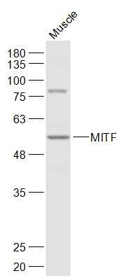 , ,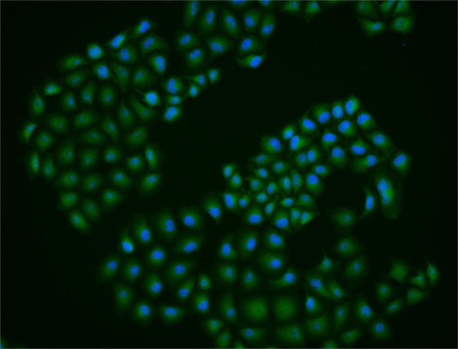 |
| Application | |
| Recommended Dose | WB: 1:500-2000; ICC/IF: 1:50-200 |
| Antibody Type | Polyclonal |
| Host Species | Rabbit |
| Subcellular Localization | Nucleus. |
| Tissue Specificity | Isoform M is exclusively expressed in melanocytes and melanoma cells. Isoform A and isoform H are widely expressed in many cell types including melanocytes and retinal pigment epithelium (RPE). Isoform C is expressed in many cell types including RPE but n |
| Construction | Polyclonal Antibody |
| Purification | Protein A purified |
| Appearance | Liquid |
| Formulation | 0.01M TBS (pH7.4) with 1% BSA, 0.02% Proclin300 and 50% Glycerol. |
| Concentration | 1 mg/mL |
| Research Background | The protein encoded by this gene is a transcription factor that contains both basic helix-loop-helix and leucine zipper structural features. The encoded protein regulates melanocyte development and is responsible for pigment cell-specific transcription of the melanogenesis enzyme genes. Heterozygous mutations in the this gene cause auditory-pigmentary syndromes, such as Waardenburg syndrome type 2 and Tietz syndrome. [provided by RefSeq, Aug 2017] |
| Immunogen | KLH conjugated synthetic peptide: human MITF |
| Antigen Species | Human |
| Gene Name | MITF |
| Gene ID | |
| Protein Name | Microphthalmia-associated transcription factor |
| Uniprot ID | |
| Biology Area | HLH / Leucine Zipper,HLH,HLH / Leucine Zipper,Mast Cells |
| Function | Transcription factor for tyrosinase (TYR) and tyrosinase-related protein 1 (TYRP1) that plays a key role in melanocyte development. Binds to a symmetrical DNA sequence (E-boxes) (5'-CACGTG-3') found in the tyrosinase promoter. Plays a critical role in the differentiation of various cell types as neural crest-derived melanocytes, mast cells, osteoclasts and optic cup-derived retinal pigment epithelium. |
| Molecular Weight | Theoretical: 59 kDa. Actual: 57 kDa. |
| Stability & Storage | Store at -20°C or -80°C for 12 months. Avoid repeated freeze-thaw cycles. |
| Transport | Shipping with blue ice. |
| Size | Quantity | Unit Price | Amount | Operation |
|---|
 Hello! How can I help you today?
 Copyright © 2015-2026 TargetMol Chemicals Inc. All Rights Reserved.
