
Shopping Cart
Remove All Your shopping cart is currently empty

Synonyms:
Anti-MAPK3 Antibody
(1K596)
| Pack Size | Price | USA Stock | Global Stock | Quantity |
|---|---|---|---|---|
| 50 µL | $297 | 7-10 days | 7-10 days | |
| 100 µL | $497 | 7-10 days | 7-10 days |
| Description | Anti-MAPK3 Antibody (1K596) is a Rabbit antibody targeting MAPK3. Anti-MAPK3 Antibody (1K596) can be used in FCM,ICC/IF,IHC,IP,WB. |
| Ig Type | IgG |
| Clone | 1K596 |
| Reactivity | Human,Mouse |
| Verified Activity | 1. Western blot analysis of ERK1 on different lysates using anti-ERK1 antibody at 1/1,000 dilution. Positive control: Lane 1: Hela, Lane 2: Jurkat, Lane 3: K562. 2. Immunohistochemical analysis of paraffin-embedded human breast tissue using anti-ERK1 antibody. Counter stained with hematoxylin. 3. Immunohistochemical analysis of paraffin-embedded mouse stomach tissue using anti-ERK1 antibody. Counter stained with hematoxylin. 4. Immunohistochemical analysis of paraffin-embedded human colon cancer tissue using anti-ERK1 antibody. Counter stained with hematoxylin. 5. ICC staining ERK1 in Hela cells (green). The nuclear counter stain is DAPI (blue). Cells were fixed in paraformaldehyde, permeabilised with 0.25% Triton X100/PBS. 6. ICC staining ERK1 in MCF-7 cells (green). The nuclear counter stain is DAPI (blue). Cells were fixed in paraformaldehyde, permeabilised with 0.25% Triton X100/PBS. 7. ICC staining ERK1 in NIH/3T3 cells (green). The nuclear counter stain is DAPI (blue). Cells were fixed in paraformaldehyde, permeabilised with 0.25% Triton X100/PBS. 8. Flow cytometric analysis of Jurkat cells with ERK1 antibody at 1/50 dilution (red) compared with an unlabelled control (cells without incubation with primary antibody; black). Alexa Fluor 488-conjugated goat anti rabbit IgG was used as the secondary antibody. 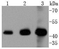 , , , ,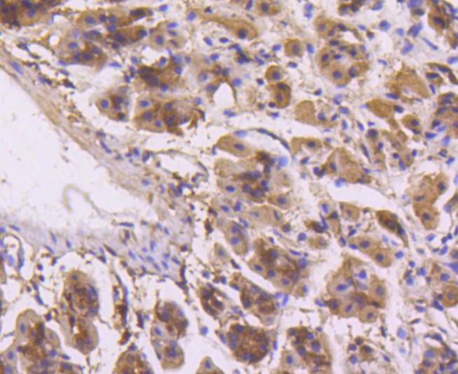 , ,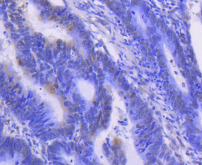 , ,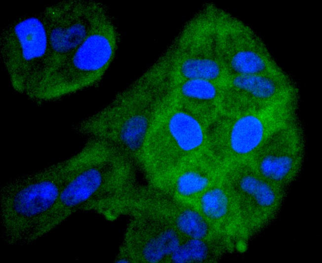 , ,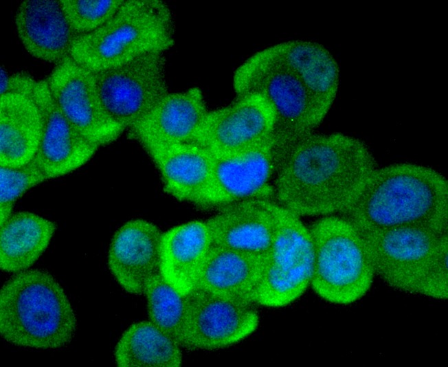 , ,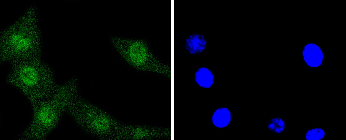 , ,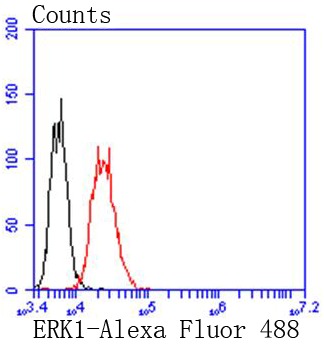 |
| Application | |
| Recommended Dose | WB: 1:1000-5000; IHC: 1:50-200; ICC/IF: 1:100-500; FCM: 1:50-100 |
| Antibody Type | Monoclonal |
| Host Species | Rabbit |
| Construction | Recombinant Antibody |
| Purification | ProA affinity purified |
| Appearance | Liquid |
| Formulation | 1*TBS (pH7.4), 1%BSA, 40%Glycerol. Preservative: 0.05% Sodium Azide. |
| Research Background | Mitogen-activated protein kinase (MAPK) signaling pathways involve two closely related MAP kinases, known as extracellular-signal-related kinase 1 (ERK 1, p44) and 2 (ERK 2, p42). Growth factors, steroid hormones, G protein-coupled receptor ligands and neurotransmitters can initiate MAPK signaling pathways. Activation of ERK 1 and ERK 2 requires phosphorylation by upstream kinases such as MAP kinasekinase (MEK), MEK kinase and Raf-1. ERK 1 and ERK 2 phosphorylation can occur at specific tyrosine and threonine sites mapping within consensus motifs that include the threonine-glutamate-tyrosine motif. ERK activation leads to dimerization with other ERKs and subsequent localization to the nucleus. Active ERK dimers phosphorylate serine and threonine residues on nuclear proteins and influence a host of responses that include proliferation, differentiation, transcription regulation and development. The human ERK 1 gene maps to chromosome 16p12-p11.2 and encodes a 379 amino acid protein that shares 83% sequence identity to ERK 2. |
| Conjucates | Unconjugated |
| Immunogen | Recombinant Protein |
| Uniprot ID |
| Molecular Weight | Theoretical: 43 kDa. |
| Stability & Storage | Store at -20°C or -80°C for 12 months. Avoid repeated freeze-thaw cycles. |
| Transport | Shipping with blue ice. |
| Size | Quantity | Unit Price | Amount | Operation |
|---|
 Hello! How can I help you today?
 Copyright © 2015-2026 TargetMol Chemicals Inc. All Rights Reserved.
