
Shopping Cart
Remove All Your shopping cart is currently empty

Synonyms: LIMK-2, LIMK2, LIMK-1, LIMK, LIM motif-containing protein kinase, LIM domain kinase 2, LIM domain kinase 1, EC 2.7.11.1
Anti-LIMK1 Polyclonal Antibody
| Pack Size | Price | USA Stock | Global Stock | Quantity |
|---|---|---|---|---|
| 50 µL | $222 | 7-10 days | 7-10 days | |
| 100 µL | $374 | 7-10 days | 7-10 days | |
| 200 µL | $528 | 7-10 days | 7-10 days |
| Description | Anti-LIMK1 Polyclonal Antibody is a Rabbit antibody targeting LIMK1. Anti-LIMK1 Polyclonal Antibody can be used in FCM,IF,IHC-Fr,IHC-P,WB. |
| Synonyms | LIMK-2, LIMK2, LIMK-1, LIMK, LIM motif-containing protein kinase, LIM domain kinase 2, LIM domain kinase 1, EC 2.7.11.1 |
| Ig Type | IgG |
| Reactivity | Human,Mouse,Rat (predicted:Cow,Rabbit) |
| Verified Activity | 1. Tissue/cell: rat brain tissue; 4% Paraformaldehyde-fixed and paraffin-embedded; Antigen retrieval: citrate buffer (0.01M, pH6.0), Boiling bathing for 15 min; Block endogenous peroxidase by 3% Hydrogen peroxide for 30 min; Blocking buffer (normal goat serum) at 37°C for 20 min; Incubation: Anti-LIMK1 Polyclonal Antibody, Unconjugated (TMAB-01070) 1:200, overnight at 4°C, followed by conjugation to the secondary antibody and DAb staining. 2. Paraformaldehyde-fixed, paraffin embedded (mouse brain); Antigen retrieval by boiling in sodium citrate buffer (pH6.0) for 15 min; Block endogenous peroxidase by 3% hydrogen peroxide for 20 min; Blocking buffer (normal goat serum) at 37°C for 30 min; Antibody incubation with (LIMK1) Polyclonal Antibody, Unconjugated (TMAB-01070) at 1:200 overnight at 4°C, followed by operating according to SP Kit (Rabbit) instructionsand DAB staining. 3. Paraformaldehyde-fixed, paraffin embedded (rat brain); Antigen retrieval by boiling in sodium citrate buffer (pH6.0) for 15 min; Block endogenous peroxidase by 3% hydrogen peroxide for 20 min; Blocking buffer (normal goat serum) at 37°C for 30 min; Antibody incubation with (LIMK1) Polyclonal Antibody, Unconjugated (TMAB-01070) at 1:200 overnight at 4°C, followed by operating according to SP Kit (Rabbit) instructionsand DAB staining. 4. Paraformaldehyde-fixed, paraffin embedded (Mouse brain); Antigen retrieval by boiling in sodium citrate buffer (pH6.0) for 15 min; Block endogenous peroxidase by 3% hydrogen peroxide for 20 min; Blocking buffer (normal goat serum) at 37°C for 30 min; Antibody incubation with (LIMK1) Polyclonal Antibody, Unconjugated (TMAB-01070) at 1:400 overnight at 4°C, followed by a conjugated secondary antibody for 20 min and DAB staining. 5. Paraformaldehyde-fixed, paraffin embedded (Rat brain); Antigen retrieval by boiling in sodium citrate buffer (pH6.0) for 15 min; Block endogenous peroxidase by 3% hydrogen peroxide for 20 min; Blocking buffer (normal goat serum) at 37°C for 30 min; Antibody incubation with (LIMK1) Polyclonal Antibody, Unconjugated (TMAB-01070) at 1:400 overnight at 4°C, followed by a conjugated secondary antibody for 20 min and DAB staining. 6. Sample: Spinal cord (Mouse) Lysate at 40 μg Primary: Anti-LIMK1 (TMAB-01070) at 1/1000 dilution Secondary: IRDye800CW Goat Anti-Rabbit IgG at 1/20000 dilution Predicted band size: 71 kDa Observed band size: 75 kDa 7. Blank control (black line): Hela. Primary Antibody (green line): Rabbit Anti-LIMK1 antibody (TMAB-01070) Dilution: 1 μg/Test; Secondary Antibody (white blue line): Goat anti-rabbit IgG-FITC Dilution: 0.5 μg/Test. Isotype control (orange line): Normal Rabbit IgG Protocol The cells were fixed with 4% PFA (10 min at room temperature) and then permeabilized with 90% ice-cold methanol for 20 min at-20°C, The cells were then incubated in 5% BSA to block non-specific protein-protein interactions for 30 min at room temperature. Cells stained with Primary Antibody for 30 min at room temperature. The secondary antibody used for 40 min at room temperature. 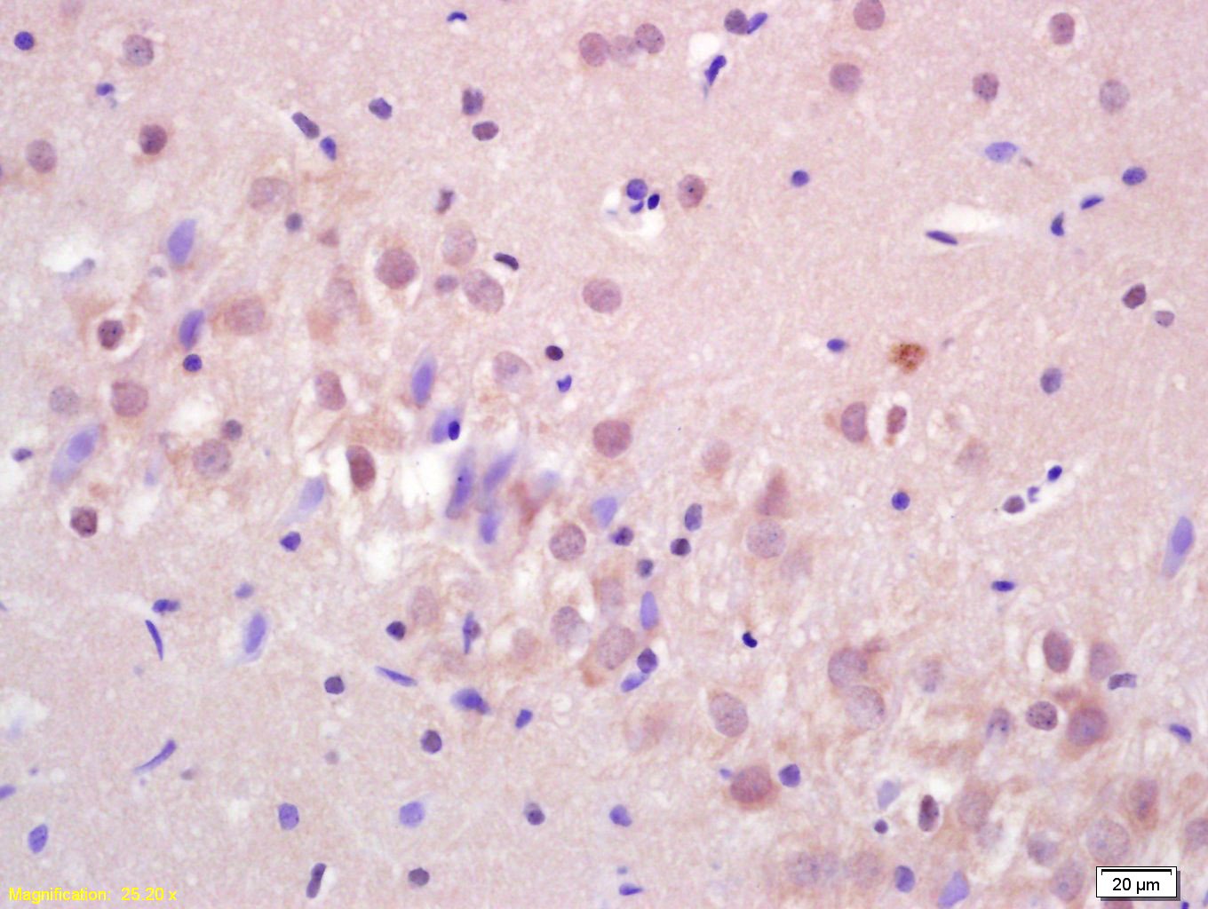 , ,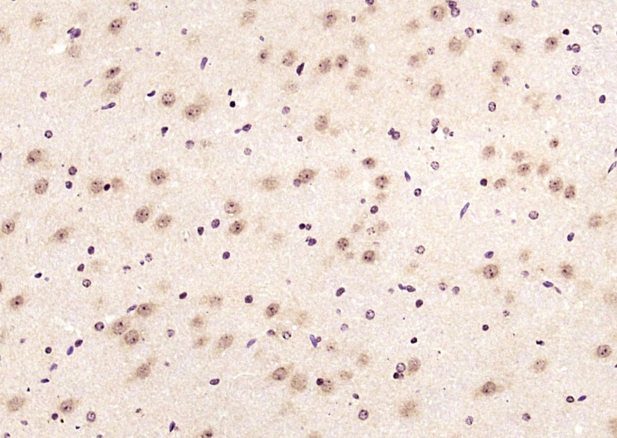 , ,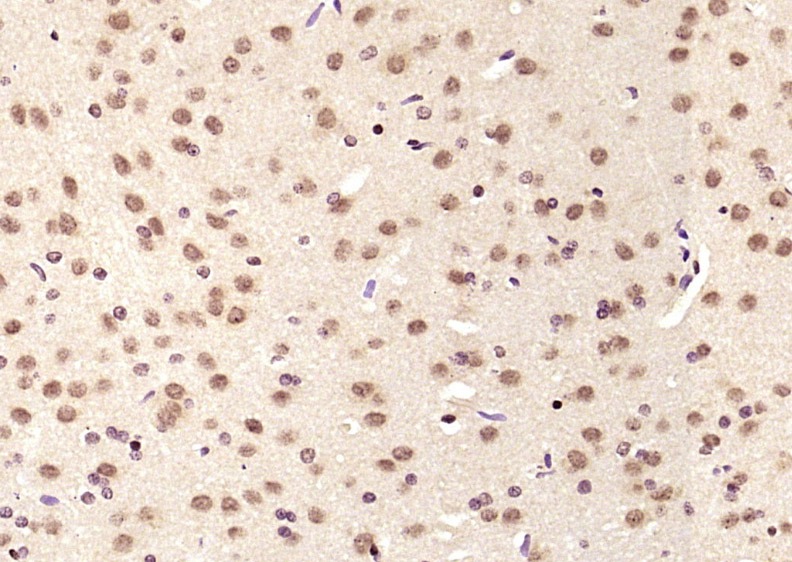 , ,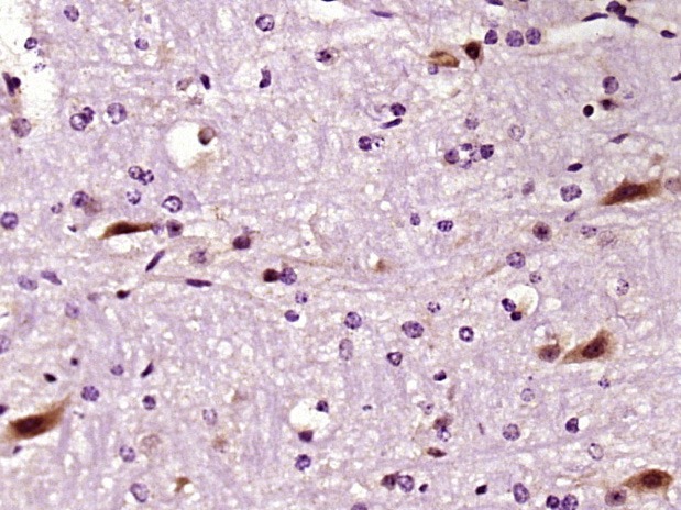 , ,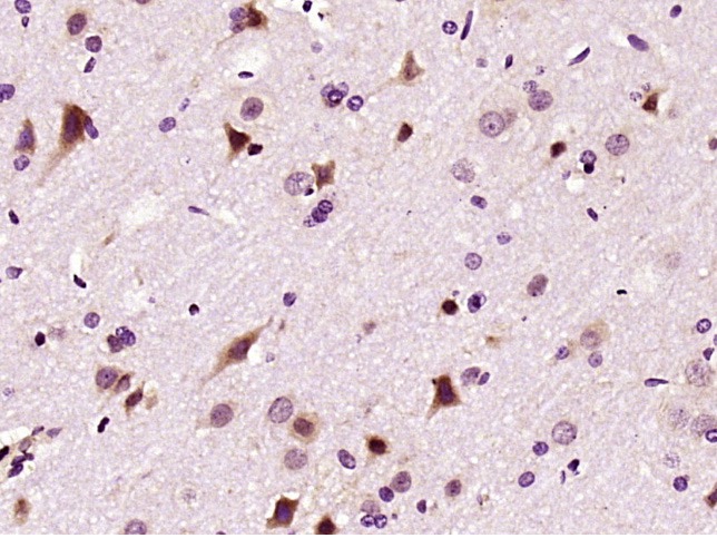 , ,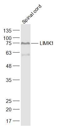 , ,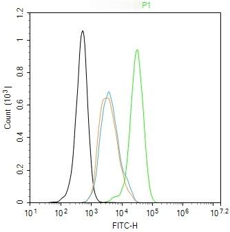 |
| Application | |
| Recommended Dose | WB: 1:500-2000; IHC-P: 1:20-100; IHC-Fr: 1:20-100; IF: 1:20-100; FCM: 1ug/Test |
| Antibody Type | Polyclonal |
| Host Species | Rabbit |
| Subcellular Localization | Cytoplasm. Nucleus. Note=Predominantly found in the cytoplasm. |
| Tissue Specificity | Highest expression in both adult and fetal nervous system. Detected ubiquitously throughout the different regions of adult brain, with highest levels in the cerebral cortex. Expressed to a lesser extent in heart and skeletal muscle. |
| Construction | Polyclonal Antibody |
| Purification | Protein A purified |
| Appearance | Liquid |
| Formulation | 0.01M TBS (pH7.4) with 1% BSA, 0.02% Proclin300 and 50% Glycerol. |
| Concentration | 1 mg/mL |
| Research Background | LIMK1 is a protein kinase which regulates actin filament dynamics. Phosphorylates and inactivates the actin binding/depolymerizing factor cofilin, thereby stabilizing the actin cytoskeleton. LIMK1 may be involved in brain development; it is highly expressed in both adult and fetal nervous system. Detected ubiquitously throughout the different regions of adult brain, with highest levels in the cerebral cortex. Expressed to a lesser extent in heart and skeletal muscle. |
| Immunogen | KLH conjugated synthetic peptide: human LIMK1 |
| Antigen Species | Human |
| Gene Name | LIMK1 |
| Gene ID | |
| Protein Name | LIM domain kinase 2 |
| Uniprot ID | |
| Biology Area | Signal transduction,LIM,Neural Signal Transduction |
| Function | Serine/threonine-protein kinase that plays an essential role in the regulation of actin filament dynamics. Acts downstream of several Rho family GTPase signal transduction pathways. Activated by upstream kinases including ROCK1, PAK1 and PAK4, which phosphorylate LIMK1 on a threonine residue located in its activation loop. LIMK1 subsequently phosphorylates and inactivates the actin binding/depolymerizing factors cofilin-1/CFL1, cofilin-2/CFL2 and destrin/DSTN, thereby preventing the cleavage of filamentous actin (F-actin), and stabilizing the actin cytoskeleton. In this way LIMK1 regulates several actin-dependent biological processes including cell motility, cell cycle progression, and differentiation. Phosphorylates TPPP on serine residues, thereby promoting microtubule disassembly. Stimulates axonal outgrowth and may be involved in brain development. Isoform 3 has a dominant negative effect on actin cytoskeletal changes. |
| Molecular Weight | Theoretical: 71 kDa. Actual: 75 kDa. |
| Stability & Storage | Store at -20°C or -80°C for 12 months. Avoid repeated freeze-thaw cycles. |
| Transport | Shipping with blue ice. |
| Size | Quantity | Unit Price | Amount | Operation |
|---|
 Hello! How can I help you today?
 Copyright © 2015-2026 TargetMol Chemicals Inc. All Rights Reserved.
