
Shopping Cart
Remove All Your shopping cart is currently empty

Synonyms: selectin L, Ly-m22, Lyam-1, Lyam1, Ly-22, L-selectin, Lnhr, LECAM-1, CD62L, AI528707
Anti-L-selectin Polyclonal Antibody
| Pack Size | Price | USA Stock | Global Stock | Quantity |
|---|---|---|---|---|
| 50 µL | $221 | 7-10 days | 7-10 days | |
| 100 µL | $374 | 7-10 days | 7-10 days | |
| 200 µL | $528 | 7-10 days | 7-10 days |
| Description | Anti-L-selectin Polyclonal Antibody is a Rabbit antibody targeting L-selectin. Anti-L-selectin Polyclonal Antibody can be used in FCM,IHC-Fr,WB. |
| Synonyms | selectin L, Ly-m22, Lyam-1, Lyam1, Ly-22, L-selectin, Lnhr, LECAM-1, CD62L, AI528707 |
| Ig Type | IgG |
| Reactivity | Mouse,Rat (predicted:Cow,Rabbit) |
| Verified Activity | 1. Blank control: mouse spleen cells (blue). Primary Antibody: Rabbit Anti-CD62L antibody (TMAB-01084), Dilution: 1 μg in 100 μL 1X PBS containing 0.5% BSA; Isotype Control Antibody: Rabbit Igg (orange),used under the same conditions); Secondary Antibody: Goat anti-rabbit IgG-Pe (white blue), Dilution: 1:200 in 1 X PBS containing 0.5% BSA. Protocol The cells were fixed with 2% paraformaldehyde (10 min). Primary antibody (TMAB-01084, 1 μg/1x10^6 cells) were incubated for 30 min on the ice, followed by 1 X PBS containing 0.5% BSA + 10% goat serum (15 min) to block non-specific protein-protein interactions. Then the Goat Anti-rabbit IgG/PE antibody was added into the blocking buffer mentioned above to react with the primary antibody at 1/200 dilution for 30 min on ice. 2. Sample: LympHnode (Mouse) Lysate at 40 μg LympHnode (Rat) Lysate at 40 μg Primary: Anti-CD62L (TMAB-01084) at 1/300 dilution Secondary: IRDye800CW Goat Anti-Rabbit IgG at 1/20000 dilution Predicted band size: 37 kDa Observed band size: 43 kDa 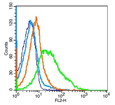 , , |
| Application | |
| Recommended Dose | WB: 1:500-2000; IHC-Fr: 1:100-500; FCM: 1μg/Test |
| Antibody Type | Polyclonal |
| Host Species | Rabbit |
| Subcellular Localization | Membrane; Single-pass type I membrane protein. |
| Tissue Specificity | Expressed in B-cell lines and T-lymphocytes. |
| Construction | Polyclonal Antibody |
| Purification | Protein A purified |
| Appearance | Liquid |
| Formulation | 0.01M TBS (pH7.4) with 1% BSA, 0.02% Proclin300 and 50% Glycerol. |
| Concentration | 1 mg/mL |
| Research Background | This gene encodes a cell surface adhesion molecule that belongs to a family of adhesion/homing receptors. The encoded protein contains a C-type lectin-like domain, a calcium-binding epidermal growth factor-like domain, and two short complement-like repeats. The gene product is required for binding and subsequent rolling of leucocytes on endothelial cells, facilitating their migration into secondary lymphoid organs and inflammation sites. Single-nucleotide polymorphisms in this gene have been associated with various diseases including immunoglobulin A nephropathy. Alternatively spliced transcript variants have been found for this gene. [provided by RefSeq, Oct 2009]. |
| Immunogen | KLH conjugated synthetic peptide: mouse CD62L |
| Antigen Species | Mouse |
| Gene Name | SELL |
| Gene ID | |
| Protein Name | L-selectin |
| Uniprot ID | |
| Biology Area | Blood,Regulatory T Cells,CD,T Lymphocytic Lineage,Neutrophil Lineage |
| Function | Cell surface adhesion protein. Mediates the adherence of lymphocytes to endothelial cells of high endothelial venules in peripheral lymph nodes. Promotes initial tethering and rolling of leukocytes in endothelia. |
| Molecular Weight | Theoretical: 37 kDa. Actual: 43 kDa. |
| Stability & Storage | Store at -20°C or -80°C for 12 months. Avoid repeated freeze-thaw cycles. |
| Transport | Shipping with blue ice. |
| Size | Quantity | Unit Price | Amount | Operation |
|---|
 Hello! How can I help you today?
 Copyright © 2015-2026 TargetMol Chemicals Inc. All Rights Reserved.
