
Shopping Cart
Remove All Your shopping cart is currently empty

Synonyms: Tyrosine-protein kinase JAK3, L-JAK, Leukocyte janus kinase, Janus kinase 3, JAK-3, EC 2.7.10.2
Anti-JAK3 Polyclonal Antibody
| Pack Size | Price | USA Stock | Global Stock | Quantity |
|---|---|---|---|---|
| 50 µL | $220 | 7-10 days | 7-10 days | |
| 100 µL | $372 | 7-10 days | 7-10 days | |
| 200 µL | $528 | 7-10 days | 7-10 days |
| Description | Anti-JAK3 Polyclonal Antibody is a Rabbit antibody targeting JAK3. Anti-JAK3 Polyclonal Antibody can be used in FCM,IF,IHC-Fr,IHC-P,WB. |
| Synonyms | Tyrosine-protein kinase JAK3, L-JAK, Leukocyte janus kinase, Janus kinase 3, JAK-3, EC 2.7.10.2 |
| Ig Type | IgG |
| Reactivity | Human,Mouse,Rat (predicted:Dog,Pig,Cow,Horse) |
| Verified Activity | 1. Tissue/cell: human lung carcinoma; 4% Paraformaldehyde-fixed and paraffin-embedded; Antigen retrieval: citrate buffer (0.01M, pH6.0), Boiling bathing for 15 min; Block endogenous peroxidase by 3% Hydrogen peroxide for 30 min; Blocking buffer (normal goat serum) at 37°C for 20 min; Incubation: Anti-Jak3 Polyclonal Antibody, Unconjugated (TMAB-01005) 1:200, overnight at 4°C, followed by conjugation to the secondary antibody and DAb staining. 2. Sample: SP2/0 Cell (Mouse) Lysate at 40 μg Jurkat Cell (Human) Lysate at 40 μg K562 Cell (Human) Lysate at 40 μg Primary: Anti-Jak3 (TMAB-01005) at 1/300 dilution Secondary: IRDye800CW Goat Anti-Rabbit IgG at 1/20000 dilution Predicted band size: 125 kDa Observed band size: 125 kDa 3. Blank control: Molt-4. Primary Antibody (green line): Rabbit Anti-Jak3 antibody (TMAB-01005) Dilution: 0.2 μg/10^6 cells; Isotype Control Antibody (orange line): Rabbit IgG. Secondary Antibody: Goat anti-rabbit IgG-PE Dilution: 0.2 μg/test. Protocol The cells were fixed with 4% PFA (10 min at room temperature) and then permeabilized with 20% PBST for 20 min at-20°C. The cells were then incubated in 5% BSA to block non-specific protein-protein interactions for 30 min at at room temperature. Cells stained with Primary Antibody for 30 min at room temperature. The secondary antibody used for 40 min at room temperature. 4. Sample: Lane 1: Mouse Thymus tissue lysates Lane 2: Rat LympHnode tissue lysates Lane 3: Human Jurkat cell lysates Lane 4: Human Hela cell lysates Lane 5: Human Molt-4 cell lysates Primary: Anti-Jak3 (TMAB-01005) at 1/1000 dilution Secondary: IRDye800CW Goat Anti-Rabbit IgG at 1/20000 dilution Predicted band size: 125 kDa Observed band size: 115 kDa 5. Blank control (black line): Molt4. Primary Antibody (green line): Rabbit Anti-Jak3 antibody (TMAB-01005) Dilution: 1 μg/Test; Secondary Antibody (white blue line): Goat anti-rabbit IgG-AF488 Dilution: 0.5 μg/Test. Isotype control (orange line): Normal Rabbit IgG Protocol The cells were fixed with 4% PFA (10 min at room temperature) and then permeabilized with 90% ice-cold methanol for 20 min at-20°C, The cells were then incubated in 5% BSA to block non-specific protein-protein interactions for 30 min at room temperature. Cells stained with Primary Antibody for 30 min at room temperature. The secondary antibody used for 40 min at room temperature. 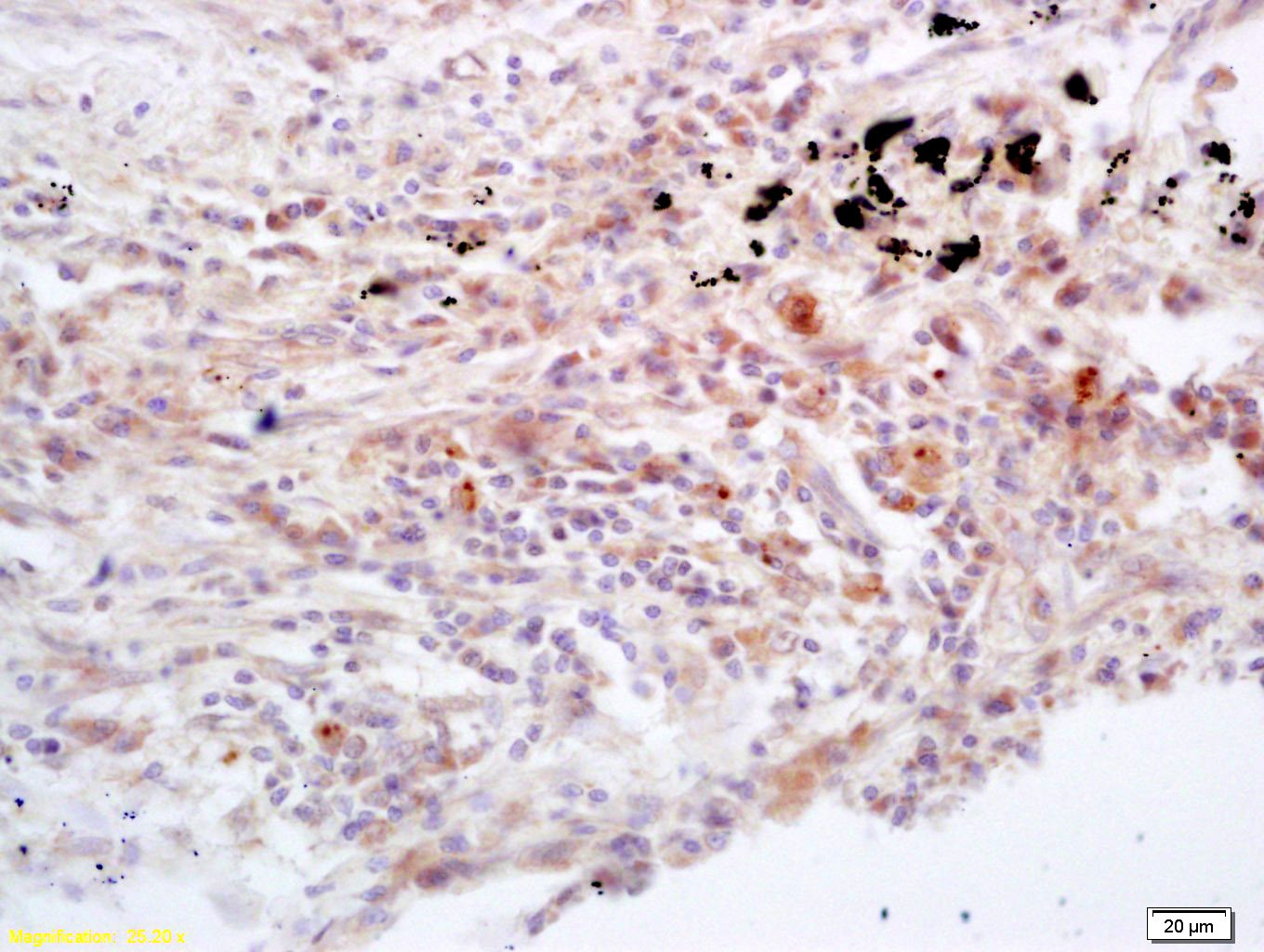 , ,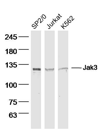 , ,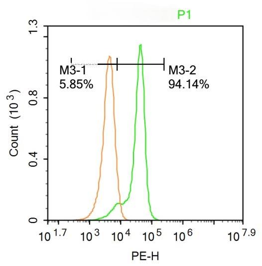 , ,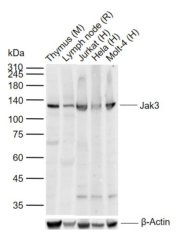 , ,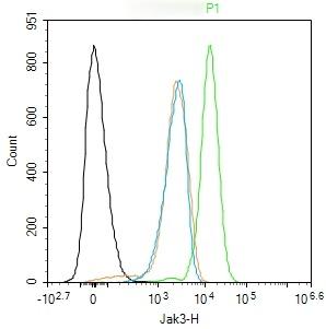 |
| Application | |
| Recommended Dose | WB: 1:500-2000; IHC-P: 1:100-500; IHC-Fr: 1:100-500; IF: 1:100-500; FCM: 1ug/test |
| Antibody Type | Polyclonal |
| Host Species | Rabbit |
| Subcellular Localization | Endomembrane system; Peripheral membrane protein. Cytoplasm. |
| Tissue Specificity | In NK cells and an NK-like cell line but not in resting T-cells or in other tissues. The S-form is more commonly seen in hematopoietic lines, whereas the B-form is detected in cells both of hematopoietic and epithelial origins. |
| Construction | Polyclonal Antibody |
| Purification | Protein A purified |
| Appearance | Liquid |
| Formulation | 0.01M TBS (pH7.4) with 1% BSA, 0.02% Proclin300 and 50% Glycerol. |
| Concentration | 1 mg/mL |
| Research Background | The protein encoded by this gene is a member of the Janus kinase (JAK) family of tyrosine kinases involved in cytokine receptor-mediated intracellular signal transduction. It is predominantly expressed in immune cells and transduces a signal in response to its activation via tyrosine phosphorylation by interleukin receptors. Mutations in this gene are associated with autosomal SCID (severe combined immunodeficiency disease). [provided by RefSeq, Jul 2008] |
| Immunogen | KLH conjugated synthetic peptide: human Jak3 |
| Antigen Species | Human |
| Gene Name | JAK3 |
| Gene ID | |
| Protein Name | Tyrosine-protein kinase JAK3 |
| Uniprot ID | |
| Biology Area | Signal transducers,Tyrosine kinases,STATs,Transcription Factors,Tyrosine Kinases,STATs |
| Function | Non-receptor tyrosine kinase involved in various processes such as cell growth, development, or differentiation. Mediates essential signaling events in both innate and adaptive immunity and plays a crucial role in hematopoiesis during T-cells development. In the cytoplasm, plays a pivotal role in signal transduction via its association with type I receptors sharing the common subunit gamma such as IL2R, IL4R, IL7R, IL9R, IL15R and IL21R. Following ligand binding to cell surface receptors, phosphorylates specific tyrosine residues on the cytoplasmic tails of the receptor, creating docking sites for STATs proteins. Subsequently, phosphorylates the STATs proteins once they are recruited to the receptor. Phosphorylated STATs then form homodimer or heterodimers and translocate to the nucleus to activate gene transcription. For example, upon IL2R activation by IL2, JAK1 and JAK3 molecules bind to IL2R beta (IL2RB) and gamma chain (IL2RG) subunits inducing the tyrosine phosphorylation of both receptor subunits on their cytoplasmic domain. Then, STAT5A AND STAT5B are recruited, phosphorylated and activated by JAK1 and JAK3. Once activated, dimerized STAT5 translocates to the nucleus and promotes the transcription of specific target genes in a cytokine-specific fashion. |
| Molecular Weight | Theoretical: 125 kDa. Actual: 125 kDa. |
| Stability & Storage | Store at -20°C or -80°C for 12 months. Avoid repeated freeze-thaw cycles. |
| Transport | Shipping with blue ice. |
| Size | Quantity | Unit Price | Amount | Operation |
|---|
 Hello! How can I help you today?
 Copyright © 2015-2026 TargetMol Chemicals Inc. All Rights Reserved.
